Journal of Medical Microbiology (2006), 55, 1229–1235
DOI 10.1099/jmm.0.46552-0
Inter-laboratory comparison of three different real-time PCR assays for the detection of Pneumocystis jiroveci in bronchoalveolar lavage fluid samples Catharina F. M. Linssen,1 Jan A. Jacobs,1 Pieter Beckers,2 Kate E. Templeton,3 Judith Bakkers,2 Ed J. Kuijper,3 Willem J. G. Melchers,2 Marjolein Drent4 and Cornelis Vink1 1
Department of Medical Microbiology, Maastricht Infection Center (MINC), University Hospital Maastricht, PO Box 5800, 6202 AZ Maastricht, The Netherlands
Correspondence Catharina F. M. Linssen
2
[email protected]
Department of Medical Microbiology, Radboud University Nijmegen Medical Centre (RUNMC), PO Box 9101, 6500 HB Nijmegen, The Netherlands
3
Department of Medical Microbiology, Leiden University Medical Center (LUMC), PO Box 9600, 2300 RC Leiden, The Netherlands
4
Department of Respiratory Medicine, University Hospital Maastricht, PO Box 5800, 6202 AZ Maastricht, The Netherlands
Received 3 February 2006 Accepted 15 June 2006
Pneumocystis jiroveci pneumonia (PCP) is an opportunistic infection affecting immunocompromised patients. While conventional diagnosis of PCP by microscopy is cumbersome, the use of PCR to diagnose PCP has great potential. Nevertheless, inter-laboratory validation and standardization of PCR assays is lacking. The aim of this study was to evaluate the inter-laboratory agreement of three independently developed real-time PCR assays for the detection of P. jiroveci in bronchoalveolar lavage fluid samples. Therefore, 124 samples were collected in three tertiary care laboratories (Leiden University Medical Center, Maastricht Infection Center and Radboud University Nijmegen Medical Centre) and were tested by both microscopy and real-time PCR. Of 41 samples positive for P. jiroveci by microscopy, 40 were positive in all three PCR assays. The remaining sample was positive in a single assay only. Out of 83 microscopy-negative samples, 69 were negative in all three PCR assays. The other 14 samples were found positive, either in all three assays (n=5), in two (n=2) or in one of the assays (n=7). The data demonstrate high inter-laboratory agreement among real-time PCR assays for the detection of P. jiroveci.
INTRODUCTION Pneumocystis carinii is an opportunistic pathogen that was classified as a fungus in 1988 (Edman et al., 1988). Although human-derived P. carinii (P. carinii f. sp. hominis) has recently been renamed Pneumocystis jiroveci, the abbreviation PCP (now referring to Pneumocystis pneumonia) remains in use (Stringer et al., 2001). Patients at risk for P. jiroveci pneumonia can be divided into two categories: HIV positive and HIV negative. Several risk factors have been identified in the HIV-negative group, such as immunosuppressive medication, or an inherited or acquired immunodeficiency (Kovacs et al., 2001). Since untreated PCP is Abbreviations: BAL, bronchoalveolar lavage; DHPS, dihydroperoate synthase; LUMC, Leiden University Medical Center; MINC, Maastricht Infection Center; MSG, major surface glycoprotein; PCP, Pneumocystis jiroveci pneumonia; RUNMC, Radboud University Nijmegen Medical Centre.
46552 G 2006 SGM
Printed in Great Britain
associated with a high morbidity and mortality (Sepkowitz, 2002; Yale & Limper, 1996), especially in HIV-negative patients, a rapid and reliable diagnosis is mandatory. Current diagnosis of PCP relies on tinctorial and/or immunofluorescent staining of induced sputum or bronchoalveolar lavage (BAL) fluid samples (Djamin et al., 1998; Thomas & Limper, 2004). Using these methods, sensitivity and specificity rates are reached which exceed 95 % (Amin et al., 1992; Chandra et al., 1988; Elvin et al., 1988; Kovacs et al., 2001). Major drawbacks of microscopy, however, are that it is cumbersome and requires trained microscopists. The latter makes it essential that positive samples are encountered regularly in order to maintain an expertise in microscopy. In recent years, the incidence of PCP has declined significantly, especially in HIV-positive patients. Due to the introduction of highly active anti-retroviral therapy, PCP chemoprophylaxis, and an increase of patients receiving chemotherapy, the future trend will be towards 1229
C. F. M. Linssen and others
samples with relatively low P. jiroveci burdens, making the diagnosis of PCP even more difficult (Limper et al., 1989; Miller, 1999). Therefore, a rapid diagnostic technique which can identify the presence of a low number of cysts is needed. Nucleic acid amplification tests, such as PCR, play an increasing role in the detection of P. jiroveci (Larsen et al., 2002b; Wakefield et al., 1990). In particular, real-time PCR is highly suitable for the diagnosis of PCP, since this technique allows the generation of quantitative results. This is crucial, as P. jiroveci may be present in low quantities in some asymptomatic individuals (Maskell et al., 2003; Nevez et al., 2001). Consequently, it is of utmost importance to be able to discriminate between asymptomatic carriership and clinically relevant infection. In recent years, molecular tests to detect P. jiroveci have shifted from research to diagnostic applications (Flori et al., 2004; Larsen et al., 2004; Olsson et al., 2001). Nevertheless, quality control panels for validation and standardization of such tests are currently lacking. Therefore, a study was initiated to compare the performance of three independently developed real-time PCR assays for the detection of P. jiroveci in three different tertiary care centres in The Netherlands. This retrospective study assessed the routine diagnostic performance of these assays on a collection of BAL fluid samples of which the microscopic evaluation for the presence of P. jiroveci had already been performed.
METHODS Design of the study. Three diagnostic medical microbiology
laboratories participated in this study, i.e. Maastricht Infection Center (MINC), Radboud University Nijmegen Medical Center (RUNMC) and Leiden University Medical Center (LUMC). All three laboratories are university tertiary care centres located in the Netherlands. Each of these laboratories supplied P. jiroveci-positive and -negative samples. Diagnosis was based on microscopy. The materials were collected by one of the authors (C. F. M. Linssen),
encoded, and distributed to the other laboratories. Each laboratory received a set of samples and performed its own real-time PCR in a blinded fashion. BAL fluid sampling and conventional diagnosis of PCP.
Samples obtained from patients suspected of PCP were collected over the period August 1999 to April 2004. Bronchoscopy with BAL was performed with sterile saline, in four aliquots of 50 ml (MINC), three aliquots of 20 ml (LUMC) and three aliquots of 50 ml (RUNMC). Samples were immediately transported to the laboratory and processed upon arrival. At the LUMC and RUNMC, aliquots of 10 ml were centrifuged at 3000 g and smears were made from the sediment. At the MINC, cytocentrifuged monolayer preparations were made as previously described (Jacobs et al., 2001). Subsequently, slides were subjected to Giemsa and methenamine silver staining (LUMC), Giemsa and direct immunofluorescence staining (PneumoCel, CeLLabs) (RUNMC), or May–Gru¨nwald Giemsa and methenamine silver staining (MINC). Nucleic acid extraction and real-time PCR analysis. At the MINC, 200 ml BAL fluid was used for DNA isolation using the
Wizard Genomic DNA Purification kit (Promega). Purified DNA was resuspended in a final volume of 120 ml. A real-time PCR was designed that targeted the major surface glycoprotein (MSG) gene (Larsen et al., 2002a). Assays were performed in 96-well Optical Reaction plates [Applied Biosystems (ABI)] in a 50 ml volume containing 0?6 mM each of primers PCPFor and PCPRev (Table 1), 0?15 mM probe PCPProbe, 16 TaqMan Universal Master Mix (ABI) and 20 ml purified DNA. Thermal cycling was carried out on an ABI PRISM 7000 Sequence Detection system (ABI) as follows: 2 min at 50 uC, 10 min at 95 uC, followed by 42 cycles of 15 s at 95 uC and 1 min at 60 uC. Quantification was performed using the ABI PRISM software and was based on extrapolation of data to standard curves, which were generated by amplification of quantified dilutions of the plasmid pPCP, which contains the P. jiroveci PCR fragment. At the RUNMC, the MagnaPure LC Isolation station (Roche Applied Science) was used for nucleic acid isolation. A 100 ml aliquot was isolated using the Total Nucleic Acid isolation kit (Roche Applied Science). Nucleic acids were resuspended in 50 ml H2O. Real-time PCR for P. jiroveci was performed as described by Larsen et al. (2002a) using the MSG gene as target. All reactions were performed in a volume of 20 ml, consisting of 16 LightCycler FastStart DNA Master
Table 1. Description of primers and probes used by the three laboratories The sequences for primers and probes used by MINC and RUNMC were obtained from Larsen et al. (2002). Laboratory
Target
Primer type*
Sequence (5§–3§) (name)
GenBank accession no.
Position on gene
MINC
MSG
DHPS
RUNMC
MSG
CAAAAATAACAYTSACATCAACRAGG (PCPFor) FAM-TGCAAACCAACCAAGTGTACGACAGG-TAMRA (PCPProbe) AAATCATGAACGAAATAACCATTGC (PCPRev) ATGATTCTATATTAATGGATGTGGAG (PJIRs) MB-CGCGCTGGGCGACGATAATTGATATTGGTGGAGCGCG-FAM (578PJIR) AGATATTTTATAGCAGGAATAACTCG (PJIRas) GAATGCAAATCYTTACAGACAACA (JKK114/15) CAAAAATAACAYTSACATCAACRAGGC (PCMSGFRET1U) TGCAAACCAACCAAGTGTACGACAGG (PCMSGFRET1D) AAATCATGAACGAAATAACCATTGC (JKK17)
AF372980
LUMC
F P R F P R F P1 P2 R
223–248 252–277 378–354 148–173 190–214 290–268 135–158 223–248 252–277 378–354
AJ586567
AF37298
*F, forward primer; P, probe; R, reverse primer. 1230
Journal of Medical Microbiology 55
Real-time PCR of Pneumocystis jiroveci
Table 2. Origin of BAL fluid samples included in this study Microscopy result for P. jiroveci
Patients at risk*
Patients not at risk
Total
HIV positive
HIV negative
Ventilator-associated pneumonia
SarcoidosisD
21 10
20 33
0 20
0 20
Positive Negative
41 83
*HIV-infected patients, or patients with a known risk factor for PCP, such as haematological malignancy, bone marrow or organ transplantation, Wegener’s granulomatosis, and immunosuppressive or corticosteroid therapy. DSarcoidosis patients were not receiving immunosuppressive therapy or corticosteroids.
Hybridization Probes reaction mixture (Roche), 4 mM MgCl2, 1?0 mM each of primers JKK14/15 and JKK17 (Table 1), 0?2 mM each of probes PCMSGFRET1U and PCMSGFRET1D, and 5 ml template. The PCR thermal profile consisted of an initial incubation at 95 uC for 10 min, followed by a touch-down procedure, consisting of 11 cycles of 5 s at 95 uC and 10 s at temperatures decreasing from 65 to 50 uC. This was followed by 35 cycles of 5 s at 95 uC and 10 s at 50 uC, and a final step of 15 s at 72 uC. Amplification, detection and data analysis were executed using the LightCycler v2.0 system (Roche). At the LUMC, nucleic acids were extracted from 200 ml samples using the Qiagen whole blood DNA extraction kit (Qiagen). Each sample was eluted using 200 ml elution buffer. Real-time PCR for P. jiroveci was targeted at the dihydroperoate synthase (DHPS) gene, and was performed in 50 ml reaction mixtures, consisting of 16 HotstarTaq mastermix (Qiagen), 3?5 mM MgCl2, 0?4 mM each primer, 0?34 mM molecular beacon probe and 10 ml purified DNA. The PCR thermal profile consisted of 15 min at 95 uC, followed by 50 cycles of 30 s at 95 uC, 30 s at 55 uC and 30 s at 72 uC. Amplification, detection and data analysis were performed with an iCycler IQ Real-Time Detection system (Bio-Rad). Additional analyses. Since two of the three PCR assays were based
on a multicopy target (MSG) and one assay on a single-copy target (DHPS), comparison of quantification was performed on the basis threshold cycle (Ct) values rather than absolute quantifications. In 26 samples (positive samples from the MINC), the P. jiroveci burden was also quantified microscopically (Kovacs et al., 1988). For these samples, microscopic quantification was compared to PCR quantification using the MINC assay (described above).
malignancy, bone marrow or organ transplantation, Wegener’s granulomatosis, and immunosuppressive or corticosteroid therapy (Miller, 1999; Thomas et al., 2004). In addition, a number of BAL fluid samples obtained from patients with no known risk factor for PCP were included (Table 2). These samples were obtained from patients with newly diagnosed sarcoidosis or ventilator-associated pneumonia. The samples were obtained form the LUMC (n=20), RUNMC (n=18) and MINC (n=86). As shown in Table 2, 41 samples (33?1 %) were found to be positive for P. jiroveci by microscopy. Qualitative agreement between the three P. jiroveci real-time PCR assays All 124 samples were subjected to real-time PCR at each of the three participating laboratories. For 114 (91?9 %) of the 124 samples, the three laboratories obtained identical qualitative results (Table 3). Forty out of 41 (97?6 %) microscopy-positive samples were found positive in all three PCR assays. The remaining microscopy-positive sample was
Table 3. Comparison of the three real-time PCR assays for the detection of P. jiroveci +, Positive; 2, negative.
Statistical analysis. Qualitative inter-assay agreement was assessed
by pairwise comparisons of test results from the three laboratories by calculating the crude percentage agreement and the kappa statistic. For this comparison, the results obtained on the initial run for each sample were taken and any Ct value obtained was considered as a positive result. Further pairwise correlations between the Ct values of the different laboratories for each sample were calculated using the Pearson correlation coefficient. Correlation between microscopy quantification and real-time PCR quantification was also expressed as a correlation coefficient.
RESULTS AND DISCUSSION Origin of BAL fluid samples A total of 124 BAL fluid samples collected during the period August 1999 to April 2004 were included. They were recovered either from HIV-positive or HIV-negative patients with a known risk factor for PCP, such as (haematological) http://jmm.sgmjournals.org
Number of samples
Microscopy-positive samples (n=41) 40 1 Microscopy-negative samples (n=83) 69 5 4 2 1 1 1
Real-time PCR result for: MINC
LUMC
RUNMC
+ +
+ 2
+ 2
2 + 2 + + + 2
2 + 2 2 + 2 +
2 + + 2 2 + 2
1231
C. F. M. Linssen and others
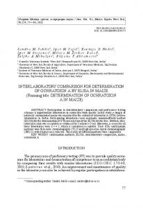
only positive in a single assay, but showed a relatively high Ct value (36?6). This sample was obtained from a patient with a low parasite burden (one cyst in one out of three investigated microscopic slides). This patient had been diagnosed with PCP a week earlier and had been treated with cotrimoxazole during the week previous to the bronchoscopy. Of the 83 microscopy-negative samples, 69 (83?1 %) were found negative in all three PCR assays (Table 3). The remaining 14 samples were PCR positive in at least one of the three assays. Five of these were positive in all three assays, two samples were positive in two of the assays, and seven samples were positive in a single assay only. The majority (12/14) of the patients with microscopy-negative/PCRpositive results were HIV-negative patients with one or more risk factors for PCP (data not shown). An additional patient was HIV-positive, and the remaining patient did not have any known risk factors. The most likely explanation for the microscopy-negative/PCR-positive results is the higher sensitivity of PCR in comparison with microscopy. The agreement between the three P. jiroveci real-time PCR assays was assessed by pairwise comparisons of the qualitative test results. The highest agreement was found between the MINC and LUMC assays, with a percentage agreement of 96?8 % and a kappa value of 0?93. The agreement between the other assays was also excellent, with a percentage agreement of 94?4 % and a kappa value of 0?88, both between the MINC and RUNMC assays and between the RUNMC and LUMC assays. Quantitative comparison between the three real-time PCR assays To compare the quantitative performance of the three realtime PCR assays, pairwise correlations between Ct values generated on samples that were scored positive in all three assays (n=45; Table 3) were calculated. As shown in Fig. 1, there was a good correlation between the Ct values produced by each of the three assays. The correlation coefficients were 0?84, 0?90 and 0?99 between the LUMC and MINC assays, the MINC and RUNMC assays, and the LUMC and RUNMC assays, respectively. While the RUNMC and LUMC assays generated similar Ct values (Fig. 1a), the Ct values produced by the MINC assay were somewhat lower than those produced by the other assays (Fig. 1a, c). Quantitative comparison between microscopy and real-time PCR To examine the correlation between microscopy and realtime PCR in quantitative detection, the results from these assays were compared for a selection of positive samples (n=26). Since a comparison with Ct values might be difficult to interpret, these values were first converted to copy-number equivalents of a plasmid (pPCP) containing the P. jiroveci amplicon. This was achieved in the MINC realtime PCR assay by generating a standard curve using this 1232
Fig. 1. Pairwise comparison between the different Ct values for all samples (n=45) that were found to be positive in all three real-time PCR assays. (a) Comparison of Ct values from the RUNMC assay (y axis) and the LUMC assay (x axis); (b) comparison of the Ct values from the MINC assay (y axis) and the RUNMC assay (x axis); (c) comparison of the Ct values from the LUMC assay (y axis) and the MINC assay (x axis).
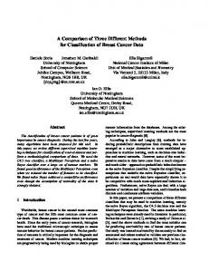
plasmid (Fig. 2a, b). Subsequently, the copy-number equivalents of the 26 P. jiroveci-positive samples were plotted against the microscopically quantified P. jiroveci burden (expressed as clusters per cytospin spot). As shown in Fig. 2(c), there was a good correlation between both methods, which was expressed as a correlation coefficient of 0?83. Journal of Medical Microbiology 55
Real-time PCR of Pneumocystis jiroveci
Fig. 2. Correlation between microscopy and the MINC real-time PCR assay in quantification of P. jiroveci. (a) Amplification plot (DRn versus cycle number) generated with dilutions of plasmid pPCP (26104, 26103, 26102, 100 and 20 copies per reaction), which contains the P. jiroveci amplicon. (b) Standard curve [Ct versus log(copy number)] generated from the amplification plot shown in (a). (c) Correlation between the number of P. jiroveci clusters per cytospin spot (foamy alveolar casts per spot) and the log of the P. jiroveci copy number [log(quantity)] for 26 PCR-positive BAL fluid samples. http://jmm.sgmjournals.org
1233
C. F. M. Linssen and others
P. jiroveci carriership versus clinically relevant infection An important issue in the diagnosis of PCP is the distinction between apparent asymptomatic P. jiroveci carriers and patients with clinically obvious PCP. In most studies, carriers have been defined as patients in whom P. jiroveci DNA can be detected in the absence of clinical signs of P. jiroveci infection, and without microscopically detectable P. jiroveci cysts in BAL fluid samples. In the present study, we found an overlap between Ct values in samples obtained from potential carriers of P. jiroveci on the one hand and some of the samples from patients with clinically and microscopically proven PCP on the other. This is in line with earlier reports, in which a reliable cut-off value for the differentiation between disease and carrier states could not be firmly established (Flori et al., 2004; Larsen et al., 2004). Nevertheless, it is possible to divide patients into three categories. The first category includes patients with clinical symptoms indicative of PCP, and with a positive PCR result and/or a positive microscopy result; these patients are diagnosed with PCP. The second category includes patients who have no clinical symptoms and do not have any indication of infection with P. jiroveci, as indicated by negative results in both microscopy and PCR. The third group is more complex, and consists of patients who do not have typical clinical symptoms of PCP, but show a positive PCR result (usually with high Ct values). We hypothesize that the patients without risk factors for PCP should be regarded as asymptomatic carriers and do not require treatment for PCP. In the case of patients belonging to the at-risk group with negative microscopy results, the microscopy should be re-evaluated and the patient should be followed clinically and receive therapy at the appearance of any clinical symptoms indicative of PCP. Previously, only immunocompromised patients, such as HIV-positive patients, were considered to be potential carriers of P. jiroveci (Weig et al., 1996, 1997). More recently, however, immunocompetent individuals have also been found to be putative carriers (Sing et al., 1999; Visconti et al., 2000). Miller and coworkers investigated health-care workers who had come into contact with patients with PCP and found among them a carrier rate of 30?5 % (Miller, 1999). In particular, health-care workers taking BAL or induced sputum samples were found to be at risk of developing a carrier status, which in one case persisted for 27 months (Miller et al., 2001). All P. jiroveci-DNA-positive patients from our study belonged to the group at risk, except for a single patient who was admitted to the intensive care unit and diagnosed with a community-acquired pneumonia caused by Streptococcus pneumoniae. In conclusion, we have compared the performance of three different in-house-developed real-time PCR assays for P. jiroveci. Interestingly, while these three assays employ different methods for nucleic acid isolation, amplification and detection, an excellent agreement in performance was found between the assays, both qualitatively (the diagnosis 1234
of PCP) and quantitatively (the P. jiroveci burden). Likewise, a good correlation was found between the P. jiroveci quantities determined by real-time PCR and microscopic quantification. A cut-off value to discriminate between disease and carrier status for P. jiroveci could not be established in the present retrospective study. A future prospective study is needed to investigate whether quantitative PCR results can be employed to differentiate between PCP and carriership of P. jiroveci. Finally, in order to monitor the performance of the different in-house PCR assays currently used in microbiology laboratories, the availability of quality control panels is of utmost importance. The assays described here could serve as reference assays in the development and maintenance of such panels.
ACKNOWLEDGEMENTS C. V. is supported by a fellowship from the Royal Netherlands Academy of Arts and Sciences (KNAW).
REFERENCES Amin, M. B., Mezger, E. & Zarbo, R. J. (1992). Detection of
Pneumocystis carinii. Comparative study of monoclonal antibody and silver staining. Am J Clin Pathol 98, 13–18. Chandra, P., Delaney, M. D. & Tuazon, C. U. (1988). Role of special
stains in the diagnosis of Pneumocystis carinii infection from bronchial washing specimens in patients with the acquired immune deficiency syndrome. Acta Cytol 32, 105–108. Djamin, R. S., Drent, M., Schreurs, A. J., Groen, E. A. & Wagenaar, S. S. (1998). Diagnosis of Pneumocystis carinii pneumonia in HIV-
positive patients. Bronchoalveolar lavage vs. bronchial brushing. Acta Cytol 42, 933–938. Edman, J. C., Kovacs, J. A., Masur, H., Santi, D., Elwood, V. H. J. & Sogin, M. L. (1988). Ribosomal RNA sequence shows Pneumocystis
carinii to be a member of the fungi. Nature 334, 519–522. Elvin, K. M., Bjorkman, A., Linder, E., Heurlin, N. & Hjerpe, A. (1988).
Pneumocystis carinii pneumonia: detection of parasites in sputum and bronchoalveolar lavage fluid by monoclonal antibodies. BMJ 297, 381–384. Flori, P., Bellete, B., Durand, F., Raberin, H., Cazorla, C., Hafid, J., Lucht, F. & Sung, R. T. (2004). Comparison between real-time PCR,
conventional PCR and different staining techniques for diagnosing Pneumocystis jiroveci pneumonia from bronchoalveolar lavage specimens. J Med Microbiol 53, 603–607. Jacobs, J. A., Dieleman, M. M., Cornelissen, E. I., Groen, E. A., Wagenaar, S. S. & Drent, M. (2001). Bronchoalveolar lavage fluid
cytology in patients with Pneumocystis carinii pneumonia. Acta Cytol 45, 317–326. Kovacs, J. A., Ng, V. L., Masur, H. & 7 other authors. (1988). Diagnosis
of Pneumocystis carinii pneumonia: improved detection in sputum with use of monoclonal antibodies. N Engl J Med 318, 589–593. Kovacs, J. A., Gill, V. J., Meshnick, S. & Masur, H. (2001). New insights
into transmission, diagnosis, and drug treatment of Pneumocystis carinii pneumonia. JAMA (J Am Med Assoc) 286, 2450–2460. Larsen, H. H., Kovacs, J. A. F., Stock, F., Vestereng, V. H., Lundgren, B., Fischer, S. H. & Gill, V. J. (2002a). Development of a rapid real-time
PCR assay for quantitation of Pneumocystis carinii f. sp. carinii. J Clin Microbiol 40, 2989–2993. Journal of Medical Microbiology 55
Real-time PCR of Pneumocystis jiroveci
Larsen, H. H., Masur, H., Kovacs, J. A. & 7 other authors (2002b).
Sepkowitz, K. A. (2002). Opportunistic infections in patients with
Development and evaluation of a quantitative, touch-down, realtime PCR assay for diagnosing Pneumocystis carinii pneumonia. J Clin Microbiol 40, 490–494.
and patients without Acquired Immunodeficiency Syndrome. Clin Infect Dis 34, 1098–1107.
Larsen, H. H., Huang, L., Kovacs, J. A. & 7 other authors (2004). A
prospective, blinded study of quantitative touch-down polymerase chain reaction using oral-wash samples for diagnosis of Pneumocystis pneumonia in HIV-infected patients. J Infect Dis 189, 1679–1683. Limper, A. H., Offord, K. P., Smith, T. F. & Martin, W. J., 2nd (1989).
Pneumocystis carinii pneumonia. Differences in lung parasite number and inflammation in patients with and without AIDS. Am Rev Respir Dis 140, 1204–1209. Maskell, N. A., Waine, D. J., Lindley, A., Pepperell, J. C., Wakefield, A. E., Miller, R. F. & Davies, R. J. (2003). Asymptomatic carriage of
Pneumocystis jiroveci in subjects undergoing bronchoscopy: a prospective study. Thorax 58, 594–597. Miller, R. F. (1999). Pneumocystis carinii infection in non-AIDS
patients. Curr Opin Infect Dis 12, 371–377. Miller, R. F., Ambrose, H. E. & Wakefield, A. E. (2001). Pneumocystis
carinii f. sp. hominis DNA in immunocompetent health care workers in contact with patients with P. carinii pneumonia. J Clin Microbiol 39, 3877–3882. Nevez, G., Guyot, K., Totet, A., Raccurt, C. & Dei-Cas, C. (2001).
Sing, A., Roggenkamp, A., Autenrieth, I. B. & Heesemann, J. (1999).
Pneumocystis carinii carriage in immunocompetent patients with primary pulmonary disorders as detected by single or nested PCR. J Clin Microbiol 37, 3409–3410. Stringer, J. R., Cushion, M. T. & Wakefield, A. E. (2001). New
nomenclature for the genus Pneumocystis. J Eukaryot Microbiol Suppl., 184S–189S. Thomas, C. F., Jr & Limper, A. H. (2004). Pneumocystis pneumonia. N
Engl J Med 350, 2487–2498. Visconti, E., Marinaci, S., Zolfo, M., Mencarini, P., Tamburrini, E., Pagliari, G., Ortona, E. & Siracusano, A. (2000). Very low frequence
of Pneumocystis carinii DNA detection by PCR in specimens from patients with lung damage. J Clin Microbiol 38, 1307–1308. Wakefield, A. E., Pixley, F. J., Banerji, S., Sinclair, K., Miller, R. F., Moxon, E. R. & Hopkin, J. M. (1990). Detection of Pneumocystis
carinii with DNA amplification. Lancet 336, 451–453. Weig, M., Klinker, H., Wilhelm, M., Lemmer, K. & Gross, U. (1996).
Correlation of Pneumocystis carinii PCR with clinical diagnosis in immunocompromised patients. Lancet 347, 1266.
Pulmonary colonisation with Pneumocystis carinii in an immunosuppressed HIV-negative patient: detection and typing of the fungus by PCR. J Med Microbiol 50, 198–200.
Weig, M., Klinker, H., Bogner, B. H., Meier, A. & Gross, U. (1997).
Olsson, M., Stralin, K. & Holmberg, H. (2001). Clinical significance of nested polymerase chain reaction and immunofluorescence for detection of Pneumocystis carinii pneumonia. Clin Microbiol Infect 7, 492–497.
Yale, S. H. & Limper, A. H. (1996). Pneumocystis carinii pneumonia in patients without acquired immunodeficiency syndrome: associated illness and prior corticosteroid therapy. Mayo Clin Proc 71, 5–13.
http://jmm.sgmjournals.org
Usefulness of PCR for diagnosis of Pneumocystis carinii pneumonia in different patient groups. J Clin Microbiol 35, 1445–1449.
1235