homogenized as described above in 0.24 M sucrose,. 0.05 M ...... sions, and John W. Daly for support and encouragement. The ... Turtle, J. R. & Kipnis, D. M. (1967) Biochem. Biopliys. ... Abe, K., Robison, G. A,, Liddle, G. W., Butcher, R. W.,.
Eur. J. Biochem. 113, 521-529 (1981) 9 FEBS 1981
Interaction of Clonidine and Clonidine Analogues with a-Adrenergic Receptors of Neuroblastoma x Glioma Hybrid Cells and Rat Brain Comparison of Ligand Binding with Inhibition of Adenylate Cyclase Daphne ATLAS and Steven L. SABOL Laboratory of Biochemical Genetics, National Heart, Lung and Blood Institute and Laboratory of Bioorganic Chemistry, National Institute of Arthritis, Metabolism and Digestive Disease, National Institutes of Health, Bethesda, Maryland (Received April 21 /September 2, 1980)
Clonidine and several analogues of clonidine are shown to be useful probes for a2-adrenergic receptors in a comparative study of ligand binding and inhibition of adenylate cyclase. The a-adrenergic properties of a new potential probe, N-(4-hydroxyphenacetyl)-4-aminoclonidinehydrochloride, are described. [3H]Clonidine binds to a-receptors of NG108-15 neuroblastoma x glioma hybrid cell membranes with & values of 1.7 and 33 nM for putative high-affinity and low-affinity sites, respectively. p-Aminoclonidine and hydroxyphenacetyl aminoclonidine displace [“Hlclonidine from the high-affinity sites with & values of 2.3 and 5.8 nM, respectively. Rat brain a2-receptors also exhibit high affinity toward clonidine, p-aminoclonidine, and hydroxyphenacetyl aminoclonidine, as determined by displacement of specifically bound [3H]clonidine. Clonidine, p-aminoclonidine, and hydroxyphenacetyl aminoclonidine elicit modest inhibition (up to 24 %) of NG108-15 adenylate cyclase by interaction with a2-receptors (&,app 300, 50, and 130 nM, respectively); these compounds also partially reverse the inhibition elicited by (-)-norepinephrine. Components of the adenylate cyclase assay mixture, particularly ATP, GTP, sodium ions, and a nucleoside-triphosphate-regenerating system, decrease the high-affinity [3H]clonidine binding to NG108-15 membranes; in the presence of these components, a-receptors possess only low affinity (& 43 nM) for [3H]clonidine. These results are consistent with the concept that certain components required for the receptor-mediated inhibition of adenylate cyclase convert a2-receptors from a highaffinity inactive state to a low-affinity active state. nanomolar concentrations selectively label receptors with a2 specificity in brain and peripheral tissues, while the a-receptor antagonists [3H]WB-4101 [5,6] or [3H]prazosin [lo- 121 at nanomolar concentrations selectively label receptors with a1 specificity. Unlike P-adrenergic receptors, which upon stimulation exert physiological responses by means of the activation of adenylate cyclase, the mechanism(s) whereby a-adrenergic receptors exert physiological responses are still obscure. Activation of a-receptors in certain tissues reduces intracellular CAMP levels or reduces the magnitude of responses elicited by compounds that elevate the CAMP concentration [13- 221. Inhibition of adenylate cyclase mediated by a-receptor activation in broken cell preparations was first reported for uterus [23] and human platelets Enzyme. Adenylate cyclase or ATP pyrophosphate-lyase (cy[16] and more recently has been characterized in clizing) (EC 4.6.1.1). human and rabbit platelets [24-271 and NG108-15 Triviul Names. Ro20-1724, 4-(3-butoxy-4-methoxyhenzyl)-2imidazolidinone; WB4101, 2-([2‘,6’-dimethoxyl-phenoxyethylami- neuroblastoma x glioma hybrid cells [28,29]. In NG108-15 cells and human platelets, the order of no)methylhenzodioxan.
Recent pharmacological and biochemical studies have led to the characterization of several classes of a-adrenergic receptors. Two classes of a-receptors were originally distinguished in the nervous system by differences in synaptic localization and function. Classical post-synaptic a-receptors that mediate smooth muscle contraction were termed a1 , while pre-synaptic Ix-receptors that mediate inhibition of neurotransmitter release were termed a2 (reviewed in [l,21). Recently this classification has been modified: sIt and a2 receptors are currently defined according to their distinct patterns of ligand specificity rather than location or function [3,4]. Ligand-binding studies have provided evidence that the a-receptor agonists [3H]clonidine [5 - 81 or p-[3H]aminoclonidine [9] at
522
potency of cx-ligands as inhibitors of adenylate cyclase resembles that of az-receptors more than that of alreceptors. This observation led to the suggestion [29] that ccz-receptors may be generally coupled to inhibition of adenylate cyclase. In view of the selective high-affinity binding of clonidine and other imidazolines to a2-receptors, analogues of clonidine may serve as useful probes to detect cxz-receptors by a variety of methods. In the present study, we assess the cx-adrenergic properties of clonidine, p-aminoclonidine [30- 311 and a novel clonidine analogue, N-(4-hydroxyphenacetyl)-4-aminoclonidine, by a comparative study of ligand binding and x2-receptor-mediated inhibition of adenylate cyclase. For this purpose, specific binding of [3H]clonidine to NG108- 15 cx-receptors, not previously reported, was characterized with respect to affinity, and the properties of the clonidine analogues were determined in direct binding studies using [3H]clonidine. Since ligand affinities calculated from displacement of [3H]clonidine from NG108-15 membranes were 22 - 176-times the apparent affinities determined by the inhibition of NG108-15 adenylate cyclase, an examination was made of the effect of adenylate cyclase assay components on the binding of [3H]clonidine to NG108-15 a-receptors.
MATERIALS AND METHODS Grot+& und Fraccionution qf NG108-15 Cells
NG108 - 15 cells (subculture 17 - 22 were grown in Falcon flasks (75 em2 surface area) as described [29]. Cells from confluent cultures (approximately 10 mg protein/flask) were harvested 3 - 6 h after the medium was replaced with fresh medium. Cells were washed twice with a buffer consisting of 167 mM NaCl, 2.7 mM KCl, 8.1 mM NaHP04, 1.5 mM KH2P04 pH 7.4, and dissociated in this buffer, then centrifuged at 250 x g for 5 min at 25 'C. Subsequent operations were carried out at 0 - 4 'C. The pellet was suspended in 0.29 M sucrose, 0.025 M Tris/HCl pH 7.5, quickly frozen in solid COZ,thawed, and homogenized with 20 strokes of a Teflon pestle rotating at 1200 rev./min. The homogenate was stored in aliquots at - 140 'C until use. For preparation of washed membranes, cells were homogenized as described above in 0.24 M sucrose, 0.05 M Tris/HCl pH 7.5, 0.005 M MgC12. Nuclei and unbroken cells were removed by centrifugation at 500 x g for 6 min, and the nuclear pellet was resuspended in homogenization buffer and centrifuged as before. The supernatant fractions were pooled and centrifuged at 28000 x g for 20 min. The pellet was suspended in buffer A (0.05 M Tris/HCI pH 7.5, 0.01 M MgClZ) and centrifuged again twice, then finally suspended in buffer A at a concentration of
Clonidine Analogues and a-Adrenergic Receptors
2.5 mg protein per ml and stored at - 140 'C. Protein was determined by the method of Lowry et al. [32] using bovine serum albumin as standard. Binding of' (3HJClonidine to NG108-15 Menibrunes
Binding reaction mixtures contained in a volume of 0.2 ml, 50 mM Tris/HCl pH 7.5, 10 m M MgC12, 0.5 - 80 nM [3H]clonidine (22.2 Ciinimol), and approximately 190 pg of NG108-15 washed membrane protein. Reaction mixtures were incubated at 25 'C for 1 h. They were subsequently diluted with 4 in1 ice-cold buffer A and filtered over Whatman GF;C filters (25 mm diameter) under gentle suction, unless otherwise indicated. Each filter was then washed and counted in 10 ml Aquasol (New England Nuclear) in a liquid scintillation counter. Specific a-receptor binding was determined by subtraction of binding in the presence of 10 pM phentolamine (non-specific binding). Under standard conditions with different membrane preparations, the fraction of total binding that was non-specific ranged between 6 - 15 %, 14- 25 and 21 -34% at 2, 10, and 20 nM [3H]clonidine, respectively. Calculation of dissociation constants and maximum binding capacities for [3H]clonidine-receptor interaction was performed by computer modeling with the SCATFIT program of V. B. Faden and D. Rodbard [33] on the IBM 370 computer of the National Institutes of Health Computer Center. Utilizing weighted non-linear least-squares analysis, this program fits data relating specifically bound ligand and total ligand concentration to Scatchard models having 2 to 5 parameters defining 1, 2 or 3 classes of binding sites, the third of which is non-saturable. The program computes the closeness of fit for each model and chooses the statistically most favored model. For studies described here, binding data were weighted to correct for the observed non-homogeneity of the variance; the weighting function was l/var(Y) = I / (0.0001 Y". Dissociation constants (Kdl values) of unlabeled ligands for high-affinity (site 1) receptors were approximated by the equation [34] &I
=
[1]50/(1
+ [s]/&i)
where [I]50 is the concentration of unlabeled ligand eliciting half-maximal inhibition of [3H]clonidine binding, [S] is the clonidine concentration, and KSI is the dissociation constant for [3H]clonidine at site 1. Assuy of Adenylute Cyclase Activity
Basal adenylate cyclase activity was routinely determined in 100-p1 reaction mixtures containing 50 mM Tris/HCl pH 7.5, 5 mM magnesium acetate, 1 16 mM sucrose, 0.25 mM Ro20-1724 (a cyclic nucleo-
D. Atlas and S. L. Sabol
523
tide phosphodiesterase inhibitor), 1 mM [ M - ~ ~ P I A T Pwas immediately filtered onto Whatman GF/B filters (25 mm diameter) under reduced pressure. Each (sodium salt, approximately 6 x lo6 counts/min), 1 pM filter was immediately washed three times with 4 ml GTP (sodium salt), 1 mM CAMP(sodium salt), 20mM creatine phosphate (disodium salt), 5 units (34-40 pg of ice-cold buffer A and subsequently counted in Aquasol liquid scintillation fluid. protein) of creatine phosphokinase, and 90 - 150 pg Specific binding of [3H]clonidine to a-receptors NG108-15 homogenate protein. Incubation was for was defined as the 3H radioactivity that could be dis10 min at 37 "C. Formation of [32P]cAMPwas determined as described previously [29]. All assays were placed by 10 pM phentolamine. Specific binding at 20 nM [3H]clonidine (total concentration) was 70 performed in duplicate ; duplicates generally were of the total binding. within 3 % of the mean. Dissociation constants for [3H]clonidine binding Apparent Kd values (&,a&,,,) for ligands affecting adenylate cyclase activity were calculated as follows. were estimated as described for NG108-15 receptors. For agonists (inhibitors of adenylate cyclase) the conTo determine dissociation constants K d l and K d 2 of centrations eliciting half-maximal inhibition, [I]~o, unlabeled ligands at putative high-affinity and lowaffinity sites (sites 1 and 2, respectively), displacement were equated with the Kd,app values, while for antagstudies were performed at 1 nM and 20 nM [3H]clonionists Kd,app values were calculated from the equadine. For displacement at 1 nM, at which concentration [34] tion most binding should be to the high-affinity site, &app = [EI50/(1 + [ s l / K % a ~ ~ ) Kdl values were approximated as described for NG108-15 receptors. To determine Kd2 values for where (El50 is the concentration of antagonist resulting unlabeled ligands, [I150values were obtained at 20 nM in half-maximal reversal of inhibition by agonist, [S] is the agonist concentration, and is the Kd,app and 1 nM [3H]clonidine, and K d 2 values were calculated according to equations derived directly from the for agonist (0.4 pM for (-)-norepinephrine [29]}. The Michaelis-Menten formulation describing one reacabove equation can be shown to be valid for antagtion catalyzed by two enzymes with distinct affinities onism of a pure agonist by a partial agonist, as it can for substrate and competitive inhibitors (see for be derived from the Michaelis-Menten formulation example [35]). for one enzyme reacting with two substrates having different V and K, values. The resultant Kd,app values do not necessarily approximate true dissociation conChenzicals stants for ligand-receptor interaction. The synthesis of hydroxyphenacetyl aminoclonidine has been reported ; the preparation was judged Prepurution of Rut Bruin Membranes to be nearly homogeneous by thin-layer chromatogFemale Sprague-Dawley rats (approximately 200 g) raphy, high-pressure liquid chromatography, as well were killed by decapitation. The brains were rapidly as nitrogen content [36]. p-Aminoclonidine was obremoved, dissected free of cerebella and placed in tained as a generous gift from Dr G. Leclerc (Univer20 vol. of ice-cold buffer A. The tissue was homogsity of Strasbourg, France). The following were kind enized in a glass tube by 15 strokes of a motor-driven gifts : phentolamine hydrochloride, Ciba-Geigy ;cloniTeflon pestle rotating at 1000 rev./min, and the homogdine hydrochloride, Boehringer-Ingelheim ; and nalenate was then centrifuged for 5 min at 500 x g at oxone hydrochloride, Endo Laboratories. [4-3H]4°C. The pellet, which consisted of nuclei and other Clonidine and [ U - ~ ~ P ] Awere T P purchased from New debris, was discarded. The supernatant fraction was England Nuclear. ATP (prepared by phosphorylation centrifuged at 50000 x g for 20 min at 4 "C. The superof adenosine), GTP, yohimbine hydrochloride, and natant fraction was discarded and the pellet (Pz) was (-)-norepinephrine hydrochloride were from Sigma. suspended in cold buffer A and centrifuged once more Guanosine 5'-[P,y-imido]triphosphate(sodium salt) as before. The washed pellet was suspended in buffer A was from I.C.N. Other chemicals were from sources at approximately 3 mg protein/ml. indicated previously [29] or were reagent grade.
Binding of [3HjClonidine to Rut Bruin Membranes Binding reaction mixtures contained, in a final volume of 0.4 ml, 50 mM Tris/HCl pH 7.5, 10 mM MgC12, 20 nM [4-3H]clonidine (22.2 Ci/mmol), approximately 300 pg washed membrane protein, ligand to be tested as indicated, or 10 pM phentolamine to determine non-specific binding. Reaction mixtures were incubated for 30 min at 25 "C. 4 ml ice-cold buffer A were added to each reaction mixture, which
RESULTS
Binding of [3H]Clonidine to NG108-15 Washed Memhrunes and Displucenzent by Clonidine Derivatives The structures of clonidine, p-aminoclonidine, and hydroxyphenacetyl-aminoclonidineare shown in Fig. 1.
524
Clonidine Analogues and a-Adrenergic Receptors
u -arninoc!onidine
10
?I ~
Fig. 1. Tlie siruciures of c,lonidine. p-uminoclonidine, und lzydroxyphenucelrjl urninoc,loniciine
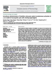
Binding of [3H]clonidine to NG108-15 a-receptors was determined both in the absence and presence of components required for the adenylate cyclase assay. As shown in Fig.2, binding as a function of [3H]clonidine concentration in Trisimagnesium chloride buffer was greater in the absence than in the presence of adenylate cyclase assay components. A Scatchard plot of data for binding in buffer alone, shown in Fig. 3 , is concave: such a shape is consistent with the presence of either multiple classes of receptors with different affinities. or negative cooperativity. The data were fitted by computer to a model of two classes of non-cooperative binding sites, a 'high-affinity' class of sites with k; 1.7 nM and a capacity of 80 fmol/mg membrane protein, and a 'low-affinity' class of sites of Kd 33 nM and a capacity of 155 fmol/mg protein. The concentration of high-affinity sites varied from 40- 140 fmol mg protein (3600- 17000 receptors/ cell) in membrane preparations from different batches of cells; however, the Kd obtained by computer fitting was consistently 1.7 nM. In order to determine whether components of the adenylate cyclase assay system that are not present in the standard receptor binding assay (namely ATP, GTP, CAMP, Ro20-1724, creatine phosphate, creatine phosphokinase, and Na' ions) affect binding of ligands to x-receptors, binding of [3H]clonidine to washed membranes was determined in the presence of all components of the adenylate cyclase assay (Fig.2, H) in parallel with binding in buffer alone (Fig. 2, 0 ) .The Scatchard plot of binding in the presence of adenylate cyclase assay components (Fig. 3, a) is consistent with a single class of receptors with relatively low affinity (Kd 43 nM) and a capacity of 150 fmol/'mg protein. Little or no high-affinity binding is evident in the presence of these components. Thus, under conditions in which adenylate cyclase is rou-
30
40
50
60
[[3H]Clonidine](nM)
H
N , 4 - ,hydrox ). p h en a c e t y I 1 - 4 - amino cio n idin e
20
Fig. 2. Binding of f 3H]clonidine to N G 108-15 ~i.usliedmernhrune.r under standard binding conditions ( 0 )or in thepresence of components of the udenylate cyclase reaction mixture )I. Each reaction mixture contained 190 pg NG108-15 membrane protein and other components described in Materials and Methods. Where indicated, reaction mixtures were supplemented with 1 mM ATP (unlabeled, sodium salt), 1 pM GTP (sodium salt), 1 miM CAMP(sodium salt), 20 mM creatine phosphate (disodium salt), 50 unitsjml creatine phosphokinase, and 0.25 mM Ro20-1724; the Tris:kICI and MgC12 concentrations were the same as those of the standard binding mixture. Binding was performed in duplicate determinations for 1 h at 25 "C in the presence and absence of 10 pM phentolamine. Results are expressed as mean specific binding vs. total [3H]clonidine concentration
: . 0.04 n
f
I
I
\;,.standard
0
-0
40 80 120 160 200 240 [WIClonidine bound (frnollrng protein)
Fig. 3 . Scutcliurd unulysis of' specific [3H]clonidinr hinding to NG108-15 washed membranes. Data are identical to those shown in Fig.2. The best fits, determined by computer modeling as described in Materials and Methods, are indicated by solid lines. For binding under standard conditions ( 0 ) .the computer chose a two-site model with dissociation constants & I = 1.7 nM and Kd2 = 33 nM, and corresponding binding capacities RI = 80 fmol mg protein, and RZ = 155 fmol/mg protein, respectively. For binding in the presence of adenylate cyclase components (=), the computer chose a one-site model with Kdl = 43 nM and R1 = 150 finol:' mg protein
tinely assayed, the high-affinity sc-receptors of the NG108-15 membranes appear to be essentially absent, and the available a-receptors possess relatively low affinity. An alteration, if any, in the receptor concentration by the presence of adenylate cyclase assay com-
D. Atlas and S. L. Sabol
525
phenacetyl aminoclonidine ([I150 40 nM), and phentolamine ([I150 56 nM). Estimates of the dissociation constants are given in Table 1. The & for phentolamine (6.5 nM) is of the expected magnitude for interaction of this classical a-blocker with a-receptors.
ponents cannot be determined with confidence, because of uncertainty in the intercept of the Scatchard plot for low-affinity binding. The displacement of [3H]clonidine by p-aminoclonidine, hydroxyphenacetyl aminoclonidine, and phentolamine was examined at 10 nM [3H]clonidine, a concentration at which most binding is to the highaffinity sites (Fig.4). The order of affinities observed is as follows :p-aminoclonidine ( [I]5016 nM), hydroxy-
Binding of Clonidine Analogues to a-Adrenergic Receptors of Rut Bruin Membranes The affinities of the clonidine analogues for rat brain membrane a2-receptors were determined in order to compare them with those determined in the less well characterized NG108-15 system. Specific binding of [3H]clonidine to a-receptors of a typical preparation of brain membranes (Pz pellet) was determined to be approximately 160 fmol/mg protein at 20 nM [3H]clonidine.The Scatchard plots (not shown) of specific [3H]clonidine binding at varied [3H]clonidine concentrations are concave and are consistent with either negative cooperativity or the presence of multiple classes of binding sites with different affinities. The data were fitted by computer to a model of two classes of non-cooperative sites, a 'very-high affinity' class (site 1, Kd 0.8 nM, 47 fmol/mg protein) and a 'high-affinity' class (site 2, Kd 4.4 nM, 105 fmol/mg protein). These results are in essential agreement with those reported by U'Prichard et al. [7]. To determine the binding affinities of the clonidine analogues to the two putative classes of brain [3H]clonidine receptors, competition studies were performed at 1 nM [3H]clonidine, which should label mainly site 1, and 20 nM [3H]clonidine (Fig. 5), which should label both sites 1 and 2. The calculated K d l
-1
0
o
10-10
lo= 10-8 10-7 10-6 Concentration of ligand (M)
10-5
10-4
Fig. 4. Displacement of [3H]clonidinefrom NG108-15 washed membranes by p-aminoclonidine (A), lzydroxyplzenacetyl aminoclonidine (O), and phentolamine (0). Binding was carried out in reaction mixtures containing 190 pg NG108-15 washed membrane protein, 10 nM [3H]clonidine, and unlabeled ligands as indicated, in standard buffer, as described in Materials and Methods. Binding in the presence of 10 pM phentolamine was subtracted from total binding to obtain specific binding. Results are expressed as a percentage of control specific binding, which was 95 fmol/mg protein. Each point is the mean of duplicate determinations, except for control binding which was determined in quadruplicate
Table 1. Summary of dissociation constants in @-receptor binding and apparent dissociation constants in adenylate cyclase inhibition for clonidine and analogues Dissociation constants (Kdl and Kd2 at putative high-affinity and low-affinity sites, respectively) and apparent dissociation constants (Kd,app) were calculated from relevant concentration curves (Fig. 2 - 7) as described in Materials and Methods. For clonidine and analogues, the values are the averages of values obtained from inhibition of adenylate cyclase (agonist effect) and values obtained from reversal listed Kd,app of (-)-norepinephrine-elicited inhibition (antagonist effect). Relative efficacy is defined as the ratio of the maximal extent of inhibition of adenylate cyclase by the tested compound relative to the inhibition obtained by 10 pM (-)-norepinephrine. n.d., not determined Rat brain [3H]clonidine binding
Compound
NG108-15 cells ~
~
Kdi
Kd2
[3H]clonidine bin din g Kd 1
adenylate cyclase
KaaPP
relative efficacy
1.7 (33") 2.3 5.8 6.5 16 3.3
300 50 130 200 70 400
0.4b 0.3 0.3 0" 0" (1)
-~~- -
nM 0.8 0.4 1.3 12 110 n.d.
Clonidine p -Aminoclonidine
Hydroxyphenacetylaminoclonidine Phentolamine Yohimbine (-)-Norephinephrine -~
4.4 0.3 3.2 2.9 30 n.d.
~~
Low-affinity site Concentration curves yielded Kd,app values varying between 70 and 400 nM and relative efficacies of 0-0.4. From [29] n.d. = not determined a
Clonidine Analogues and a-Adrenergic Receptors
526
i 14 16+ !-+-+-%T
I o
10-10
10-8 10-7 104 Concentration of ligand (M)
10-9
10-5
i
10-4
Fig. 5. Di,vpluc,crnentqf'.ypwijIc (3H]clonidine,frorn rat-brain waslied
(m),
rnemhranrs by p-uminoclonidine hydroxyphenacetyl aminoc h i d i n e (@), plieiitolaminc ( O ) , and yoliimhine ( 0 ) .Each reaction
mixture contained 20 nM [3H]clonidine, and 300 pg protein in buffer A. Total binding for the preparation shown was 240 fmol/mg protein: non-specific binding was 80 fmol/mg protein. Results are expressed as a percentage of uncompeted specific [3H]clonidine binding, which was 160 fmol,mg protein. The dissociation constants are lihted in Table 1
and Kd2 values are given in Table 1. At both putative sites 1 and 2, the affinities of p-aminoclonidine are greater than those of clonidine (2-fold and 15-fold, respectively), while the affinities of hydroxyphenacetyl aminoclonidine are similar to those of clonidine. The two sites exhibit different ligand specificities; compared to the affinities of site 2, site 1 has higher affinities for clonidine and hydroxyphenacetyl aminoclonidine but lower affinities for the a-receptor antagonists phentolamine and yohimbine. Similar differences between the two putative sites in the affinities for agonists and antagonists have been reported by U'Prichard et al. [?I. 4ffect.y of Clonirlinc und Clonidine Anulogues on NG108-15 A4dcn.vbteCycluse
The effects of clonidine and p-aminoclonidine on basal NGl08-15 adenylate cyclase activity were tested both in the absence and presence of 10 pM (-)-norepinephrine, a full cc-receptor agonist in this system (Fig. 6). Clonidine and p-aminoclonidine inhibit adenylate cyclase to a maximum extent of 10-24%,, in contrast to the 40 - 60 ":; inhibition elicited by maximally effective concentrations of (-)-norepinephrine. In addition, both clonidine and p-aminoclonidine partially reverse the inhibition by norepinephrine. Thus, these compounds behave as mixed a-receptor agonists-antagonists with respect to their effects of NG108-15 adenylate cyclase. Hydroxyphenacetyl aminoclonidine inhibits NG108-15 adenylate cyclase with an [I150 of 0.5- 1.0 pM. However, unlike its parent compounds, it possess a
'!-.-I 6 4
I
1
+ + L U L - . . .~
o
10-5 10-4 (Clonidine] or [p-Aminoclonidine] (M) 10-8
10-7
10-6
10-3
Fig. 6. Efect of ( A ) c,lonidine and ( B ) p-uniinoclonirliizr o i l hasd and norepinephrine-inhibited NG108-15 adenylate cycluse uctivity. Basal aclcnylate cyclase activity was determined in the absence (0) or presence (0) of I0 pM (-)-norepinephrine. Reaction mixtures contained 130 bg or 138 bg total homogenate protein for A a n d B. respectively
relative efficacy equal to that of (-)-norepinephrine (Fig.7A), but only about 10-20"/; of this inhibiton is reversed by the a-antagonists dihydroergotamine or yohimbine [36]. In addition to a2-receptors, NG108-15 cells possess opiate and muscarinic cholinergic receptors linked to adenylate cyclase inhibition [37,38]. Naloxone, an opiate-receptor antagonist, reverses most (70 - 90 %) of the inhibition of adenylate cyclase by hydroxyphenacetyl aminoclonidine ; thus, hydroxyphenacetyl aminoclonidine possess strong opiate agonist activity [36]. In the presence of 50 pM naloxone to saturate the opiate receptors, 0.1 - 3 pM hydroxyphenacetyl aminoclonidine behaves, like its parent compounds, as a mixed a-receptor agonist-antagonist, inhibiting adenylate cyclase 15 and reversing the inhibition by the full agonist norepinephrine (Fig. 7 B). The small inhibition in the presence of naloxone can be fully reversed by 100 pM phentolamine (data not shown). The apparent Kd values for the clonidine analogues are given in Table 1. Alteration of the [3H]Clonidine Binding to NG108-15 Membranes by Conzponents o j the Adenylute Cycluse System As shown in Table 1, clonidine. clonidine analogues, phentolamine, yohimbine, and (-)-norepinephrine possess apparent Kd values for inhibition of NG108-15 adenylate cyclase that are 22- 176 times
D. Atlas and S. L. Sabol
521
._
c
4
$I4 0 [ ; , .o 20 m
c
M i
4 '
o
'
1
':
'
I
'
I
10-5 [Hydroxyphenacetyl aminoclonidine] (M) 10-9
10-8
10-7
10-6
-
10-4
Fig Eflect of Izydroxyphenacetyl aminoclonidine on hasa and norephinephrine-inhibitedadenylate cyclase activity in the absence ( A ) or presence ( B ) of naloxone to block opiate receptors. Reaction mixtures contained 116 pg homogenate protein, SO pM naloxone (in B), and indicated concentrations of hydroxyphenacetyl aminoclonidine. (0) Without (-)-norepinephrine; ).( with 3 pM (-)norepinephrine
the Kd values estimated for high-affinity binding to the NG108-15 a-receptor. As shown previously in Fig. 2 and 3, components of the adenylate cyclase assay system reduce or eliminate high-affinity binding, but apparently not low-affinity binding of [3H]clonidine to a-receptors. In order to determine which component(s) of the adenylate cyclase assay mixture inhibit the high-affinity binding, the effect of each component was investigated (Fig. 8). Strong inhibition of binding is elicited by GTP (50% inhibition at 0.2 mM), ATP (50% inhibition at 1 mM), disodium creatine phosphate (50% inhibition at 7 mM in the presence of creatine phosphokinase to complete the nucleosidetriphosphate-regenerating system), or sodium ions (as NaCl, 50% inhibition at 60 mM). The inhibition by NaCl at concentrations of 50 mM or lower appears to be due to the Na' ion, since additional Tris/HCl buffer pH 7.5 inhibits binding with a potency (based on Tris molarity) of 115 of that of NaCl. The other components of the adenylate cyclase assay (creatine phosphokinase, cycle AMP, and Ro20-1724) are not strongly inhibitory at the concentrations tested. The non-hydrolyzable GTP analogue, guanosine 5'-[p,yimidoltriphosphate, tested for comparison with GTP, strongly inhibits binding (50 % inhibition at 0.03 mM). When creatine phosphate (sodium salt), creatine phosphokinase, ATP, and GTP are present together at concentrations used in the adenylate cyclase assay, binding of [3H]clonidine is inhibited 76 %; this degree
2
0
, o
) \.
P"HlppG
k
10-6
,
,
GTP CP
(+:q
c;!,k]
10-5 10-4 10-3 Concentration of addition (M)
10-1
Fig. 8. Effect of components of the adenylate cyclase assay on higkaflinity binding of [3H]clonidine to NGIO8-15 membranes. Reaction mixtures (0.2 ml) contained, in buffer A, 10 nM [3H]clonidine, 328 pg "3108-15 membrane (Pz pellet) protein, and the following compounds at indicated concentration: guanosine S'-[/j',y-imido]triphosphate (p[NH]ppG), sodium salt (A);GTP, sodium salt (m); ATP, sodium salt (O), cyclic AMP, sodium salt (0);creatine phosphate (CP), disodium salt in the presence of SO units/ml creatine phosphokinase (CPK) (0);20 mM creatine phosphate in the presence of SO units/ml creatine phosphokinase, 1 mM ATP, and 1 pM GTP (A); NaCl (v); or additional Tris/HCI buffer Reaction mixtures were incubated for 60 min at 25 "C pH 7.5 (0). and were filtered over GF/B rather than GF/C filters. A relatively large amount of membrane protein was used in this experiment because of the relatively low amount of high-affinity binding by the preparation used. Results (means of duplicates) are expressed as percentages of the control specific [3H]clonidine binding, which was 39 fmol/mg protein. In other experiments 0.2.5 mM Ro20-1724, an adenylate cyclase assay component, did not inhibit binding
of inhibition is similar to that found for the complete adenylate cyclase system (Fig. 2). The inhibition by creatine phosphate is not entirely due to the accompanying sodium counterion, because the Tris salt of creatine phosphate is also strongly inhibitory (data not shown). Unfractionated homogenates of NG108-15 cells, normally used for the adenylate cyclase assays, were generally found to possess mainly low-affinity specific [3H]clonidine-bindingsites. For a typical homogenate, phentolamine-displaced [3H]clonidine binding consisted of a small component of high-affinity (& 3.4 nM, 10 fmol/mg protein) and a component that did not begin to saturate within the [3H]clonidine concentration range tested (up to 80 nM) (data not shown). Thus a-receptors in unfractionated homogenates appear to be predominantly in a state characterized by an affinity for clonidine too low to be reliably measured by the filtration binding method. This result is consistent with those of Fig. 8, because of the presence of GTP and ATP in the unfractionated homogenate.
DISCUSSION A goal of the present study is to develop ligands that bind with high affinity to specific classes of
528
x-receptors. We demonstrate that clonidine, p-aminoclonidine, and a derivative of the latter compound, hydroxyphenacetyl aminoclonidine, are potent a2receptor ligands. They possess partial a-receptor agonist activity with respect to the inhibition of adenylate cyclase of NG108-15 cell homogenates and bind with high affinity to receptor sites on NG108-15 and rat brain membranes. The results of this paper also extend the evidence for the validity of the NG108-15 system as an assay for x2-adrenergic activity by the demonstration of high-affinity phentolamine-sensitive [3H]clonidine binding to NG108-15 membranes and the apparent modulation of this binding by compounds required for receptor-mediated inhibition of adenylate cyclase. Clonidine and its analogues are partial agonists with respect to the NG108-15 adenylate cyclase, in that they elicit less maximal inhibition than that of a full agonist such as (-)-norepinephrine. These results are consistent with reports that clonidine and other imidazolines are partial a-receptor agonists in inhibition of platelet adenylate cyclase [20,27] and at presynaptic and post-synaptic receptors of smooth muscle [39]. In contrast, clonidine has been reported to be a full antagonist with respect to a-receptor-stimulated accumulation of CAMP in brain slices [40] and a-receptor-induced potassium release in rat parotid slices [41]. Hydroxyphenacetyl aminoclonidine, which contains a hydroxyphenacetyl group that is a potential site for radioiodination, possesses both partial a-adrenergic and full opiate agonist activities in the NG108-15 adenylate cyclase system (Fig. 7). This compound, however, is more potent as an a-agonist ( K d , a p p 125 nM) than as an opiate agonist (&,app 5001000 nM). The possible future use of hydroxyphenacetyl aminoclonidine as an a-receptor probe in tissues which possess also opiate receptors may necessitate the use of an opiate antagonist to block these receptors. The &,app values calculated for inhibition of NG108-15 adenylate cyclase by clonidine and the analogues tested were many times the Kd values calculated for high-affinity binding at the NG108-15 receptors (Table 1). This discrepancy was apparently not specific for agonists, because it was found also for the antagonists phentolamine and yohimbine. While some of the discrepancy may be due to the different incubation temperatures and times (25 "C and 60 min for binding; 37 ' C and 10 min for adenylate cyclase), much of the discrepancy is due to inhibition of highaffinity binding by specific components of the standard adenylate cyclase assay system. The characteristics of the binding of [3H]clonidine in the presence of these components (a single class of site with Kd 43 nM and capacity 150 fmol/mg protein) are similar to those of the low-affinity site found in the absence of adenylate cyclase components (& 33 nM, 155 fmol/
Clonidine Analogues and x-Adrenergic Receptors
mg protein). It is not possible to decide from the present study whether adenylate cyclase assay components eliminate high-affinity sites altogether, or alternatively convert high-affinity sites to low-affinity sites, because of uncertainty in the determination of the number of low-affinity receptors by the filtration binding method. Clearly, the affinities of the a-receptors under standard adenylate cyclase conditions are relatively low and would approach the apparent affinities obtained for adenylate cyclase inhibition. Similarly, affinities of opiate receptors of NG108-15 homogenates in the presence of adenylate cyclase assay components were reported to be 13-667-fold less than the affinities obtained with washed membranes alone [37]. Guanyl nucleotides and N a t ions have been reported to reduce high-affinity binding of agonists to a-receptors (probably a2-receptors) in brain [8, 42,431 and platelet membranes [27,44]. GTP (0.1 - 1 pM) is required for az-receptor-mediated inhibition of adenylate cyclase of NG108-15 cells [29] and human platelets [45]. GTP is also required for receptor-mediated inhibitions of adenylate cyclase by opiates, cholinergic agents, and adenosine [46-491, as well as for receptor-mediated activation of the enzyme [50]. Also, sodium ions (half-maximally effective at 20 - 40 mM), enhance the maximal inhibition of NGlO8-15 adenylate cyclase by opiates [46], cholinergic agents [47], and a2-receptor agonists (S.L.S., unpublished observation). The results of this study demonstrate that GTP (or guanosine 5'-[p,y-imido]triphosphate) and Na' ions inhibit [3H]clonidinebinding to NG108-15 a-receptors; the characteristics of this inhibition resemble those reported for brain membranes [8]. The concentration of GTP (100 yM) required to inhibit strongly [3H]clonidine binding is higher than that (1 pM) required for maximal receptor-mediated inhibition of adenylate cyclase [29,45 - 491 ; this discrepancy might be explained by GTP-phosphohydrolase activity in membrane preparations and a nucleoside-triphosphateregenerating system in the adenylate cyclase assay. In several studies of P-adrenergic and glucagon receptors, GTP, which is required for activation of adenylate cyclase via these receptors, was found to reduce the affinity of agonists for the receptor [51- 531. Models have been proposed suggesting that GTP and agonist, in concert, convert these receptors from a high-affinity state that is not effectively coupled to adenylate cyclase to a low-affinity state that is associated with increased adenylate cyclase activity [44, 51 - 531. The results of the present study indicate that compounds required for effective coupling of inhibitory receptors to adenylate cyclase (i.e. GTP, Na' ions, and possibly ATP) also inhibit high-affinity binding of agonists to az-receptors. Thus, the mechanisms by which inhibitory and stimulatory receptors are coupled to adenylate cyclase may in certain respects be similar.
529
D. Atlas and S. L. Sabol We thank Scott Davis (Montgomery County Heart Association Student Fellowship Winner) for helpful assistance, David Rodbard and Peter Munson for help with curve-fitting programs, and Alex Levitzki, Werner Klee, and Marshall Nirenberg for helpful discussions, and John W. Daly for support and encouragement. The SCATFIT manual (1976, Faden, V. B. and Rodbard, D.) was kindly povided by Dr David Rodbard, Reproduction Research Branch, NICHD, NIH, Bethesda, M D 20205.
REFERENCES 1. Langer, S. Z. (1974) Biochem. Pharmacol. 23, 1793-1800. 2. Starke, K. (1977) Rev. Physiol. Biochem. Pharmacol. 77, 1- 124. 3. Berthelsen, S. & Pettinger, W. A. (1977) Life Sci. 21, 595-606. 4. Starke, K . & Langer, S. Z. (1979) in Presynaptic Receptors (Langer, S. Z., Starke, K. & Dubocovich, M. L., eds) pp. 1-4, Pergamon Press, Oxford. 5. U’Prichard, D. C., Greenberg, D. A. & Snyder, S. H. (1977) Mol. Pharmacol. 13,454-473. 6. U’Prichard, D. C. & Snyder, S. H. (1979) Life Sci. 24, 79-88. 7. U’Prichard, D. C., Bechtel, W. D., Rouot, B. M. & Snyder, S. H. (1979) Mol. Pharmacol. 16, 47-60. 8. Glossmann, H. & Presek, P. (1979) Naunyn-Schmiedeberg’s Arch. Pharnzacol. 306,67 - 73. 9. Rouot, B. R. & Snyder, S. H . (1979) Life Sci. 25, 769-774. 10. Raisman, R., Briley. M. & Langer, S. Z. (1979) NaunynSchmiedeberg’s Arch. Pharmacol. 307, 223 - 226. 11. Karliner, J. S., Barnes, P., Hamilton, C. A. & Dollery, C. T. (1979) Biochem. Biophys. Res. Commun. 90, 142- 149. 12. Hornung, R., Presek, P . & Glossman, II. (1979) NaunynSchmiedeberg’s Arch. Pharmacol. 308, 223 - 230. 13. Turtle, J. R. & Kipnis, D. M. (1967) Biochem. Biopliys. Res. Commun. 28, 797 - 802. 14. Handler, J. S., Bensinger, R. & Orloff, J. (1968) Am. J . Physiol. 215, 1024-1031. 15. Abe, K., Robison, G . A,, Liddle, G. W., Butcher, R. W., Nicholson, W. E. & Baird, C. E. (1969) Endocrinology, 85, 674 - 682. 16. Moskowitz, J., Harwood, J . P., Reid, W. D. & Krishna, G. (1971) Biocliem. Biophys. Acta, 230, 279-285. 17. Field, M., Sheerin, H. E., Henderson, A. & Smith, P. L. (1975) Am. J . Physiol. 229, 86 - 92. 18. Traber, J., Reiser, G., Fischer, K. & Hamprecht, B. (1975) FEBS Lett. 52, 321- 332. 19. Guder, W. G . & Rupprecht, A. (1975) Pfligers Arch. 354, 177- 186. 20. Kafka, M . S., Tallman, J. F. & Smith, C. C. (1977) Life Sci. 21, 1429-1438. 21. McCarthy, K. D. & De Vellis, J. (1978) J . Cyclic Nucleotide Res. 4, 15 - 26. 22. Brown, E. M., Hurwitz, S. H. & Aurbach, G. D. (1978) Endocrinology, 103, 893 899. 23. Triner, L., Vulliemoz, Y., Verosky, M. & Nahas, G. G . (1970) Life Sci. 9, 707-712. 24. Jakobs, K. H., Saur, W. & Schultz, G. (1976) J . Cyclic Nucleotide Res. 2, 381 - 392. 25. Newman, K. D., Williams, L. T., Bishopric, N. H. & Lefkowitz, R. J. (1978) J . Clin. Invest. 61, 395-402. -
26. Steer, M. L. & Wood, A. (1979) J . Biol. Chem. 254, 10791 10797. 27. Tsai, B. S. & Lefkowitz, R. J. (1978) Mol. Pharmacol. 14, 540- 548. 28. Sabol, S . L. & Nirenberg, M. (1977) Fed. Proc. 36, 736. 29. Sabol, S. L. & Nirenberg, M. (1979)J. Biol. Chem. 254, 19131920. 30. Rouot, B. & Leclerc, G. (1978) Chim. Ther. 6 , 521 -526. 31. Rouot, B., Leclerc, G., Bieth, H., Wermouth, C. A. & Schwartz, J . (1978) C.R. Hehd. Seances Acad. Sci. Ser. D , Sci. Nut. 280, 909-910. 32. Lowry, 0 . H., Rosebrough, N. J., Farr, A. L. & Randall, R. J. (1951) J . Biol. Chem. 193, 265-275. 33. Rodbard, D., Munson, P. J. & DeIxan, A. (1977) in International Symposium on Radioimmunoassay and Related Procedures in Medicine, pp. 469- 504, International Atomic Energy Commission, Vienna. 34. Cheng, Y.-C. & Prusoff, W. H. (1973) Biochem. Pharmacol. 22, 3099 - 3108. 35. Segel, I. H. (1975) Enzyme Kinetics, p. 64, Wiley-Interscience, New York. 36. Atlas, D. & Sabol, S. L. (1980) Biochem. Biophys. Res. Commun. 94, 924-931. 37. Sharma, S . K., Nirenberg, M. & Klee, W. A. (1975) Proc. Natl Acad. Sci. USA, 72, 590 - 594. 38. Nathanson, N. M., Klein, W. L. & Nirenberg, M. (1978) Proc. Nut1 Acad. Sci. USA, 75, 1788-1791. 39. Medgett, I. C., McCulloch, M. W. &Rand, M. J. (1978) NaunynSchmiedeberg’s Arch. Pharmacol. 304, 21 5 - 222. 40. Skolnick, P. & Daly, J. W. (1975) Mol. Pliarmacol. 11, 545551. 41. Davis, J. N. & Maury, W. (1978) J . Pharmacol. Exp. Ther. 207, 425 - 432. 42. U’Prichard, D. C. & Snyder, S. H. (1977) J . Biol. Cliem. 252, 6450 - 6463. 43. U’Prichard, D. C. & Snyder, S. H. (1978) J . Biol. Chem. 253, 3444- 3452. 44. Tsai, B. S. & Lefkowitz, R. J. (1979) Mol. Pharmacol. 16, 61 -68. 45. Jakobs, K. H., Saur, W. & Schultz, G . (1978) FEBS Lett. 85, 167-170. 46. Blume, A. J., Lichtshtein, D. & Boone, G. (1979) Proc. Nut1 Acad. Sci. USA, 76, 5626- 5630. 47. Lichtshtein, D., Boone, G. & Blume, A. (1979) J. Cyclic Nucleotide Res. 5, 367 - 375. 48. Cooper, D. M. F., Schlegel, W., Lin, M. C. & Rodbell, M. (1979) J. Biol. Chem. 254, 8927-8931. 49. Wilkening, D., Sabol, S. & Nirenberg, M. (1980) Brain Res. 189, 459 - 466. 50. Rodbell, M., Lin, M. C., Salomon, Y., Londos, C., Harwood, J. P., Martin, B. R., Rendell, M. & Berman, M. (1975) Adv. Cyclic Nucleotide Res. 5, 3 - 29. 51. Maguire, M. E., Van Arsdale, P. M. & Gilman, A. G. (1976) Mol. Pharmucol. 12, 335- 339. 52. Williams, L. T. & Lefkowitz, R. J. (1977) J . Biol. Chem. 252, 7207 - 7213. 53. Lad, P. M., Welton, A. F. & Rodbell, M. (1977) J . B i d . Chem. 252, 5942 - 5946.
S. L. Sabol, Laboratory of Biochemical Genetics, National Heart, Lung, and Blood Institute, National Institutes of Health, Building 36, Room 4C-16, Bethesda, Maryland, USA 20205 D. Atlas, Department of Biological Chemistry, Hebrew University of Jerusalem, Institute of Life Sciences, Givat Ram, 94149 Jerusalem, Israel