A
R
T
I
C
L
E
S
Interaction of Lumican with Aggrecan in the Aging Human Sclera Jane R. Dunlevy1 and Jody A. Summers Rada2 PURPOSE. Lumican is a keratan sulfate proteoglycan originally identified in cornea, but present in a variety of connective tissues where it presumably regulates collagen fibril formation and organization. The present study was designed to describe the chemical nature of lumican core protein in the aging human sclera. METHODS. Western blot analyses, immunohistochemistry, and immunoaffinity chromatography were used to detect and purify the lumican core protein from tissue extracts from human donors 6 to 89 years of age. Treatment of lumican with chondroitinase ABC, keratanase-I and -II, and/or endo--galactosidase was used to determine the degree of glycosylation of the lumican core protein. RESULTS. Lumican was present in the human sclera as a 70- to 80-kDa core protein with short unsulfated lactosaminoglycan side chains. In addition, on Western blots, a larger ⬎200-kDa species was apparent that was immunologically related to lumican. This high-molecular-weight material increased in scleral extracts with increasing age. The complex was most abundant in unreduced samples, and approximately two thirds of the 70- to 80-kDa lumican core protein was released from the complex on reduction of the scleral extract. Further characterization of the ⬎200-kDa lumican-immunopurified complex indicated that aggrecan (the cartilage proteoglycan) was covalently associated with lumican. CONCLUSIONS. Reducible and nonreducible lumican-aggrecan interactions occur in the scleral extracellular matrix and result in the formation of high-molecular-weight complexes that increase with age. These results represent the first report demonstrating lumican-aggrecan interactions and suggest they may play a role in age-related scleral extracellular matrix changes. (Invest Ophthalmol Vis Sci. 2004;45:3849 –3856) DOI: 10.1167/iovs.04-0496
From the 1Department of Anatomy and Cell Biology, University of North Dakota School of Medicine and Health Sciences, Grand Forks, North Dakota; and the 2Department of Cell Biology, University of Oklahoma Health Sciences Center, Oklahoma City, Oklahoma. Supported by Macula Vision Research Foundation, Bala Cynwyd, Pennsylvania (JASR), National Eye Institute Grant EY09391 and core Grant EY121291 (JASR); National Center for Research Resources (NCRR) Center of Bioresearch Excellence (COBRE) grant RR017703 (JASR); and ND Experimental Program to Stimulate Competitive Research (EPSCoR) Grant EPS-0132289 (JRD). Submitted for publication May 4, 2004; revised July 20, 2004; accepted July 23, 2004. Disclosure: J.R. Dunlevy, None; J.A. Summers Rada, None The publication costs of this article were defrayed in part by page charge payment. This article must therefore be marked “advertisement” in accordance with 18 U.S.C. §1734 solely to indicate this fact. Corresponding author: Jane R. Dunlevy, Department of Anatomy and Cell Biology, University of North Dakota School of Medicine and Health Sciences, 510 North Columbia Road, Grand Forks, ND 58203;
[email protected]. Investigative Ophthalmology & Visual Science, November 2004, Vol. 45, No. 11 Copyright © Association for Research in Vision and Ophthalmology
T
he sclera is a typical connective tissue that provides the structural framework that defines the shape and therefore the axial length of the eye. The biomechanical properties of the sclera are determined by the extracellular matrix, which consists of interwoven collagen fibrils in close association with proteoglycans. Although the number, arrangement, and type of collagen fibrils determine the mechanical strength of a connective tissue, proteoglycans have been shown to serve several biological functions that include regulation of hydration, regulation of collagen fibrillogenesis, maintenance of structural integrity, growth regulation, matrix organization, and cell adhesion.1 Alterations in the composition of the scleral extracellular matrix (ECM) would be likely to lead to changes in the biomechanical properties of the sclera, alter scleral shape, and severely affect vision. The major sulfated proteoglycans (PGs) of the sclera include aggrecan, decorin, and biglycan.2,3 Aggrecan consists of a core protein covalently linked to more than 100 chondroitin sulfate (CS) side chains. The negative charge associated with the glycosaminoglycan (GAG) chains of aggrecan acts to sequester water molecules within the scleral ECM, which is key to proper tissue hydration and elasticity during mechanical stress. In contrast to decorin and biglycan, which are evenly dispersed throughout all regions of the sclera, aggrecan has been found in highest concentration in the posterior sclera, where it may be responsible for maintaining the pliancy observed in the posterior regions of the sclera.3 In the ECM of cartilage, aggrecan associates with hyaluronan through linker proteins to form large hydrated macromolecules4; however, this has yet to be demonstrated for sclera. The human sclera has also been shown to contain the core proteins of several members of a family of related proteins termed the SLRPs, small leucine-rich PGs5,6 (Johnson JM, et al. IOVS 2002;43:ARVO E-Abstract 1100). Decorin and biglycan are the best-known and most-studied members of the SLRP family, and these PGs have been shown to exist in the human sclera as PGs, containing one or two chondroitin/dermatan sulfate side chains, respectively.2 In addition, the human sclera contains the SLRP core proteins lumican, proline-arginine-rich and leucine-rich repeat protein (PRELP), keratocan, fibromodulin, dermatan sulfate PG-3 (DSPG-3; also known as PG-Lb, epiphycan), chondroadherin, and osteoglycin (Johnson JM, et al. IOVS 2002;43:ARVO E-Abstract 1100).7 All SLRPs contain a common central domain, consisting of ⬃10 leucine-rich repeats, which have been shown to be involved in high-affinity protein–protein interactions.8 Lumican is a member of the SLRP gene family that has been shown to be a potent regulator of collagen fibril formation,9 –11 and lumican-null mice exhibit defects in collagen fibril structure and packing in the cornea12 and sclera.13 In addition, lumican–fibromodulin double-null mice have a 10% increase in axial length of the eye, and retinal detachments are common in these mice.14 Lumican was first identified in the cornea as a keratan sulfate (KS) PG,15–18 but has also been demonstrated in bovine 3849
3850
Dunlevy and Summers Rada
aorta and in articular cartilage as a glycoprotein (GP) of 70 to 80 kDa, lacking the long sulfated keratan sulfate side chains observed in corneal lumican.19,20 In the present study we identified the 70- to 80-kDa lumican core protein in the sclera, as well as a ⬎200-kDa lumicancontaining, high-molecular-weight complex. Through a combination of affinity chromatography and Western blot analyses, we demonstrate that aggrecan participates in covalent and nonreducible interactions with lumican in this high-molecularweight complex in an age-dependent manner. The present study was therefore designed to characterize the chemical nature of this high-molecular-weight complex in the human sclera.
MATERIALS
AND
METHODS
Extraction Human donor eyes of ages 6 to 89 years were obtained from the National Disease Research Interchange (NDRI) within 48 hours after death. Human tissue was handled according to the tenets of the Declaration of Helsinki, and the research was approved by the University of North Dakota’s and University of Oklahoma’s Institutional Review Boards. On receipt, eyes were immediately frozen at ⫺80°C. To extract PGs, adherent muscle, fat lamina cribrosa, and optic nerve head were removed from the sclera and the entire sclera (anterior, equatorial, and posterior regions) was minced into small (⬍2 mm3) pieces with a razor blade. The minced tissue was extracted in 10 volumes of 4 M guanidine-HCl containing 0.01 M sodium acetate, 0.01 M sodium EDTA, 0.005 M benzamidine-HCl, and 0.1 M ⑀-amino-n-caproic acid at 4°C, as described previously.2 Scleral extracts were dialyzed exhaustively against water and lyophilized. In some experiments, human corneas and corneal–scleral junctions were obtained, minced, and extracted as just described. Lyophilized scleral extracts were reconstituted in distilled water based on the wet weight of the starting material at a ratio of 0.74 g/500 L distilled (d)H2O before enzyme digestion and electrophoresis. In extracts of older tissues, an insoluble residue was present in scleral extracts. To compare the PG composition between soluble and insoluble fractions, we collected the residue by centrifugation at 14,000 rpm for 10 minutes, reconstituted it in the starting volume of dH2O, and ran it on SDS-PAGE, with and without prior digestion with chondroitinase ABC, keratanase II, and endo--galactosidase. For initial characterizations of lumican in human sclera, aliquots of scleral extracts were loaded on gels based on the starting wet weight of the tissue, because the PG content of human sclera has been shown to vary with age and scleral region.3 For subsequent evaluation of the amounts of the high-molecular-weight lumican complex relative to that of the uncomplexed lumican core protein, total protein concentrations were determined for each scleral extract by the Bradford assay (Bio-Rad Laboratories, Hercules, CA) and 30 g of total protein from each extract was applied to SDS-PAGE for Western blot analysis.
DEAE Chromatography Lyophilized scleral extracts were reconstituted in 6.0 M urea containing 0.15 M NaCl, 0.05 M Tris (pH 6.8) and 0.1% CHAPS (3-([3-cholamidopropyl]dimethylammonio-1-propanesulfonate) and applied to 6 mL diethylaminoethyl (DEAE) Sepharose step columns (3.7 ⫻ 1.5 cm) equilibrated in the same solvent. Scleral GPs passed through the column in the flow-through fraction, whereas scleral PGs were eluted from the column with 6.0 M urea containing 1.15 M NaCl. The flowthrough and eluted fractions were dialyzed against water and lyophilized.
Affinity Chromatography Affinity purification of lumican was performed by covalently crosslinking the Fc region of the purified anti-lumican C-terminal peptide
IOVS, November 2004, Vol. 45, No. 11 antibody21 (provided by Peter J. Roughley, PhD, Shriners Hospitals for Children, Montreal, Que´bec, Canada) to immobilized protein A using dimethyl pimelimidate (DMP). Remaining active sites on protein A were blocked with 0.1 M ethanolamine (pH 8.2). After equilibration with 10 mM Tris buffer, approximately 60 mg of lyophilized scleral extracts were reconstituted in 10 mM Tris buffer and bound to the affinity substrate using a batch protocol. After antigen binding, proteins were washed from the affinity substrate in a column format until a baseline absorbance at 280 nm was reached. Lumican was eluted from the column with 100 mM triethylamine (pH 11.5), dialyzed against dH2O and lyophilized.
Western Blot Analysis Scleral extracts, DEAE-purified PG fractions, and affinity-purified lumican were electrophoresed on 10% SDS-polyacrylamide gels according to the method of Laemmli22 under standard reducing (97.2 mM dithiothreitol [DTT]) or nonreducing conditions. In some cases, samples were digested with chondroitinase ABC, endo--galactosidase, and/or keratanase II (Seikagaku America, Ijamsville, MD),2 before electrophoresis. Electrophoresed proteins were transferred to nitrocellulose, and Western detection was performed using the following antisera: rabbit anti-human C-terminal lumican peptide21; rabbit anti-human aggrecan (R114 antibody), characterized in human sclera,2,3 from Robin Poole, PhD (Joint Diseases Laboratory, McGill University, Montre´al, Que´bec, Canada); and rabbit anti-aggrecan G1 domain23,24 from John Sandy, PhD (Shriners Hospitals for Children, Tampa, FL). Immunoreactive proteins were detected with chemiluminescent substrate (Western Star; Tropix, Bedford, MA). When necessary, blots were stripped by incubating them for 30 minutes at 70°C in 100 mM -mercaptoethanol and 2% SDS in 62.5 mM Tris-HCl (pH 6.8). Removal of the detection antibodies was confirmed by repeating the Western blot protocol without the primary antibody incubation.
Immunohistochemistry Immunohistochemistry was performed on 10-m-thick frozen human scleral tissue from the anterior pole of a 49-year-old donor eye. The tissue was sectioned on a cryostat microtome (Shandon-Lipshaw, Pittsburgh, PA) and stained with the purified anti-lumican C-terminal peptide antibody21 diluted 1:500 in PBS, and the detection of lumican protein distribution in human sclera was performed according to the manufacturer’s directions in the avidin-biotin complex (ABC) kit (Vectastain ABC; Vector Laboratories Inc., Burlingame, CA).
RESULTS Identification of Lumican in the Human Sclera Scleral extracts were obtained from a 32-year-old human donor, separated into GP (DEAE flow-through) and PG (DEAEbound) fractions and subjected to SDS-PAGE and Western blot analyses with antibodies specific for the core protein of human lumican. For comparison, corneas were also extracted, GP and PG fractions were obtained, and Western blot analyses were performed. In undigested corneal PG fractions (C), lumican appeared as a smear (⬎100 –70 kDa), whereas lumican appeared as an ⬃70-kDa band from the corneal–scleral junction (Fig. 1, C/S), as well as the anterior, equatorial, and posterior sclera (Fig. 1, A, E, and P, respectively). After digestion with keratanase II and endo--galactosidase, the lumican core protein appeared as an ⬃50-kDa band in all regions. Based on the intensity of the 50-kDa lumican core protein, lumican appeared in highest concentration in cornea and in lowest concentration in the anterior sclera. In addition, lumican was found to contain keratan sulfate chains in the cornea but only short, unsulfated lactosaminoglycan chains in the sclera. Proteins in the flow-through fraction from DEAE (GP fraction) were similarly examined by Western blot analyses. The
IOVS, November 2004, Vol. 45, No. 11
Interaction of Lumican with Aggrecan in Aging Sclera
FIGURE 1. Western detection of lumican in the human cornea and sclera from a 32-year-old donor. Guanidine HCl extracts of cornea (C), corneoscleral junction (C/S), anterior sclera (A), equatorial sclera (E) and posterior sclera (P) were run on 10% SDS-polyacrylamide gels and subjected to Western blot analyses using the anti-lumican C-terminal peptide antibody with and without prior digestion with keratanase II and endo--galactosidase.
undigested GP migrated at ⬃66 kDa in corneal and scleral extracts. After digestion with keratanase II and endo--galactosidase, the lumican core protein was identified as an ⬃50-kDa band in all regions. Based on the intensity of the 50-kDa lumican core protein, the GP form of lumican was in greatest concentrations in the cornea and posterior sclera and at similar but somewhat lower levels in the corneal–scleral region, anterior sclera, and equatorial sclera.
Immunohistochemistry Sclera from a 49-year-old donor eye was stained with the lumican C-terminal peptide antibody followed by peroxidaseconjugated secondary antibody and color development with diaminobenzidine (DAB) to localize lumican on sections of human sclera (Fig. 2). Lumican was distributed throughout the thickness of the sclera in anterior (Fig. 2A), equatorial (Fig. 2B), and posterior (Fig. 2C) regions of the eye, with the most intense labeling on the retinal side of the sclera, particularly in equatorial and posterior scleral regions. Minimal staining was detected on paraffin sections when nonimmune rabbit IgG was substituted for the primary antibody (Fig. 2D).
Identification of High-Molecular-Weight Lumican Complexes in the Human Sclera After dialysis in dH2O, both the GP (GP) and PG fractions of scleral extracts contained insoluble (i) and soluble (s) material. Western blot analysis indicated that lumican was present in both the soluble and insoluble fractions, with most of the lumican-positive material in the insoluble fraction of the PG fraction (Fig. 3). In addition to the expected ⬃70- to 80-kDa lumican core protein, high-molecular-weight, lumican-positive bands (⬎200 kDa) were apparent in the insoluble PG fractions. After digestion with chondroitinase ABC, keratanase II, and endo--galactosidase, the high-molecular-weight material became reduced in size to approximately 100 kDa and a highermolecular-weight, lumican-positive band became apparent at the top of the gel (⬎200 kDa, arrow) and the 80-kDa lumican core was reduced to approximately 50 kDa, as expected (Fig. 3, Dig).
Changes with Aging Lumican-immunopositive material was compared in scleral extracts of donors aged 6 to 89 years. The 70- to 80-kDa lumican
3851
FIGURE 2. Immunodetection of lumican in the human sclera. Paraffinembedded sections of sclera from a 49-year-old human donor eye were stained with the anti-lumican C-terminal peptide antibody, followed by peroxidase-conjugated secondary antibody and color development with DAB. (A) Anterior sclera, (B) equatorial sclera, (C) posterior sclera, and (D) negative control (rabbit IgG). OS, outer sclera. Bar, 100 m.
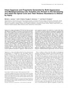
band was present in all samples, but was found to increase in concentration in donors aged 6 to 18 years (Fig. 4, left). Lumican concentrations were then found to decrease in scleral extracts from donors aged 39 to 89 years. In addition, highmolecular-weight, lumican-positive material was apparent at ⬃118, ⬃200, ⬃210, and ⬃220 kDa, with higher-molecularweight forms increasing in concentration in donors aged 39 to 89 years (Fig. 4, right).
Characterization of High-Molecular-Weight Lumican Complexes Human sclera were extracted and lumican was affinity purified using an anti-lumican antibody column. To characterize the high-molecular-weight lumican complex, affinity-purified lumican was passed through a 100-kDa nominal-molecular-weight cutoff centrifugal filter, and the retentate was used for subsequent analyses. In unreduced samples, all affinity-purified, lu-
FIGURE 3. Western blot of insoluble and soluble proteins extracted from human sclera and lumican immunodetection. After DEAE chromatography and water dialysis, the GP and PG fractions contained insoluble (i) and soluble (s) material. Most of the lumican-positive material in the PG fraction was insoluble and contained a high-molecular-weight protein that appeared to be reduced in size after digestion with keratanase II and endo--galactosidase (Dig). Undigested (Und) and enzyme alone (E) samples are also shown. (A) Five-minute exposure and (B) 30-second exposure of the same blot as in (A).
3852
Dunlevy and Summers Rada
IOVS, November 2004, Vol. 45, No. 11
DISCUSSION
FIGURE 4. Identification of high-molecular-weight, lumican-positive material in the aging sclera. Thirty micrograms of total protein, extracted from sclera of donor eyes aged 6, 8, 16, 18, 30, 39, 48, 54, 82, and 89 years was separated on a 7.5% polyacrylamide gel in nonreducing conditions, and lumican-positive material was detected by Western blot with the anti-lumican C-terminal peptide antibody.
mican-positive material was present as high-molecular-weight bands migrating at ⬃117 kDa and ⬎200-kDa (Fig. 5, Lum). Duplicate Western blot analyses with antisera specific for the core protein of aggrecan indicated that aggrecan-positive material was also present as high-molecular-weight bands in unreduced samples (Fig. 5, Agg). No bands were detected when primary antibodies were omitted (Fig. 5, Con). Digestion of affinity-purified, high-molecular-weight lumican with chondroitinase and keratanase did not alter the migration of the high-molecular-weight, aggrecan-positive complexes in nonreducing conditions, with the exception of the appearance of an ⬃97-kDa band after chondroitinase digestion (Fig. 6A, lane 2). After reduction with DTT, large amounts of two distinct aggrecan-immunopositive bands, migrating at ⬃50 and ⬃60 kDa were detected after digestion with chondroitinase and keratanase (Fig. 6A, lanes 5, 6). When the same blots were stripped and reprobed with the antibody to lumican, lumican-positive, high-molecular-weight complexes were present in unreduced samples, which appeared to comigrate with those of aggrecan (Fig. 6B, lanes 1–3). After reduction, the lumican core protein was identified at 50 to 70 kDa in undigested samples (Fig. 6B, lane 4). Digestion with chondroitinase or chondroitinase⫹keratanase resulted in increased levels of the 50- to 70-kDa band and resulted in an increase in high-molecular-weight, lumican-positive material at the top of the gel (Fig. 6B, lanes 5, 6). Thus, apparently three different pools of lumican were observed from 4 M guanidine HCl sclera extracts: a pool of soluble 70- to 80-kDa core protein present in nonreducing conditions, a second pool of 50- to 70-kDa lumican core protein present as a high-molecular-weight complex but released on reduction with dithiothreitol, and a third pool of lumican core protein that remained covalently cross-linked as a high-molecularweight complex in reducing conditions. Treatment with chondroitinase increased the fraction of free lumican core protein in reducing conditions, indicating that the GAG side chains on aggrecan may play a role in lumican–aggrecan interaction. The presence of aggrecan in the high-molecular-weight, affinity-purified lumican fraction was verified on Western blots, with antibodies specific for the G1 domain of aggrecan (Fig. 7). After digestion with chondroitinase, bands migrating at ⬎200kDa and 50 kDa could be identified, which represent fragments of aggrecan containing the N-terminal G1 domain (Fig. 7, lane 2). Digestion with chondroitinase⫹keratanase resulted in a slight increase in the intensity of the 50-kDa band (Fig. 7, lane 3) over that seen with chondroitinase alone.
In the present study, we identified lumican (corneal keratan sulfate PG) in the human sclera. Lumican is present in the sclera as a 50-kDa core protein with short, unsulfated lactosaminoglycan side chains. Lumican was recovered in both the flow-through and eluted fractions from DEAE suggesting that lumican is present in sclera with various charge densities. Data from our laboratory suggest that the variation in charge density is due to various amounts of tyrosine sulfation on N-terminal tyrosines on the lumican core protein (manuscript in preparation). The more highly charged form of lumican formed most of the high-molecular-weight complexes. These complexes were stable after 4 M guanidine HCl extraction, incubation in 2% SDS, and reduction with DTT, suggesting a covalent, nonreducible interaction with other proteins. Treatment of the high-molecular-weight complexes with chondroitinase ABC, keratanase II, and endo--galactosidase resulted in a reduction in size of the complex, as well as the visualization of multiple bands including the 50- to 70-kDa lumican, 50- to 60-kDa G1-containing fragments of aggrecan, a 150-kDa lumican-aggrecan complex and the ⬎200-kDa lumican-aggrecan complexes suggesting that GAGs play a role in the formation of the large protein complex. We speculate that hyaluronic acid (HA) is a GAG involved in the large lumican-aggrecan complex, because (1) this GAG is sensitive to digestion with chondroitinase ABC, (2) HA is present in the human sclera,25,26 and (3) a HA binding region is present within the G1 domain of aggrecan.27 The amount of lumican-containing, high-molecular-weight material increased with donor age, whereas the amount of uncomplexed lumican appeared to decline with age. Several high-molecular-weight, lumican-positive bands were present that were replaced with the ⬎200-kDa band with advanced age. This suggests that the formation of high-molecular-weight lumican-containing complexes involved the assembly of intermediates which participate in further aggregation in an agedependent manner.
FIGURE 5. Affinity purification of high-molecular-weight lumican complexes. Twenty nanograms of affinity-purified, high-molecular-weight lumican (⬎100,000 Da) was applied to a 7.5% gel and subjected to Western blot analysis with the anti-lumican C-terminal peptide antibody (Lum). The blot was stripped and reprobed with anti-aggrecan (R114) antiserum (Agg). Negative control blot (Con) was incubated with goat-anti-rabbit IgG conjugated to alkaline phosphatase only.
IOVS, November 2004, Vol. 45, No. 11
Interaction of Lumican with Aggrecan in Aging Sclera
3853
FIGURE 6. Characterization of high-molecular-weight lumican–aggrecan complexes. Twenty nanograms of lumican of ⬎100,000 in molecular weight was run, reduced or unreduced and undigested (U, lanes 1, 4), or digested with chondroitinase ABC (C, lanes 2, 5), chondroitinase ABC/keratanase I/ keratanase II (C/K, lanes 3, 6). Samples were run on 7.5% minigels and detected with anti-aggrecan (R114) antibody (A) or anti-lumican antibody (B). Western blot analysis of chondroitinase ABC/keratanase I/ keratanase II enzymes alone (E, lane 7) are also shown.
Scleral lumican was purified using affinity chromatography and high-molecular-weight lumican-containing complexes were separated and characterized. Western blot analyses using aggrecan-specific antibodies indicated that aggrecan-positive material comigrated with lumican-containing, high-molecularweight complexes. Digestion of affinity-purified, high-molecular-weight lumican complexes with chondroitinase and keratanase had little effect on the migration of the complexes under nonreducing conditions; however, after reduction with DTT, large amounts of two aggrecan-immunopositive bands, 50- and 60-kDa, were detected after digestion with chondroitinase and keratanase. From these studies, we conclude that in the sclera, lumican exists in three pools: (1) A pool of soluble 70- to 80-kDa core GP that is present in nonreducing conditions; (2) a second pool that is present as a high-molecular-weight complex with aggrecan fragments but is released upon reduction and digestion with chondroitinase ABC, and (3) a third pool that remains covalently cross-linked as a high-molecular-weight
FIGURE 7. Identification of aggrecan G1-domain fragments in highmolecular-weight lumican complex. Twenty nanograms of lumican with a molecular weight of ⬎100,000 was electrophoresed under reducing conditions on 7.5% polyacrylamide gels either undigested (U, lane 1) or after digestion with chondroitinase (C, lane 2) or chondroitinase/keratanase I/keratanase II (C/K, lane 3). Chondroitinase ABC alone (E(c); lane 4) and chondroitinase ABC/keratanase I/ keratanase II alone (E(c/k); lane 5) are also shown.
lumican–aggrecan-containing complex after glycosidase treatment and reduction. Although aggrecan has been identified in the human sclera,2,3 an interaction between aggrecan and lumican has never been described. Western blot analyses indicated that 50to 60-kDa fragments of the aggrecan core protein were present in the high-molecular-weight, lumican-containing complexes. The 50-kDa fragment, as well as a ⬎200-kDa protein were shown to contain the G1 domain of aggrecan, (the N-terminal globular, hyaluronate-binding domain),28,29 confirming the presence of aggrecan in the affinity-purified lumican complexes. In addition, treatment with chondroitinase ABC increased the fraction of free lumican core protein and uncomplexed aggrecan fragments under reducing conditions, indicating that chondroitin/dermatan sulfate– containing GAGs may play a role in the lumican-aggrecan aggregates. These GAGs may be derived from additional fragments of the aggrecan core protein or may be derived from other chondroitin/ dermatan sulfate PGs, such as biglycan and decorin, both of which have been identified in the human sclera.2,3 In human cartilage, proteolytic modification of the aggrecan core protein results in increasing amounts of smaller G1 domain-containing fragments with increasing age.30,31 We speculate that scleral aggrecan may undergo a similar age-dependent proteolytic process which generates aggrecan fragments available for complex formation with lumican. Aggrecanase-1 and -2, also known as ADAMTS-4 and -5, respectively, are proteolytic enzymes that belong to the ADAM family of metalloproteases, which have a critical role in degrading aggrecan in human cartilage, particularly in aging and osteoarthritis.32–34 Both aggrecanase-1 and -2 have been shown to generate 50- to 90-kDa G1-containing fragments of aggrecan that correspond to a cleavage site in the interglobular domain.33,35 Matrix metalloproteinases (MMPs) have also been shown to cleave aggrecan within the interglobular domain of aggrecan to generate similar sized fragments.36,37 In addition, aggrecanases have four cleavage sites within the CS2 domain of aggrecan that have been shown to generate larger, ⬎200-kDa G1-containing fragments.32,33,35 This study shows that both 50- and ⬎200-kDa aggrecan fragments were present in lumican-aggrecan complexes that and it is likely that aggrecanases and/or MMPs are involved in generating these aggrecan fragments. The mechanism responsible for the formation of the lumican-containing, high-molecular-weight complexes in the sclera is unknown. Results of this study indicate that these highmolecular-weight complexes increase in amount and in molec-
3854
Dunlevy and Summers Rada
IOVS, November 2004, Vol. 45, No. 11
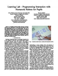
FIGURE 8. Theoretical model of lumican–aggrecan complexes found in human sclera. All complexes contained lumican and the G1 domain of aggrecan and were dependent on chondroitinase ABC digestion to be visible on the gel, most likely indicating binding to hyaluronic acid (HA). The estimated sizes of immunoblot bands generated after reduction and chondroitinase ABC digestion are also listed. (A) Lumican is covalently linked to the aggrecan through both disulfide bonding and the transglutaminase linkage of Gln-Lys (Q-K). On reduction and chondroitinase ABC digestion, this complex would produce a ⬎250-kDa lumican–aggrecan complex and the 50- to 70-kDa lumican core protein. (B) Aggrecan fragments generated from aggrecanase cleavage within the CS2 region of aggrecan are covalently bound to lumican through both disulfide bonding and the Gln-Lys transglutaminase linkage. On reduction and chondroitinase ABC digestion, this complex would produce a 220-kDa lumican-aggrecan complex and the 50- to 70-kDa lumican core protein. (C) Aggrecan G1– containing fragments generated from MMP or aggrecanase cleavage within the interglobular domain of aggrecan are covalently associated with lumican by disulfide bonding. On reduction and chondroitinase ABC digestion, this complex would produce 50- or 60-kDa aggrecan G1 fragments and the 50- to 70-kDa lumican core protein. (D) Aggrecan G1-containing fragments are covalently associated with one or more lumican molecules by a Gln-Lys bond. On reduction and chondroitinase ABC digestion, this complex would produce the 150-kDa lumican–aggrecan G1 fragment complex.
IOVS, November 2004, Vol. 45, No. 11
Interaction of Lumican with Aggrecan in Aging Sclera
ular size with age. Aging has been associated with the crosslinking of molecules through a variety of mechanisms, including carbonylation,38 non-enzymatic glycosylation,39,40 and transglutamination.41,42 Transglutaminases, which catalyze Ca2⫹-dependent cross-linking reactions between proteins through an N⑀(␥ -glutamyl)lysine isopeptide bond formation, have been shown to be involved in several age-related processes, such as cataract formation and Alzheimer’s disease.42– 45 Moreover, the ubiquitously expressed transglutaminase C has been shown to increase in activity and protein levels in an age-dependent manner.41 Therefore, it is possible that the increased levels of high-molecular-weight, nondisulfide covalent linkages of lumican–aggrecan complexes in the aging human sclera are the result of transglutaminase activity. The multiple lumican-aggrecan complexes that are found in human sclera are shown in a theoretical model and are based on results from this study as well as previous models of aggrecan and its fragments27,32,46 (Fig. 8). A similar age-dependent accumulation of high-molecularweight protein aggregates has been well characterized in the aging human lens.47,48 In the lens, the high-molecular-weight aggregates consisting of ␣-, -, and ␥-crystallins and are thought to be a contributing factor to cataract formation. It is speculated that these high-molecular-weight aggregates are formed as a result of oxidation of cysteine sulfhydryl groups and subsequent intramolecular disulfide bond formation.49 Common to both lens and sclera are proteins (crystallins and PGs, respectively) that have relatively low turnover rates and are subjected to post-translational modifications, which can greatly affect tissue architecture and function. Although virtually nothing is known about the functional consequences of the accumulation of high-molecular-weight lumican–aggrecan complexes in the sclera, we predict that the formation of these protein aggregates will contribute to the decrease in scleral pliancy,50 decrease in uveoscleral outflow,51 and increase in scleral rigidity and stiffness associated with aging.52,53
Acknowledgments The authors thank Victoria Swift for outstanding graphics assistance with the lumican–aggrecan model.
References 1. Hassell JR, Blochberger TC, Rada JA, Chakravarti S, Noonan D. Proteoglycan gene families. In: Bittar EE, Kleinman HK. Advances in Molecular and Cell Biology: Extracellular Matrix. Vol. 6. Greenwich, CT: JAI Press; 1993:69 –113. 2. Rada JA, Achen VR, Perry CA, Fox PW. Proteoglycans in the human sclera: evidence for the presence of aggrecan. Invest Ophthalmol Vis Sci. 1997;38:1740 –1751. 3. Rada JA, Achen VR, Penagonda S, Mount BA. Proteoglycan composition in the human sclera during growth and aging. Invest Ophthalmol Vis Sci. 2000;41:1639 –1648. 4. Muir H. Proteoglycans as organizers of the intercellular matrix. Seventeenth CIBA Medical Lecture. Biochem Soc Trans 1982;11: 613– 622. 5. Hocking AM, Shinomura T, McQuillan DJ. Leucine-rich repeat glycoproteins of the extracellular matrix. Matrix Biol. 1998;17:1– 19. 6. Iozzo RV. The family of the small leucine-rich proteoglycans: key regulators of matrix assembly and cellular growth. Crit Rev Biochem Mol Biol. 1997;32:141–174. 7. Young TL, Guo XD, King RA, Johnson JM, Rada JA. Identification of genes expressed in a human scleral cDNA library. Mol Vis. 2003;9:508 –514. 8. Iozzo RV. The biology of the small leucine-rich proteoglycans. J Biol Chem. 1999;274:18843–18846. 9. Rada JA, Cornuet PK, Hassell JR. Regulation of corneal collagen fibrillogenesis in vitro by corneal keratan sulfate proteoglycan (lumican) and decorin core proteins. Exp Eye Res. 1993;56:635– 648.
3855
10. Neame PJ, Kay CJ, McQuillan DJ, Beales MP, Hassell JR. Independent modulation of collagen fibrillogenesis by decorin and lumican. Cell Mol Life Sci. 2000;57:859 – 863. 11. Ezura Y, Chakravarti S, Oldberg A, Chervoneva I, Birk DE. Differential expression of lumican and fibromodulin regulate collagen fibrillogenesis in developing mouse tendons. J Cell Biol. 2000;151: 779 –788. 12. Chakravarti S, Petroll WM, Hassell JR, et al. Corneal opacity in lumican-null mice: defects in collagen fibril structure and packing in the posterior stroma. Invest Ophthalmol Vis Sci. 2000;41:3365– 3373. 13. Austin BA, Coulon C, Liu C-Y, Kao WW-Y, Rada JA. Altered collagen fibril formation in the sclera of lumican-deficient mice. Invest Ophthalmol Vis Sci. 2002;43:1695–1701. 14. Chakravarti S, Paul J, Roberts L, Chervoneva I, Oldberg A, Birk DE. Ocular and scleral alterations in gene-targeted lumican-fibromodulin double-null mice. Invest Ophthalmol Vis Sci. 2003;44:2422–2432. 15. Nakazawa K, Newsome DA, Nilsson B, Hascall VC, Hassell JR. Purification of keratan sulfate proteoglycan from monkey cornea. J Biol Chem. 1983;258:6051– 6055. 16. Oeben M, Keller R, Stuhlsatz HW, Greiling H. Constant and variable domains of different disaccharide structure in corneal keratan sulphate chains. Biochem J. 1987;248:85–93. 17. Nilsson B, Nakazawa K, Hassell JR, Newsome DA, Hascall VC. Structure of oligosaccharides and the linkage region between keratan sulfate and the core protein on proteoglycans from monkey cornea. J Biol Chem. 1983;258:6056 – 6063. 18. Dunlevy JR, Neame PJ, Vergnes JP, Hassell JR. Identification of the N-linked oligosaccharide sites in chick corneal lumican and keratocan that receive keratan sulfate. J Biol Chem. 1998;273:9615–9621. 19. Funderburgh JL, Funderburgh ML, Mann MM, Conrad GW. Arterial lumican. Properties of a corneal-type keratan sulfate proteoglycan from bovine aorta. J Biol Chem. 1991;266:24773–24777. 20. Melching LI, Roughley PJ. Modulation of keratan sulfate synthesis on lumican by the action of cytokines on human articular chondrocytes. Matrix Biol. 1999;18:381–390. 21. Grover J, Chen XN, Korenberg JR, Roughley PJ. The human lumican gene: organization, chromosomal location, and expression in articular cartilage. J Biol Chem. 1995;270:21942–21949. 22. Laemmli UK. Cleavage of structural proteins during the assembly of the head of bacteriophage T4. Nature. 1970;227:680 – 685. 23. Sandy JD, Plaas AHK, Koob TJ. Pathways of aggrecan processing in joint tissues. Acta Orthop Scand. 1995;66:26 –32. 24. Sandy JD, Gamett D, Thompson V, Verscharen C. Chondrocytemediated catabolism of aggrecan : aggrecanase-dependent cleavage induced by interleukin-1 or retinoic acid can be inhibited by glucosamine. Biochem J. 1998;335:59 – 66. 25. Gong H, Ye W, Freddo TF, Hernandez MR. Hyaluronic acid in the normal and glaucomatous optic nerve. Exp Eye Res. 1997;64:587– 595. 26. Brown CT, Vural M, Johnson M, Trinkaus-Randall V. Age-related changes of scleral hydration and sulfated glycosaminoglycans. Mech Ageing Dev. 1994;77:97–107. 27. Doege KJ, Sasaki M, Kimura T and Yamada Y. Complete coding sequence and deduced primary structure of the human cartilage large aggregating proteoglycan, aggrecan human specific repeats, and additional alternatively spliced forms. J Biol Chem. 1991;266: 894 –902. 28. Morgelin M, Paulsson M, Hardingham TE, Heinegard D, Engel J. Cartilage proteoglycans: assembly with hyaluronate and link protein as studied by electron microscopy. Biochem J. 1988;253: 175–185. 29. Paulsson M, Morgelin M, Wiedemann H, et al. Extended and globular protein domains in cartilage proteoglycans. Biochem J. 1987;245:763–772. 30. Wells T, Davidson C, Morgelin M, Bird JL, Bayliss MT, Dudhia J. Age-related changes in the composition, the molecular stoichiometry and the stability of proteoglycan aggregates extracted from human articular cartilage. Biochem J. 2003;370:69 –79. 31. Bayliss MT. Proteoglycan structure and metabolism during maturation and ageing of human articular cartilage. Biochem Soc Trans. 1990;18:799 – 802.
3856
Dunlevy and Summers Rada
32. Arner EC. Aggrecanase-mediated cartilage degradation. Curr Opin Pharmacol. 2002;2:322–329. 33. Roughley PJ, Barnett J, Zuo F, Mort JS. Variations in aggrecan structure modulate its susceptibility to aggrecanases. Biochem J. 2003;375:183–189. 34. Sandy JD, Flannery CR, Neame PJ, Lohmander LS. The structure of aggrecan fragments in human synovial fluid: evidence for the involvement in osteoarthritis of a novel proteinase which cleaves the Glu 373-Ala 374 bond of the interglobular domain. J Clin Invest. 1992;89:1512–1516. 35. Little CB, Flannery CR, Hughes CE, et al. Aggrecanase versus matrix metalloproteinases in the catabolism of the interglobular domain of aggrecan in vitro. Biochem J. 1999;344:61– 68. 36. Flannery CR, Lark MW, Sandy JD. Identification of a stromelysin cleavage site within the interglobular domain of human aggrecan. J Biol Chem. 1992;267:1008 –1014. 37. Fosang AJ, Neame PJ, Last K, Hardingham TE, Murphy G, Hamilton JA. The interglobular domain of cartilage aggrecan is cleaved by PUMP, gelatinases, and cathepsin B. J Biol Chem. 1992;267:19470 –19474. 38. Rabek JP, Boylston WH III, Papaconstantinou J. Carbonylation of ER chaperone proteins in aged mouse liver. Biochem Biophys Res Commun. 2003;305:566 –572. 39. Sullivan R. Contributions to senescence: non-enzymatic glycosylation of proteins. Arch Physiol Biochem. 1996;104:797– 806. 40. Aronson D. Cross-linking of glycated collagen in the pathogenesis of arterial and myocardial stiffening of aging and diabetes. J Hypertens. 2003;21:3–12. 41. Park SC, Yeo EJ, Han JA, et al. Aging process is accompanied by increase of transglutaminase C. J Gerontol A Biol Sci Med Sci. 1999;54:B78 –B83. 42. Rosenthal AK, Derfus BA, Henry LA. Transglutaminase activity in aging articular chondrocytes and articular cartilage vesicles. Arthritis Rheum. 1997;40:966 –970.
IOVS, November 2004, Vol. 45, No. 11 43. Shin DM, Jeon JH, Kim CW, et al. Cell-type specific activation of intracellular transglutaminase 2 by oxidative stress or UV irradiation: implications of transglutaminase 2 in age-related cataractogenesis. J Biol Chem. 2004;279:15032–15039. 44. Kim SY, Jeitner TM, Steinert PM. Transglutaminases in disease. Neurochem Int. 2002;40:85–103. 45. Lorand L, Graham RM. Transglutaminases: crosslinking enzymeswith pleiotropic functions. Nat Rev Mol Cell Biol. 2003;4:140 – 156. 46. Vertel BM, Ratcliffe A. Aggrecan. In: Iozza RV, ed. Proteoglycans: Structure, Biology and Molecular Interactions. New York, NY: Marcel Dekker, Inc. 2000;343–377. 47. Spector A, Freund T, Li LK, Augusteyn RC. Age-dependent changes in the structure of alpha crystallin. Invest Ophthalmol. 1971;10: 677– 686. 48. Jedziniak JA, Kinoshita JH, Yates EM, Hocker LO, Benedek GB. On the presence and mechanism of formation of heavy molecular weight aggregates in human normal and cataractous lenses. Exp Eye Res. 1973;15:185–192. 49. Takemoto L. Increase in the intramolecular disulfide bonding ofalpha-A crystalline during aging of the human lens. Exp Eye Res. 1996;63:585–590. 50. Friedman E, Ivry M, Ebert E, Glynn R, Gragoudas E, Seddon J. Increased scleral rigidity and age-related macular degeneration. Ophthalmology. 1989;96:104 –108. 51. Toris CB, Yablonski ME, Wang YL, Camras CB. Aqueous humor dynamics in the aging human eye. Am J Ophthalmol. 1999;127: 407– 412. 52. Friberg TR, Lace JW. A comparison of the elastic properties of human choroid and sclera. Exp Eye Res. 1988;47:429 – 436. 53. Lam AK, Chan ST, Chan H, Chan B. The effect of age on ocular blood supply determined by pulsatile ocular blood flow and color Doppler ultrasonography. Optom Vis Sci. 2003;80:305–311.