the nonspecific DNA-binding and tetramerization domains, consistently bind .... segments of p53 (100 ng) were mixed in this order into DNA-binding buffer.
MOLECULAR AND CELLULAR BIOLOGY, Apr. 1995, p. 2157–2165 0270-7306/95/$04.0010 Copyright q 1995, American Society for Microbiology
Vol. 15, No. 4
Interaction of p53 with Its Consensus DNA-Binding Site YUN WANG,1 JOHN F. SCHWEDES,1 DOROTHY PARKS,2 KRISTINE MANN,2 1 AND PETER TEGTMEYER * Department of Molecular Genetics and Microbiology, State University of New York, Stony Brook, New York 11794,1 and Biology Department, University of Alaska, Anchorage, Alaska 995082 Received 7 November 1994/Returned for modification 20 December 1994/Accepted 2 January 1995
We have analyzed the specific interaction of murine p53 with the consensus DNA-binding sequence 5*AGACATGCCT-AGACATGCCT-3*. We used segments of p53 lacking the C-terminal, nonspecific DNA-binding domain because the presence of an autonomous nonspecific DNA-binding domain in wild-type p53 would complicate analysis of site-specific DNA binding. p53 amino acids 1 to 360 bind the consensus sequence as tetramers, and DNA binding promotes tetramer-tetramer interactions. p53 amino acids 80 to 290, lacking both the nonspecific DNA-binding and tetramerization domains, consistently bind consensus DNA as four monomers and only as four monomers. The virtual absence of stable binding by fewer than four monomers, even at low concentrations of p53, argues that binding by amino acids 80 to 290 is strongly cooperative. Because p53 tetramers and monomers do not simultaneously bind a single DNA consensus sequence, we conclude that a single tetramer of wild-type p53 engages the recognition sequences of the entire DNA consensus site. We further show that consensus DNA consists of two functional half-sites. Insertions, deletions, or rearrangements within the half-sites reduce DNA binding dramatically. In contrast, two half-sites separated by insertions bind p53 relatively efficiently. Insertions that place half-sites on opposite faces of the DNA helix reduce DNA binding more than insertions that place half-sites on the same face of the helix. Transcription studies, in vivo, strongly confirm the rotational specificity of the p53 interaction with consensus DNA. The ability of single p53 tetramers to bind separated DNA half-sites argues that p53 has a flexible tetramerization region.
selected fragments have two copies of the sequence 59-PuPu PuC(A/T)(T/A)GPyPyPy-39 separated by 0 to 13 bp. Funk et al. (11) used p53 to select the recognition site 59-GGACAT GCCCGGGCATGTCC-39 from a mixture of random sequences synthesized in vitro. The latter sequence is a subset of the consensus sequence identified by El-Deiry et al. and has no DNA between the consensus repeats. The absence of inserted sequences may have reflected the sizes of the synthetic oligonucleotides used in the selection procedure. The importance of the consensus sequence for DNA binding has been further confirmed by base substitution mutagenesis at every position in the sequence (12). A systematic analysis of potential substructures within DNA consensus sequences has not been reported. In our present studies, we used segments of p53 and mutants of the consensus sequence to investigate the interaction of p53 subunits with consensus site subcomponents.
Specific DNA binding and transcriptional activation play central roles in the suppression of cellular proliferation by p53 (6, 29, 32). Most p53 mutations that are associated with human cancer affect the central conserved region of p53 (13) that is necessary and sufficient for specific DNA binding (2, 17, 31). Indeed, many mutations severely reduce site-specific DNA binding (1, 15) and transactivation in vivo (9, 16, 20). In model systems, there is also an excellent correlation between the transactivation and suppression functions of p53 (19, 21). Removal of the natural transactivation domain of p53 blocks suppression of transformation by other oncogenes. Replacement of the transactivation domain with the heterologous VP16 transactivation domain restores transactivation and suppression. Studies of human and murine p53 have identified a number of autonomous functional domains. The N-terminal, acidic region strongly activates transcription when positioned at a promoter either as part of p53 or as a chimera with the GAL4 DNA-binding domain (10, 20). The large central conserved region of p53 binds specific DNA sequences (2, 17, 31) and forms unstable oligomers without a preference for a particular oligomeric form (30). The C-terminal region assembles stable tetramers (30) and binds to DNA without apparent specificity (31). Surprisingly, C-terminal-truncation mutants lacking the tetramerization and nonspecific DNA-binding domains retain reduced but significant levels of transactivation and growth suppressor activities (22, 23, 28, 31). The roles of p53 tetramerization and nontetrameric oligomerization in DNA binding have not been analyzed in detail. A number of investigators have identified a consensus DNAbinding sequence for wild-type p53. El-Deiry et al. (8) used p53 to select DNA fragments from genomic DNA and found that
MATERIALS AND METHODS Expression vectors. We have previously described the construction of baculovirus vectors for the expression of p53 or segments of p53 in insect cells (21). The vectors overproduce p53s with 22-amino-acid N-terminal tags, which include six histidines to facilitate purification by metal affinity chromatography (see below). The full-length p53 corresponds to the wild-type, murine p53 described by Pennica et al. (18). All p53 segments were derived from wild-type p53. The entire sequence of wild-type p53 and junctions of all cloned segments in expression plasmids were verified by sequencing. Overexpression and metal affinity purification of p53. Sf9 insect cells at 80% confluency in Grace’s medium with 10% fetal bovine serum were infected with high-titer recombinant baculoviruses expressing wild-type p53 or segments of p53. Three days after infection, cell monolayers were removed from a plastic 175-cm2 flask by vigorous pipetting and were pelleted by low-speed centrifugation. After being washed four times with phosphate-buffered saline (PBS) at 48C, the pellets were lysed in a solution containing 0.6 ml of 150 mM Tris-HCl (pH 9.0), 150 mM NaCl, 0.5% Nonidet P-40, 10% glycerol, 2 mM phenylmethylsulfonyl fluoride, 50 mg of aprotinin per ml, 50 mg of leupeptin per ml, 10 mg of pepstatin A per ml, 2 mM benzamidine, and 1 mM b-mercaptoethanol for 30 min on ice. The lysates were cleared by centrifugation at 20,000 rpm in a Sorvall SS-34 rotor at 48C for 30 min. Wild-type p53 or p53 segments with histidine tags were bound to 0.4 ml of Ni-nitrilotriacetic acid-agarose (Qiagen Inc.) and washed
* Corresponding author. Phone: (516) 632-8799. Fax: (516) 6328891. 2157
2158
WANG ET AL.
MOL. CELL. BIOL. presence or absence of DNA for 40 min on ice in 20 ml of DNA binding buffer. Increasing concentrations of freshly diluted glutaraldehyde (Sigma) were added for 50 min on ice. p53 oligomers, partially cross-linked in the absence of DNA, were dissociated by boiling in SDS. These forms served as markers for oligomers of p53. p53-DNA complexes were similarly cross-linked but were not treated with SDS and did not dissociate. p53 markers and p53-DNA complexes were compared by electrophoresis in the same 4% native polyacrylamide gels as described for the gel shift assay. Following electrophoresis, the gels were analyzed by immunoblotting. The p53 markers and p53-DNA complexes were transferred by the semidry blotting method with a Graphite Electroblotter II (Millipore) onto two stacked membranes which differentially bound proteins and DNA (5). The first membrane, a nitrocellulose membrane, bound proteins but not doublestranded DNA, and the second, a DE-81 membrane, bound DNA. p53 oligomers were identified by enhanced chemiluminescence (Amersham) with monoclonal antibodies 240 and 246, while the DNA was detected by autoradiography. Luciferase assay. Mouse embryo fibroblasts that express no endogenous p53 (7) were grown to 20% confluency in 10-cm-diameter dishes. The cells were transfected with 5 mg each of reporter and expresser plasmids by liposomemediated transfection as described previously (21). The reporter plasmid pLB contains a single wild-type or mutant consensus DNA-binding site 25 to 30 bp upstream from an E1B TATA sequence and the luciferase reporter gene. This plasmid includes the following sequences: a BamHI-to-XbaI restriction fragment (nucleotides 3943 to 2279) from plasmid pCAB-TATA (25), the double-stranded linker CGCCTAGGCCCGGGTCGACGGTACCTCGCGACCCTGCG with multiple cloning sites, and an NheI-to-BamHI restriction fragment (nucleotides 28 to 2738) from plasmid pGL2-Basic (Promega). Wild-type and mutant consensus DNA-binding sequences were directionally cloned into the AvrII and AccI sites (creating deletion and rearrangement mutants) or the SmaI site (creating insertion mutants) of pLB upstream from the E1B TATA box. Following sequence verification, reporter plasmids were purified by cesium chloride centrifugation. The pCMH6K plasmid (31) was used to express wild-type p53. After 30 h, transfected cells were pelleted and washed extensively with PBS. The 30-ml cell pellets were lysed in 150 ml of cell culture lysis reagent from Promega for 15 min at room temperature and cleared by centrifugation. The protein concentration in the lysate was determined by the Bradford protein assay. Lysate containing 50 mg of total protein was used in each reaction. A total of 100 ml of luciferase assay reagent (Promega) was added immediately before measurement of the mixture for 20 s in a luminometer. Each assay was repeated four times; there was less than 20% variation among the four samples.
FIG. 1. DNA binding by wild-type p53 and segments of p53. (A) Domain structure of p53. The approximate locations of the N-terminal acidic transactivation domain, the central specific DNA-binding domain, the tetramerization domain (tetra), the basic C terminus, and the C-terminal nonspecific DNAbinding domain are shown. (B) Wild-type p53 or segments of p53 (100 ng) were incubated with radiolabeled consensus site DNA (2 ng) in the presence of a 30-fold excess of nonspecific competing DNA. p53-DNA complexes were identified by the gel retardation assay as described in Materials and Methods. WT, wild type.
extensively with lysis buffers (pH 9.0 and pH 7.0). The columns were step eluted with lysis buffer (pH 7.0) containing increasing concentrations of imidazole (25, 50, 100, and 250 mM). p53-containing fractions were dialyzed overnight at 48C against 20 mM Tris-HCl (pH 8.0)–100 mM NaCl–50% glycerol (dialysis buffer). The proteins were stored in aliquots at 2708C. Purified p53 segments were the only bands seen in sodium dodecyl sulfate (SDS)-polyacrylamide gels stained with Coomassie blue. Gel shift assay for DNA binding by p53. All DNAs were synthesized in vitro and purified as previously described (31). The sequences of the wild-type and mutant p53 DNA recognition sites are given in Results. Completely doublestranded DNA probes were made by annealing complementary, synthetic oligonucleotides and filling their 3- to 4-nucleotide overhangs with DNA polymerase. Equal amounts of complementary oligonucleotides were annealed in 20 mM Tris (pH 7.4)–2 mM MgCl2–50 mM NaCl at 808C for 10 min and were cooled slowly to room temperature. The annealed oligonucleotides were diluted to 1 mg/ml in TE buffer (10 mM Tris [pH 8.0], 1 mM EDTA). The probes (1 mg) were end labeled with [32P]deoxynucleoside triphosphates by using the Klenow fragment of DNA polymerase I and were ethanol precipitated. Annealed radiolabeled oligonucleotides (2 ng), unlabeled competitor DNA (60 ng), and wild-type p53 or segments of p53 (100 ng) were mixed in this order into DNA-binding buffer containing (final concentrations) 25 mM HEPES (N-2-hydroxyethylpiperazineN9-2-ethanesulfonic acid) (pH 7.4), 50 mM KCl, 20% glycerol, 0.1% Nonidet P-40, 1 mM dithiothreitol, and 1 mg of bovine serum albumin per ml. After incubation on ice or at room temperature for 40 min, the samples were loaded onto 4% polyacrylamide gels and subjected to electrophoresis in 0.33 Trisborate-EDTA at 48C for 2 h. The gels were dried and exposed to X-ray film overnight at 2708C. Cross-linking and immunoblotting of p53 oligomers. The quaternary structures of p53-DNA complexes were analyzed by protein cross-linking in conjunction with gel electrophoresis (24, 30). Purified p53 (2 mg) was incubated in the
RESULTS DNA binding by wild-type p53 and segments of p53. We wanted to investigate the role of p53 oligomerization in site-
FIG. 2. Identification of p53 in the oligomeric state bound to consensus DNA recognition sites. The oligomeric structure of p53 segment 1–360 with or without denaturation by SDS, in the presence of a variety of glutaraldehyde concentrations (GA%), and in the absence or presence of DNA was determined by native gel electrophoresis and immunoblotting as described in Materials and Methods. p53 segment 1–360 (100 ng) was incubated with radiolabeled consensus site DNA (2 ng) in the presence of a 30-fold excess of nonspecific competing DNA. p53 and p53-DNA complexes were analyzed by gel electrophoresis and transfer to two membranes as described in Materials and Methods. Free probe is not shown. The relative amounts of bound and free probe are similar to those shown in Fig. 1. (A) Immunoblot of p53 bound to a nitrocellulose membrane. The first four lanes contain size markers for p53. (B) Autoradiograph of DNA bound to a DE-81 membrane.
VOL. 15, 1995
FIG. 3. DNA binding by mixtures of p53 segments. Segments of p53 (100 ng) were incubated with radiolabeled consensus site DNA (2 ng) in the presence of a 30-fold excess of nonspecific competing DNA. p53-DNA complexes were identified by the gel retardation assay described in Materials and Methods. (A) Mixtures of p53 segments 80–290 and 1–320. (B) Mixtures of p53 segments 80–290 and 1–360. The relative amounts of each segment used in each reaction are indicated. Free probe is not shown. The relative amounts of bound and free probe are similar to those shown in Fig. 1.
specific DNA binding to develop a better understanding of how p53 interacts with DNA and how these interactions might activate transcription. For this purpose, we used an existing collection of p53 segments whose oligomerization and DNA binding functions have been defined in previous studies (30, 31). The p53 segments (Fig. 1A) are identified by their amino acid components, which are numbered according to the method of Pennica et al. (18). Wild-type p53 forms stable tetramers and multiples of tetramers, binds to DNA both specifically and nonspecifically, and strongly induces transcription by binding to consensus DNA. The p53 segment from positions 1 to 360 (segment 1–360) forms tetramers and multiples of tetramers and binds consensus DNA but has no C-terminal nonspecific DNA-binding domain. p53 segments 1–320 and 80–290 lack the stable tetramerization domain within amino acids 315 to 360 but form unstable oligomers without a preference for any particular oligomeric form and bind DNA with site specificity. With the exception of segment 80–290, which lacks the transactivation domain, these segments activate transcription in vivo at 10 to 50% of the level of wild-type p53. p53 and segments of p53 were overexpressed in insect cells and were purified by metal affinity chromatography. We compared the levels of binding by wild-type p53 and p53
p53 BINDING TO DNA
2159
segments to the p53 recognition sequence 59-AGACATGC CTAGACATGCCT-39 within a 30-bp synthetic doublestranded DNA by a gel retardation assay (Fig. 1B). Unlabeled, 30-bp competitor DNA without a consensus sequence was also added in a 30-fold excess to suppress nonspecific DNA binding. We have previously demonstrated the specificity of these p53-DNA interactions under identical conditions (31). Wildtype p53 tetramers, in a sixfold molar excess over the amount of DNA, retarded the migration of the labeled specific probe to multiple positions in the gel. Because purified wild-type p53 consists of stable tetrameric forms, we assumed that the multiple retarded species represented tetramers and multiples of tetramers bound to DNA fragments. This assumption was confirmed as described below. Segment 1–360, missing the Cterminal nonspecific DNA-binding domain, had DNA binding properties similar to those of wild-type p53. The nonspecific DNA-binding domain of wild-type p53, therefore, does not have a significant effect on DNA migration in the gel retardation assay in the presence of an excess of nonspecific competitor DNA. Segment 1–320 bound to the labeled specific probe mostly as a single species. At higher protein concentrations, it also retards DNA as indistinct multiple species (data not shown). The similarity of the lower-band gel shift by tetramers of wild-type p53 to the gel shift by segment 1–320 suggests that four monomers of segment 1–320 bind a consensus DNA sequence. Segment 80–290 also bound specific DNA mostly as a single species. In this case, however, the position of the retarded band was significantly lower than those of DNAs retarded by the other p53s. Qualitative differences in the positions of retarded gel bands could reflect differences either in the oligomerization or in the size, shape, or charge of the p53 components in the complexes. These p53-DNA complexes are characterized below. p53 binds a single consensus site as tetrameric species. We directly examined the oligomeric state of p53 segment 1–360 in the retarded gel species described above. After DNA binding and gel electrophoresis, p53-DNA complexes were transferred to stacked nitrocellulose and DE-81 membranes. p53 was identified by immunostaining of the former membrane, and DNA was identified by autoradiography of the latter membrane. Figure 2A shows the results of the immunostaining of p53. In lanes 1 to 4, purified p53 was treated with a variety of glutaraldehyde concentrations and was dissociated with SDS before being analyzed by gel electrophoresis in the absence of SDS. At low glutaraldehyde concentrations, preexisting tetramers of p53 were partially cross-linked and, therefore, could be partially dissociated with SDS. Monomers, dimers, trimers, and tetramers were evident. When fully cross-linked by 0.1% glutaraldehyde, only tetramers and multiples of tetramers were evident, as previously reported (24, 30). Note that with increasing glutaraldehyde concentrations, the tetramers move lower in the gel because of intramolecular cross-linking and a more compact structure. In the absence of SDS (Fig. 2A, lanes 5 to 8), p53 consisted of tetramers, octamers, and dodecamers with or without complete cross-linking. Note that the addition of DNA to p53 (lanes 6 and 8) resulted in a distinct increase in the concentration of octameric species of p53 relative to the concentration of tetrameric species. This finding suggests that DNA enhances the assembly of octamers. Two tetramers could bind DNA directly and interact, or DNA could promote the stacking of a tetramer on a tetramer perpendicular to the DNA via changes in protein conformation. Because the 30-bp DNA probes have a single consensus sequence, we favor the latter possibility. Moreover, we confirmed that a single tetramer engages the entire consensus DNA-binding site, as described below. Additional tetramers would, therefore, have to bind
2160
WANG ET AL.
MOL. CELL. BIOL.
FIG. 4. Efficiency of p53 binding to partial p53 consensus DNA sequences. p53 segment 1–360 (100 ng) was incubated with radiolabeled wild-type and partial consensus site DNAs (2 ng) in the presence of a 50-fold excess of unlabeled wild-type and partial DNA consensus sites. The labeled probe used in each reaction is indicated on the left. The 5-bp wild-type consensus quarter-site repeats are shown in capital letters, and their orientations are indicated by arrows. Nonconsensus sequences are in lowercase letters. Competing DNAs are indicated at the top. Quarter-site repeats are indicated by arrows. The dashed line represents competing DNA without repeats. Free probe is not shown. The relative amounts of bound and free probe are similar to those shown in Fig. 1. We exposed each autoradiogram shown for a different length of time to normalize the amounts of the various DNAs bound in the absence of competitor DNA.
DNA nonspecifically. The presence of a 30-fold excess of nonspecific 30-bp competitor DNA would have effectively blocked such interactions with the labeled probe. Figure 2B shows the results of autoradiography of DNA in complexes. Retarded DNA migrated at the positions of p53 tetramers and octamers (lanes 2 and 4). The mobilities of the p53-DNA complexes, therefore, are determined primarily by the properties of the p53 components of the complexes. This finding is not surprising, because tetramers have approximately 10 times the mass of the oligonucleotide probes used in this assay. We conclude that segment 1–360 consists almost exclusively of tetramers or multiples of tetramers and that these are the oligomeric species that bind DNA in our gel retardation assays. Analysis of wild-type p53 complexes gave the same results (data not shown). Four monomers of p53 segments 1–320 and 80–290 or a single tetramer of segment 1–360 engages all sites within consensus DNA. Although segment 1–320 is mostly monomeric in solution (30), it retarded the migration of bound consensus DNA to the same gel position that wild-type tetramers did (Fig. 1A). This finding suggested to us that four monomers of segment 1–320 bound a single consensus site. The existence of two monomeric p53 segments that cause different shifts in the gel retardation assay allowed us to test how many monomers bind consensus DNA. Figure 3A shows DNA binding by seg-
ment 1–320, segment 80–290, and mixtures of the segments. Either segment alone bound consensus DNA as a single species; the distinct species were widely separated in the gel. If four monomers of the two segments bound four sites in the consensus sequence with equal probabilities, they would produce three intermediate species in mixing experiments. Mixtures of p53 segments 80–290 and 1–320 did indeed produce three intermediate p53-DNA complexes. The finding that four monomers of segments 1–320 and 80–290 consistently bind DNA under conditions of DNA excess argues that the binding of monomers to a consensus DNA site is strongly cooperative. Indeed, we have detected virtually no binding of one, two, or three monomers of these p53 segments to full-site consensus DNA even at very low protein concentrations; note, for example, the absence of smaller complexes of segment 80–290 with DNA at a variety of protein concentrations (Fig. 2B). In contrast, monomers, dimers, and trimers of segments 80–290 and 1–320 bind to consensus sites with deletions (see below). The finding that four and only four monomers of segments 80–290 and 1–320 bound the consensus sequence suggested that four monomers completely engage all available sites in the consensus sequence. To test this conclusion, we did similar mixing experiments with monomers of segment 80–290 and tetramers of segment 1–360. Figure 3B shows that both seg-
VOL. 15, 1995
FIG. 5. Analysis of the number of p53 subunits bound to wild-type (WT) and partial DNA consensus sites. Segments of p53 (100 ng) were incubated with radiolabeled consensus site DNA (2 ng) in the absence of competing DNA. p53-DNA complexes were identified by the gel retardation assay described in Materials and Methods. (A) DNA binding by p53 segment 1–360. (B) DNA binding by p53 segment 1–320. The autoradiogram in panel B was obtained by a longer exposure than that used to obtain the autoradiogram in panel A. The labeled probe used in each reaction is indicated at the top; arrows indicate the 5-bp quarter-site repeats. The dashed line represents competing DNA without repeats. The number of p53 subunits present in complexes is shown at the side (identified as described in the legend to Fig. 2). Free probe is not shown. The relative amounts of bound and free probe are similar to those shown in Fig. 1.
ments bound consensus DNA as a single predominant species under the conditions of the experiment. If there had been reassortment among the subunits in the tetramers of segment 1–360 and monomers of segment 80–290 during the DNA binding assay, intermediate species would have been evident; none were. Given the stability of p53 tetramers, it is not surprising that there was no reassortment among monomers and tetramers. If neither four monomers nor a single tetramer had engaged all contact sites in the consensus sequence, species larger than that produced by tetramers alone should have been evident. None were evident after interaction with mixed p53 segments. We conclude that either four monomers or one tetramer of p53 appears to saturate a p53 consensus binding site. Consensus DNA sequences consist of quarter-sites and halfsites. The DNA consensus site for p53 can be viewed either as two half-site repeats [59-PuPuPuC(A/T)(T/A)GPyPyPy-39] or as four quarter-site repeats [59-PuPuPuC(A/T)-39]. To investigate the subunit structure of the consensus sequence, we synthesized DNA probes that contain four quarter-sites or any possible combination of three, two, and one quarter-site (Fig. 4). All probes had the same number of base pairs, and the recognition sequences were located in the centers of the probes. We compared the results of the binding of p53 segment 1–360 to all the radiolabeled DNA probes by using the gel retardation assay in the presence of 50-fold excesses of each mutant DNA as the cold competitor. Figure 4 shows the ar-
p53 BINDING TO DNA
2161
rangement of the mutant consensus site DNAs in a hierarchy of relative p53 binding efficiencies as determined by competition analysis. The experiments whose results are shown in each panel of Fig. 4 were done at different times. To facilitate comparison of the competition studies, we exposed each autoradiogram shown for a different length of time to normalize the amount of DNA bound in the absence of competitor DNA. A direct comparison of binding affinities in the absence of competitor DNA is presented below. The efficiency of DNA binding was proportional to the number of repeats (i.e., four repeats yielded greater efficiency than three repeats, and so on). This finding supports the concept that the consensus site consists of four quarter-sites (34/ 34). The arrangement of the quarter-sites is also important. Three adjacent quarter-sites (34/3) compete more efficiently than three nonadjacent quarter-sites (34/. . .4). This finding suggests that protein-protein interactions among subunits of tetramers are important in DNA binding efficiency. Six of the probes have various arrangements of two quarter-sites. The two adjacent quarter-sites that correspond to half-sites (34) bound p53 better than any other combination of two quarter-sites. We conclude that the number, spacing, and orientation of quarter-site repeats in the consensus sequence are important determinants of the efficiency of p53 binding. Figure 5 shows the patterns of DNA binding by p53 segments 1–360 and 1–320 as determined by the gel retardation assay. These assays were done in the absence of nonspecific competitor DNA so that we could characterize weak interactions between p53 segments and mutated DNA sites. The expected positions of various oligomeric forms shown in Fig. 5 are based on the positions of markers like those shown in lanes 1 to 4 of Fig. 2. p53 segment 1–360 bound all radiolabeled DNA probes as tetramers or multiples of tetramers (Fig. 5A). Quantitative differences in the efficiency of binding were subtle but were consistent with those established by DNA competition as shown in Fig. 4. Interestingly, an increased efficiency of DNA binding correlated with an increased proportion of octamers and dodecamers relative to tetramers in the p53-DNA complexes. This finding supports the idea that DNA binding promotes tetramer-tetramer interactions. The efficiency and pattern of DNA binding by p53 segment 1–320 were also highly dependent on the DNA substrate (Fig. 5B). Segment 1–320 bound the DNA probes with the same hierarchy of efficiency as segment 1–360 (Fig. 4 and 5A). Segment 1–320 bound (in order of decreasing efficiency) four sites, three adjacent sites, three nonadjacent sites, and a natural half-site, with the least efficient binding occurring with any other combination of two sites, a single site, or no sites. The patterns of gel retardation were also highly dependent on the DNA substrates. Because complexes of segment 1–320 with DNA are unstable, they electrophoresed as indistinct species and required longer exposure of autoradiograms for analysis. Four monomers bound four consensus repeats well, three monomers bound three consensus repeats somewhat less efficiently, and two monomers bound to two consensus repeats weakly. Interestingly, segment 1–320 bound single consensus repeats or DNA without repeats more as dimers than as monomers. The relationship between the number of consensus site repeats and the efficiency of binding by segment 1–320 supports the argument that segment 1–320, which exists predominantly as monomers in solution, binds DNA cooperatively. Binding of p53 to consensus DNA with insertions between half- and quarter-sites. El-Deiry et al. (8) previously reported that p53 bound DNA with 0- to 13-bp insertions in the center of symmetry but did not determine the efficiency of binding to
2162
WANG ET AL.
MOL. CELL. BIOL.
FIG. 6. Binding of p53 segment 1–360 to the p53 consensus sequence with insertions. Segments of p53 (100 ng) were incubated with radiolabeled consensus site DNAs (2 ng) in the presence of a 30-fold excess of the competing DNAs indicated at the top. p53-DNA complexes were identified by the gel retardation assay described in Materials and Methods. The labeled probe used in each reaction is indicated on the left. The 5-bp consensus quarter-site repeats are shown in capital letters, and their orientations are indicated by arrows. Competing DNAs are indicated at the top. Quarter-site repeats are indicated by arrows. The dashed line represents competing DNA without repeats. Free probe is not shown. The relative amounts of bound and free probe are similar to those shown in Fig. 1. We exposed each autoradiogram shown for a different length of time to normalize the amounts of the various DNAs bound in the absence of competitor DNA.
DNA with insertions relative to the efficiency of binding to DNA without insertions. We therefore undertook a systematic analysis of the effects of insertions in a p53 consensus sequence. Figure 6 shows the sequences of the probes we used. All probes consisted of 35 bp, and the various consensus sequences were centered in the probes. p53 segment 1–360 was used to eliminate nonspecific DNA binding by the C-terminal DNA-binding domain; wild-type p53 bound in a similar manner in parallel assays (data not shown). Consensus DNA and DNA with 10-bp insertions competed with all probes more efficiently than did DNA with 5- and 15-bp insertions. All consensus DNAs with insertions between half-sites competed better than half-site DNA and better than consensus DNA with insertions between quarter-sites. Our results are in general agreement with those of El-Deiry et al. but indicate for the first time that the p53 consensus half-sites have rotational specificity on the DNA helix. p53 transactivation of wild-type and mutated consensus DNA sites in vivo. Our studies of p53 binding to consensus DNA in vitro revealed clear and consistent hierarchies of DNA binding efficiency. To exclude the unlikely possibility that our choice of DNA binding conditions may have distorted that hierarchy, we used the same reagents to quantitate consensus DNA-dependent transactivation by p53 in vivo. We cotransfected p53-null mouse embryo fibroblasts obtained from transgenic mice (7) with plasmids expressing wild-type p53 and a luciferase reporter plasmid. The reporter plasmid had single copies of wild-type or mutant consensus DNA 25 bp upstream from an E1B TATA sequence. Figure 7A shows representative results of p53 transcriptional activation of promoters consisting of subsets of the four
consensus quarter-site repeats. p53 strongly activated four consensus repeats in a wild-type configuration. p53 activated (in order of decreasing efficiency) four repeats, three adjacent repeats, three nonadjacent repeats, and two repeats arranged as a half-site, with other arrangements of two consensus repeats activated the least efficiently. This is the same hierarchy seen with competition experiments used to quantitate DNA binding in vitro. Figure 7B confirms that consensus DNA sites with insertions between half-sites but not between quarter-sites are capable of significant transactivation. Moreover, the transactivation assay in vivo further emphasizes the importance of rotational specificity in functional p53-DNA interactions. p53 response elements with 10-bp inserts between half-sites were much more efficient promoters than were response elements with 5- or 15-bp insertions between half-sites. We also conclude that our conditions for DNA binding in vitro accurately reflect events in vivo. DISCUSSION We have investigated the nature of p53 interactions with the p53 DNA consensus binding site. Although the crystal structure of a single DNA-binding domain of p53 bound to DNA has been presented by Cho et al. (3), the overall organization of p53 tetramers bound to subcomponents of the DNA recognition site has not been determined. Here, we present results that extend previous findings and establish new concepts relating to DNA binding by p53. Our findings also explain the behavior of p53-DNA complexes in the gel retardation assay. p53 tetramers are the major DNA-binding form. Purified wild-type p53 exists almost exclusively as tetramers and multi-
VOL. 15, 1995
p53 BINDING TO DNA
2163
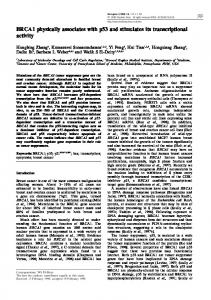
FIG. 8. Model for the interaction of p53 tetramers with consensus site DNA. The cylinders beneath the DNA-binding domains of p53 represent loop-sheethelix regions of p53 that make specific base contacts in the major grooves of a consensus DNA site (3). The arrows indicate the orientations of the globular p53 core DNA-binding domains and of the quarter-site repeats in the consensus DNA-binding sequence.
FIG. 7. Transactivation via the interaction of wild-type p53 (WT) with mutant p53 DNA consensus sites in vivo. p53-null mouse embryo fibroblasts were cotransfected with a reporter plasmid expressing the luciferase gene under the control of a variety of mutant p53 DNA consensus sites and with a plasmid expressing wild-type p53 as described in Materials and Methods. The histogram shows levels of luciferase activity. (A) The reporter plasmids contain partial p53 consensus sequences. (B) The reporter plasmids contain p53 consensus sequences with insertions. The structures of mutant consensus sites are shown at the left of the histograms. Arrows indicate quarter-site repeats of consensus DNA. The dashed line represents DNA without repeats.
ples of tetramers and binds consensus DNA which consists of four imperfect repeats. Because no smaller oligomers are evident in our preparations of purified p53, we and others have presumed that tetramers bind DNA. In the present report, we show directly that tetramers and octamers bind DNA with a single consensus site by showing that DNA comigrates with these native species during gel electrophoresis. Denatured monomers and cross-linked, denatured dimers, trimers, and tetramers served as ideal markers to demonstrate the tetrameric structure of the native p53 that bound consensus DNA. The finding that four monomers and only four monomers of p53 segment 1–320 or 80–290 bind a consensus sequence even in the absence of a tetramerization domain argues further that tetramers would be a natural DNA-binding form. Our findings are in agreement with those of Stenger et al. (25), who showed by quantitative scanning transmission electron microscopy that tetramers and multiples of tetramers bind DNA specifically, and with those of Hupp and Lane (14), who provided evidence by using antibodies that p53 binds DNA as tetramers. Our findings are not in agreement with those of Tarunina and Jenkins (26), who have argued that p53 dimers are the major DNA-binding form. Binding of p53 to a single DNA consensus sequence promotes tetramer-tetramer interactions. Stenger et al. (25) showed by electron microscopy that p53 exists as larger oligomeric forms on DNA than off DNA. They further showed that
p53 often forms oligomers in a plane perpendicular to bound DNA, and they designated these interactions ‘‘stacking’’ interactions. Electron microscopy, however, is not ideally suited to demonstrating shifts in a total population of molecules. Our present study demonstrates that the proportion of octamers in a total population of p53 is larger in the presence of DNA than in the absence of DNA. Because the DNA we used is small and has only a single DNA consensus site, it is likely that two tetramers stack perpendicularly to the plane of the DNA rather than side by side on the DNA. Furthermore, we add competitor DNA in our assays to minimize nonconsensus DNA binding. Whatever the configuration of the two tetramers bound in the region of a single DNA consensus site, the assembly of octamers induced by a single DNA consensus site would serve to increase the local concentration of p53 at promoters with p53 response elements and thereby to enhance transcription. Furthermore, each tetramer in an octamer could bind separated regions of DNA via DNA looping. Such interactions would translocate p53 bound to distal DNA to sites in promoters. Indeed, Stenger et al. have shown that p53 links separated consensus sites very efficiently (25). Four monomers of p53 bind cooperatively to a single DNA consensus sequence. p53 segment 80–320 consists mostly of monomers at low protein concentrations, but at high concentrations the segment forms unstable oligomers without a preference for a specific oligomeric form (30). The central, specific DNA-binding domain of p53, therefore, is capable of unstable monomer-monomer interactions in the absence of DNA. In contrast, four monomers of p53 consistently bind to a single DNA site even in the presence of an excess of specific and nonspecific DNA. Indeed, we can demonstrate the binding of fewer than four p53 monomers to oligonucleotides only when part of the DNA consensus site is deleted. These findings argue strongly that DNA promotes cooperative interactions among p53 monomers even in the absence of a tetramerization domain. Moreover, because p53 monomers bind very poorly to partial DNA consensus sites, these cooperative interactions clearly contribute to DNA binding efficiency in complete consensus sites. Presumably, the tetramerization domain pre-positions four monomers of p53 to enhance these cooperative interactions. p53, therefore, has three distinct protein-protein interactions that are related to DNA binding. The tetramerization domain increases the efficiency of p53 binding to DNA consensus sites. DNA binding in turn promotes cooperative
2164
WANG ET AL.
MOL. CELL. BIOL.
tetramer-tetramer and monomer-monomer interactions. Each of these interactions would increase the concentration of p53 at promoters with p53-binding sites. Tetramers bind to consensus DNA half-sites with rotational specificity. Our results with partial p53 DNA consensus sites indicate that each site consists of four quarter-site repeats and that two quarter-sites are best arranged as a half-site (34). The effects of insertions between half-sites confirm these conclusions. El-Deiry et al. (8) originally showed that p53 consensus half-sites were sometimes separated by 0 to 13 bp. Here, we show that p53 binds separated half-sites best when they are centered on the same face of the DNA helix. This finding supports the idea that a single tetramer engages the consensus site with or without an insertion. If two tetramers bound a consensus site with an insert, each tetramer would be freer to bind the half-sites simultaneously if the half-sites were on opposite faces of the helix. The role of rotational specificity in p53 DNA binding is strongly reinforced by transcriptional-activation studies done in vivo with graded insertions within p53 response elements in promoters. Tokino et al. (27) recently presented evidence that consensus sites with insertions of 4 or 14 bp in their centers had less than 10% of the activity of the contiguous consensus site. These results are similar to our present findings obtained with insertions of 5 and 15 bp. In contrast, insertions of 10 bp between half-sites have minimal effects on DNA binding and transactivation. Model for the binding of p53 tetramers to a single p53 DNA consensus sequence. Figure 8 shows a model for the specific binding of p53 to DNA. As shown by Cho et al. (3), a p53 monomer binds to a DNA quarter-site repeat and also makes contacts in adjacent quarter-sites. The unique orientation of the quarter-site determines the orientation of the bound monomer. We presume that the tetramerization domain prepositions the four central DNA-binding domains of a tetramer on one side of the helix in appropriate orientations to engage DNA readily, as originally suggested by Cho et al. (3). Cooperative interactions among monomers would further facilitate DNA binding. The nature of the tetramerization domain, perhaps in conjunction with side-by-side interactions among p53 subunits, may arrange the tetramers as a double dimer as shown by Clore et al. (4). Either the positioning of subunits within dimers or the conformation of monomers is not sufficiently flexible to accommodate insertions within DNA halfsites. In contrast, the arrangement of the C-terminal tetramerization domain as a double dimer would allow sufficient flexibility between the dimers to bind consensus DNA with 10-bp, but not 5- or 15-bp, insertions between half-sites. This idea is also consistent with the finding of Wang et al. (30) that the tetramerization region is largely responsible for the nonglobular shape of p53. Our model, which combines information from Cho et al., Clore et al., Wang et al., and our present study, will facilitate further characterization of the p53-DNA interaction and provides information important to the identification of potential p53 response elements in cellular promoters. ACKNOWLEDGMENTS This investigation was supported by Public Health Service grants NIH CA-28146, CA-18808, and CA-60089 awarded by the National Cancer Institute. REFERENCES 1. Bargonetti, J., P. N. Friedman, S. E. Kern, B. Vogelstein, and C. Prives. 1991. Wild-type but not mutant p53 immunopurified proteins bind to sequences adjacent to the SV40 origin of replication. Cell 65:1083–1091.
2. Bargonetti, J., J. Manfredi, X. Chen, D. R. Marshak, and C. Prives. 1993. A proteolytic fragment from the central region of p53 has marked sequencespecific DNA-binding activity when generated from wild-type but not from oncogenic mutant p53 protein. Genes Dev. 7:2565–2574. 3. Cho, Y. J., S. Gorina, P. D. Jeffrey, and N. P. Pavletich. 1994. Crystal structure of a p53 tumor suppressor DNA complex: understanding tumorigenic mutations. Science 265:346–355. 4. Clore, G. M., J. G. Omichinski, K. Sakaguchi, N. Zambrano, H. Sakamoto, E. Appella, and A. M. Gronenborn. 1994. High-resolution structure of the oligomerization domain of p53 by multidimensional NMR. Science 265:386– 391. 5. Demczuk, S., M. Harbers, and B. Vennstrom. 1993. Identification and analysis of all components of a gel retardation assay by combination with immunoblotting. Proc. Natl. Acad. Sci. USA 90:2574–2578. 6. Donehower, L. A., and A. Bradley. 1993. The tumor suppressor p53. Biochim. Biophys. Acta 1155:181–205. 7. Donehower, L. A., M. Harvey, B. L. Slagle, M. J. McArthur, C. A. Montgomery, J. S. Butel, and A. Bradley. 1992. Mice deficient for p53 are developmentally normal but susceptible to spontaneous tumours. Nature (London) 356:215–221. 8. El-Deiry, W. S., S. E. Kern, J. A. Pietenpol, K. W. Kinzler, and B. Vogelstein. 1992. Human genomic DNA sequences define a consensus DNA binding site for p53. Nat. Genet. 1:45–49. 9. Farmer, G., J. Bargonetti, H. Zhu, P. Friedman, R. Prywes, and C. Prives. 1992. Wild-type p53 activates transcription in vitro. Nature (London) 358: 83–86. 10. Fields, S., and S. K. Jang. 1990. Presence of a potent transcription activating sequence in the p53 protein. Science 249:1046–1049. 11. Funk, W. D., D. T. Pak, R. H. Karas, W. E. Wright, and J. W. Shay. 1992. A transcriptionally active DNA-binding site for human p53 protein complexes. Mol. Cell. Biol. 12:2866–2871. 12. Halazonetis, T. D., L. J. Davis, and A. N. Kandil. 1993. Wild-type p53 adopts a mutant-like conformation when bound to DNA. EMBO J. 12:1021–1028. 13. Hollstein, M., K. Rice, M. S. Greenblatt, T. Soussi, R. Fuchs, T. Sorlie, E. Hovig, B. Smith-Sorensen, R. Montesano, and C. C. Harris. 1994. Database of p53 gene somatic mutations in human tumors and cell lines. Nucleic Acids Res. 22:3551–3555. 14. Hupp, T. R., and D. P. Lane. 1994. Allosteric activation of latent p53 tetramers. Curr. Biol. 4:865–875. 15. Kern, S. E., K. W. Kinzler, A. Bruskin, D. Jarosz, P. Friedman, C. Prives, and B. Vogelstein. 1991. Identification of p53 as a sequence-specific DNAbinding protein. Science 252:1708–1711. 16. Kern, S. E., J. A. Pietenpol, S. Thiagalingam, A. Seymour, K. W. Kinzler, and B. Vogelstein. 1992. Oncogenic forms of p53 inhibit p53-regulated gene expression. Science 256:827–830. 17. Pavletich, N. P., K. A. Chambers, and C. O. Pabo. 1993. The DNA-binding domain of p53 contains the four conserved regions and the major mutation hot spots. Genes Dev. 7:2556–2564. 18. Pennica, D., D. V. Goeddel, J. S. Hayflick, N. C. Reich, C. W. Anderson, and A. J. Levine. 1984. The amino acid sequence of murine p53 determined from a c-DNA clone. Virology 134:477–482. 19. Pietenpol, J. A., T. Tokino, S. Thiagalingam, W. S. El-Deiry, K. W. Kinzler, and B. Vogelstein. 1994. Sequence-specific transcriptional activation is essential for growth suppression by p53. Proc. Natl. Acad. Sci. USA 91:1998– 2002. 20. Raycroft, L., H. Wu, and G. Lozano. 1990. Transcriptional activation by wild-type but not transforming mutants of the p53 anti-oncogene. Science 249:1049–1051. 21. Reed, M., Y. Wang, G. Mayr, M. E. Anderson, J. F. Schwedes, and P. Tegtmeyer. 1993. p53 domains: suppression, transformation, and transactivation. Gene Expr. 3:95–107. 22. Shaulian, E., A. Zauberman, J. Milner, E. A. Davies, and M. Oren. 1993. Tight DNA binding and oligomerization are dispensable for the ability of p53 to transactivate target genes and suppress transformation. EMBO J. 12:2789–2797. 23. Slingerland, J. M., J. R. Jenkins, and S. Benchimol. 1993. The transforming and suppressor functions of p53 alleles—effects of mutations that disrupt phosphorylation, oligomerization and nuclear translocation. EMBO J. 12: 1029–1037. 24. Stenger, J. E., G. A. Mayr, K. Mann, and P. Tegtmeyer. 1992. Formation of stable p53 homotetramers and multiples of tetramers. Mol. Carcinog. 5:102– 106. 25. Stenger, J. E., P. Tegtmeyer, G. A. Mayr, M. Reed, Y. Wang, P. Wang, and P. V. C. Hough. 1994. p53 oligomerization and DNA looping are linked with transcriptional activation. EMBO J. 13:6011–6020. 26. Tarunina, M., and J. R. Jenkins. 1993. Human p53 binds DNA as a protein homodimer but monomeric variants retain full transcription transactivation activity. Oncogene 8:3165–3173. 27. Tokino, T., S. Thiagalingam, W. S. El-Deiry, T. Waldman, K. W. Kinzler, and B. Vogelstein. 1994. p53 tagged sites from human genomic DNA. Hum. Mol. Genet. 3:1537–1542. 28. Unger, T., J. A. Mietz, M. Scheffner, C. L. Yee, and P. M. Howley. 1993.
VOL. 15, 1995 Functional domains of wild-type and mutant p53 proteins involved in transcriptional regulation, transdominant inhibition, and transformation suppression. Mol. Cell. Biol. 13:5186–5194. 29. Vogelstein, B., and K. W. Kinzler. 1992. p53 function and dysfunction. Cell 70:523–526. 30. Wang, P., M. Reed, Y. Wang, G. Mayr, J. E. Stenger, M. E. Anderson, J. F. Schwedes, and P. Tegtmeyer. 1994. p53 domains: structure, oligomerization,
p53 BINDING TO DNA
2165
and transformation. Mol. Cell. Biol. 14:5182–5191. 31. Wang, Y., M. Reed, P. Wang, J. E. Stenger, G. Mayr, M. E. Anderson, J. F. Schwedes, and P. Tegtmeyer. 1993. p53 domains: identification and characterization of two autonomous DNA-binding regions. Genes Dev. 7:2575– 2586. 32. Zambetti, G. P., and A. J. Levine. 1993. A comparison of the biological activities of wild-type and mutant p53. FASEB J. 7:855–865.