Report
Cell Cycle 9:22, 4584-4591; November 15, 2010; © 2010 Landes Bioscience
Interaction of regulators Mdm2 and Mdmx with transcription factors p53, p63 and p73 Michal Zdzalik,1 Katarzyna Pustelny,2 Sylwia Kedracka-Krok,3 Krzysztof Huben,4 Aleksandra Pecak,2 Benedykt Wladyka,2 Stefan Jankowski,4 Adam Dubin,2 Jan Potempa1 and Grzegorz Dubin1,* Department of 1Microbiology, 2Analytical Biochemistry and 3Physical Biochemistry; Faculty of Biochemistry, Biophysics and Biotechnology; Jagiellonian University; Krakow; 4 Institute of Organic Chemistry; Department of Chemistry; Technical University of Lodz; Lodz, Poland
Key words: tumor suppressor protein p53, p53/p63/p73 family of transcription factors, Mdm2/Mdmx regulators, proto-oncogene proteins, protein-protein interactions Abbreviations: TA domain, transactivation domain; NMR, nuclear magnetic resonance; FP, fluorescence polarization; ITC, isothermal titration calorimetry
The negative regulation of p53, a major human tumor suppressor, by Mdm2 and Mdmx is crucial for the survival of a cell, whereas its aberrant function is a common feature of cancer. Both Mdm proteins act through the spatial occlusion of the p53 transactivation (TA) domain and by the ubiquitination of p53, resulting in its degradation. Two p53 homologues, p63 and p73, have been described in humans. Unlike p53, these proteins regulate developmental processes rather than genome stability. Both p63 and p73 contain TA domains homologous to that of p53, but relatively little is known about their regulation by Mdm2 or Mdmx. Here, we present a detailed characterization of the interaction of Mdm2 and Mdmx with the TA domains of p63 and p73. Earlier reports of Mdm2 and Mdmx interactions with p73 are substantiated by the detailed quantitative characterization reported in this study. Most importantly, earlier contradictions concerning the presumed interaction of the Mdm proteins with p63 are convincingly resolved and for the first time, the affinities of these interactions are determined. Finally, the contribution of these findings to our understanding of the physiological role of these interactions is discussed.
Introduction The p53 family of transcription factors consists of three homologous proteins, p53, p63 and p73, encoded by three distinct genes. The key role of p53 in tumor suppression is well established. Humans carrying germline p53 gene mutations (Li-Fraumeni syndrome) and p53-null mice develop normally but are characterized by the early, spontaneous onset of different types of cancers.1,2 This manifestation is consistent with the well-documented role of p53 as a “guardian of the genome.”3 Under genotoxic stress, p53 induces cell cycle arrest and activates repair mechanisms. If the latter fail, p53 directs the cell into the apoptotic pathway.4 Aberrant p53 function results in the accumulation of genomic mutations, which ultimately leads to cancer. In fact, the majority of human tumors display the inactivation of the p53 pathway.4 Although homologous to p53, the two other family members, p63 and p73, do not participate in the control of genome stability, but instead regulate embryonic development. In humans, p63 mutations correlate with multiple syndromes, all of which are characterized by abnormal body development.5,6 p63-deficient mice have truncated limbs, no hair follicles, no teeth and no mammary, lachrymal or salivary glands. p73 plays
an important role in neurogenesis, and p73-deficient mice exhibit neurological, pheromonal, and inflammatory defects.7 The association of p73 with human developmental deficiencies has not yet been described. The primary structure similarities of p53, p63 and p73 manifest mainly in their three consecutive, conserved domains: an N-terminal transcriptional transactivation (TA) domain linked to a DNA-binding domain, followed by an oligomerization domain. The activity of p53 is tightly regulated at the level of protein function and stability by its interaction with the negative regulators Mdm2 and Mdmx. These ubiquitin ligases bind the TA domain of p53, inhibiting its transactivating function by the steric occlusion of the binding sites specific for other factors. The Mdm proteins also ubiquitinylate p53, thus directing it for proteasomal degradation.8-10 Both p63 and p73 contain TA domains homologous to that of p53. However, the interaction of p73 and especially p63 with the Mdm proteins is incompletely characterized. The data pertinent to the presumed interaction between p63 and Mdm2/Mdmx are contradictory. Kojima et al.11 observed p63-Mdmx complex formation in a yeast two-hybrid assay, and in another study, the interaction of p63 with Mdm2 was demonstrated by coimmunoprecipitation.12 However, similar
*Correspondence to: Grzegorz Dubin; Email:
[email protected] Submitted: 08/18/10; Accepted: 10/05/10 Previously published online: www.landesbioscience.com/journals/cc/article/13871 DOI: 10.4161/cc.9.22.13871 4584
Cell Cycle
Volume 9 Issue 22
report
Report
experiments performed by others did not confirm the interaction between p63 and either Mdm2 or Mdmx.13,14 The interactions of p73 with Mdm2 and Mdmx are better documented, but the affinity of the p73-Mdmx interaction has not been reported.15,16 Therefore, the effect of Mdmx on p73 activity under physiological conditions remains to be examined. The aim of this study was to precisely determine and quantitatively describe the interaction network of the p53 family of transcription factors and the Mdm ubiquitin ligases, to assess the possible significance of these interactions in vivo. Results Design of the minimal TA domain peptides. Based on reports in the literature and a computer analysis, the 15-amino-acid peptide p53 (15–29) was selected as the model of the p53 TA domain. Analysis of sequence alignments and homology modeling of the p63 and p73 TA domains in complexes with both Mdm2 and Mdmx were performed. Peptides p63 (51–65) and p73 (11–25) were chosen to model the p63 and p73 TA domains, respectively. The control peptide p63 (Ala) was designed by mutating the residues most likely responsible for its Mdm interaction, based on homology modeling. Initial assessment of the binding of the p53, p63 and p73 TA domains to Mdm2 and Mdmx. We evaluated the presumed binding of p63 and p73 to Mdm2 and Mdmx using the model peptides representing the TA domains of p63 and p73. We used an NMR approach because it provides an elegant and reliable method of assessing the interaction of two binding partners. Most importantly, the direct association and concurrent changes in the protein structure are monitored, which eliminates any possible artifacts arising from either the presence of reporter molecules or nonspecific effects, such as protein denaturation or precipitation upon ligand addition. To assess the presumed interactions, the 1H-15N TROSY spectra of Mdm2 and Mdmx in the absence and presence of the p63 and p73 TA domains were recorded. The p53 TA domain was used as the positive control. As expected, the titration of both Mdm2 and Mdmx with the p53 TA domain resulted in marked changes in the recorded chemical shifts of amide protons and nitrogens (Sup. Fig. 1 and Fig. 1), direct evidence for the TA domain binding to both the Mdm proteins. The titration of Mdm2 and Mdmx with the p73 TA domain also resulted in changes in the chemical shifts, similar in extent to those observed for p53 (15–29). This demonstrates that both Mdm2 and Mdmx are capable of binding the p73 TA domain. Similar results were obtained when Mdm2 was titrated with the p63 TA domain, showing again a tight interaction. No protein aggregation or precipitation or other nonspecific effects were observed in either case, further strengthening the observations described above. Only the titration of Mdmx with p63 (51–65) yielded results that were less clearly interpretable. The observed changes in the recorded chemical shifts were clear but not as pronounced as those for the other interactions examined. We would usually interpret such shifts as evidence of binding were it not for the concurrent reduction in the quality of the spectra, which suggests the partial precipitation or aggregation of the protein
www.landesbioscience.com
upon the addition of the TA domain peptide. To verify the specificity of the p63 (51–65) interaction with Mdmx, we used a peptide derived from the p63 TA domain in which the residues corresponding to those responsible for p53 binding to Mdmx (Table 3) were replaced with alanines. The titration of either Mdm2 or Mdmx with p63 (Ala) did not result in any detectable changes in the recorded chemical shifts of the hydrogen or amide protons, thus demonstrating the lack of any interaction and confirming the specificity of the observed p63 (51–65) -Mdmx interaction. Determination of the binding affinities of the TA domains for the Mdm proteins: FP assay. Once the specific interactions of Mdm2 and Mdmx with the p63 and p73 TA domains were confirmed, we proceeded with experiments in determining the corresponding affinity constants. A knowledge of this binding strength is important to determine the physiological relevance of the demonstrated interactions. To this end, a series of titrations was performed, in which all the tested TA domains competed with a fluorescein-labeled peptide ligand for Mdm2 and Mdmx. Fluorescence polarization (FP) was used to monitor the fraction of the labeled ligand bound. The data were analyzed according to Huang.17 The well-established FP assay confirmed the results of the NMR experiments. Moreover, the Kd values for all the tested TA domains were determined for both Mdm2 and Mdmx (Fig. 2). Taken together, these results demonstrate that the affinities of the p53 and p73 TA domains for Mdm are similar and at least one order of magnitude stronger than the binding affinities of Mdm for p63. Binding affinity of Mdm proteins for the TA domains: tryptophan fluorimetry. The FP competitive binding assay is a well-established and reliable method of assessing the affinity of different ligands of proteinaceous receptors, both qualitatively and quantitatively. However, the detection of ligand binding is only indirect and the presence of a reporter ligand may cause unwanted interference. The quantitative description of the interaction is also complex and based on several assumptions, which may result in the misestimation of the Kd values. These issues do not generally pose a serious problem, but whenever possible, confirmation based on a more straightforward method is valuable. Here, we developed a simple method that allows us to directly monitor the binding of the TA domain to the Mdm protein without the use of any secondary reporter molecule. It is well established that tryptophan fluorescence is sensitive to the polarity of the solvent. The emission wavelength shows a significant blueshift upon transfer from a polar to a nonpolar environment. The TA domains of p53, p63 and p73 all contain a single tryptophan residue. Upon Mdm binding, Trp23 of p53 is buried inside a hydrophobic pocket.18-20 Therefore, it should be possible to monitor the binding of p53, and presumably also of p63 and p73, by measuring the change in the fluorescence emission wavelength. Although the N-terminal domains of Mdm2 and Mdmx used in this study do not contain tryptophan residues, phenylalanine and tyrosine fluorescence still interferes strongly with the assay, making the classical direct titration of the TA domain with Mdm2 impossible. However, the intensity of protein fluorescence is only one third of that of the TA domain at equimolar concentrations. Therefore, a modified titration is possible, in which the
Cell Cycle
4585
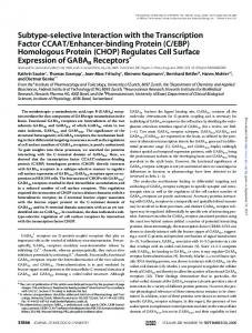
Figure 1. Titration of Mdm2 and Mdmx with the native p63(51–65) TA domain and its variant, in which the residues presumably essential for its interactions were substituted with alanines. The 1H-15N TROSY spectra of the uncomplexed Mdm protein (blue) and the spectra after the addition of the TA domain in a molar ratio of 1:1.2 (red) are shown. The observed changes in the chemical shifts upon the addition of native p63(51–65) but not the control p63(Ala) TA domain demonstrate the binding of the former to both Mdm2 and Mdmx.
molar ratio of Mdm:TA domain is kept constant (1:1) while the concentration of both agents is varied. Such a titration scheme was used in this study to determine the dissociation constants of the TA domains of p53, p63 and p73 for Mdm2 and Mdmx (Fig. 3). Our results confirmed the results of the NMR and FP assays, demonstrating interactions between all the TA domains tested and Mdm2 or Mdmx. Again, the interactions of the p63 TA domain with Mdm2 and Mdmx were weakest. The binding affinities determined for all the analyzed interactions were similar to the values obtained with FP (Table 2). Thermodynamic assessment of the TA domains—Mdm interactions. To confirm the affinity estimates described above using a method based on an independent underlying physicochemical principle and to thermodynamically characterize the force driving the observed interactions, isothermal calorimetric
4586
titrations were performed (Fig. S2). The data were analyzed according to a single-site binding model. The thermodynamic binding parameters obtained are summarized in Table 1. The determined Kd values correspond to those determined by FP and Trp fluorimetry. The equilibrium binding constants of the p53 transactivation domain for Mdm2 and Mdmx are similar, whereas the affinities of Mdm2 for both the p63 and p73 TA domains are weaker than those of Mdmx. The affinities of the p63 TA domain for both Mdm2 and Mdmx are weaker than those of either p73 or p53. The p63 (Ala) control peptide did not bind either Mdm2 or Mdmx, demonstrating again the specificity of the interactions studied. Regarding the driving force of the interactions between Mdm and the TA domains, in all cases, the binding was enthalpically driven and associated with unfavorable entropy changes.
Cell Cycle
Volume 9 Issue 22
same family, p63 and p73, regulate developmental processes. Because all three proteins contain homologous TA domains, it was speculated that p63 and p73 may be regulated by Mdm in a similar manner as has been described for p53. The ability of Mdm2 and Mdmx to bind to p73 has been relatively well documented (Sup. Table 1) and here, we have provided a detailed kinetic characterization of this interaction. The interaction of Mdm with p63 has also been studied previously, but the results were contradictory (Sup. Table 1).11-14 Here, we demonstrate that both Mdm2 and Mdmx form complexes with the p63 TA domain, although the interactions are weaker than those determined for both p53 and p73 (Table 2). The interaction of the p63 TA domain is specific and mechanistically similar to that of the p53 TA domain, because the p63 (Ala) peptide exhibited no activity in the assays performed. The observed blueshift in tryptophan fluorescence and the similar thermodynamic parameters further demonstrate that the p63 and p73 TA domains bind the Mdm proteins in a manner similar to that of the p53 TA domain. Independent estimates of the p53 binding affinity for Mdm2 range from 0.05 to 14 μM and for Mdmx from 0.2 to 6 μM (Sup. Table 1).18,21,28-35 The discrepancies between these results arise from the different lengths of the TA domain fragments evaluated by different authors, the inherent inconsistencies in concentration determinations, and the diverse methodologies and approximations used. Therefore, the data obtained in this study correspond well with the work of other authors, validating the methodology used. Although the interactions of p73 with Mdm2 and Mdmx have also been studied previously, only the affinity of p73 for Mdm2, with Kd = 0.26 μM or 1.9 μM, has been reported.29,30 To the best of our knowledge, this study is the first to report the Figure 2. Competitive displacement of a fluorescently labeled peptide ligand of affinity of Mdmx for p73. The affinity of p63 for Mdm2 (A) and Mdmx (B) by p53(15–29), p63(51–65), p73(11–25) and p63(Ala). either Mdm2 or Mdmx has not been reported previously. In fact, contradictory data made it impossible Enthalpy changes lower than those for the other interactions to infer whether these proteins were able to interact at all. This studied were observed for the binding of Mdm2 to both p63 and study provides clear evidence that both Mdm2 and Mdmx interp73. These were compensated by corresponding lower changes in act with p63 via its TA domain in vitro. However, the interaction the entropy values. The findings described above indicate that, as is one order of magnitude weaker than those of Mdm with p53 expected, the interactions of all the studied TA domains with the and p73. Mdm proteins are driven by a similar mechanism. The affinities of both Mdm2 and Mdmx for p73 are of the same order of magnitude as those for p53, which justifies the conDiscussion clusion that these proteins truly interact in cells, as has previously been suggested in several studies. The weaker interactions of both The interactions of p53 with Mdm2 and Mdmx, mediated via Mdm2 and Mdmx with p63 explain the contradictory results the TA domain of p53, are well characterized.19-22 The physi- reported by different authors on the interactions of those proological importance of the regulation of p53 by the Mdm2 and teins. Clearly, at sufficiently high concentrations, these proteins Mdmx ubiquitin ligases, as well as the role of its aberrant regu- will form a stable complex, but whether such concentrations are lation in tumors, is widely recognized.23-27 Unlike p53, which ever encountered under physiological conditions in cells remains protects genomic stability, the two homologous proteins of the a very interesting question for future studies. It is also noteworthy
www.landesbioscience.com
Cell Cycle
4587
that although the affinities of p53 for Mdm2 and Mdmx are similar, both p63 and p73 interact more strongly with Mdmx. Therefore, Mdmx, but not the more extensively studied Mdm2, may have a stronger impact on the regulation of intracellular p63 and p73. In conclusion, this study provides for the first time clear evidence of TA-domain-mediated interactions between p63 and both Mdm2 and Mdmx. It was also demonstrated that the mechanistic details of Mdm2 and Mdmx binding are similar for p53, p63 and p73. Importantly, the affinities of p63 and p73 for Mdm2 and Mdmx were comprehensively quantified. It clearly remains a challenge for future studies to determine the physiological significance of the findings described here, probably by updating our current picture of the roles of Mdm2 and Mdmx in the regulation of not only p53 but also the p73 transcription factor, and possibly even p63. Materials and Methods Protein preparation. The N-terminal domains of human Mdm2 and Mdmx were expressed and purified as previously described, with minor modifications.36 In brief, a construct encoding recombinant human Mdm2 (residues 1–118) was cloned into the pET-20 vector and expressed in Escherichia coli BL21(DE3) RIL. The cells were grown at 37°C and induced with 1 mM isopropyl β-D-1-thiogalactopyranoside (IPTG) (A&A Biotechnology) at an optical density at 600 nm (OD600) of 0.9. After induction, the cells were cultured for a further 5 h. The recombinant protein was purified from the inclusion bodies. After the inclusion bodies were washed with PBS containing 0.05% Triton X-100, they were solubilized in 6 M guanidinium hydrochloride (GuHCl) (Applichem) in 100 mM Tris-HCl (pH 8.0) conFigure 3. Binding of the p53(15–29), p63(51–65) and p73(11–25) TA domains to Mdm2 (A) and taining 1 mM EDTA (BioShop) and 10 mM Mdmx (B). Each point was determined by detecting the blueshift in the fluorescence β-mercaptoethanol (Sigma-Aldrich). The preparaemission peak of the tryptophan residue when the TA domain peptide was mixed with an equimolar amount of protein at a given concentration. A randomized, Trp-containing tion was dialyzed against 4 M GuHCl (pH 3.5) peptide was used as the negative control. and renatured by 1:100 dilution in 10 mM TrisHCl (pH 7.0) containing 1 mM EDTA and 10 mM β-mercaptoethanol. The diluted protein was incubated overnight production was induced with 0.5 mM IPTG at an OD600 of 0.6. at 4°C. Ammonium sulfate was added to a final concentration of The cells were cultured for a further 15 h at 20°C. Mdmx was 1.5 M and the protein was captured on Butyl Sepharose 4 Fast initially purified using affinity chromatography under nondenaFlow (Pharmacia). Mdm2 was eluted with 100 mM Tris-HCl turing conditions on Ni-NTA Agarose (Qiagen). The protein (pH 7.2) containing 10 mM β-mercaptoethanol and further puri- was further purified on HiLoad 16/60 Superdex 75 in 50 mM fied by gel filtration on HiLoad 16/60 Superdex 75 (Amersham KH2PO4, 50 mM Na 2HPO4, 150 mM NaCl and 5 mM DTT Biosciences) in 50 mM KH2PO4, 50 mM Na 2HPO4, 150 mM (pH 7.4). NaCl and 5 mM DTT (pH 7.4). p53, p63 and p73 transactivation domains. Peptides derived A construct encoding recombinant human Mdmx (resi- from the transactivation domains of p53, p63 and p73 and cordues 1–134) was cloned into pET-46Ek/LIC and expressed in responding to the fragment of p53 responsible for Mdm binding E. coli BL21(DE3) RIL. The cells were grown at 37°C. Protein were designed based on the analysis of the crystal structures of
4588
Cell Cycle
Volume 9 Issue 22
Table 1. Thermodynamic parameters of the binding of Mdm2 and Mdmx to the TA domains of the p53 family proteins determined by ITC Mdm protein
TA domain p53
0.48
(±0.04)
-13.0
(±0.6)
-15.02
(±0.0003)
-8.61
(±0.05)
Mdm2
p63
26
(±4)
-7.3
(±0.6)
-3.33
(±0.0137)
-6.24
(±0.09)
Mdmx
Kd (μM)
ΔH (kcal/mol)
ΔS (cal/mol*K)
ΔG25°C (kcal/mol)
p73
1.4
(±0.1)
-9.0
(±0.6)
-3.51
(±0.0005)
-7.99
(±0.05)
p53
0.42
(±0.03)
-14.9
(±0.1)
-20.60
(±0.0002)
-8.69
(±0.04)
p63
5.2
(±0.6)
-13.4
(±0.5)
-20.70
(±0.0021)
-7.20
(±0.06)
p73
0.22
(±0.02)
-13.4
(±0.1)
-14.51
(±0.0002)
-9.07
(±0.05)
Table 2. Affinities (Kd values) of Mdm2 and Mdmx for the TA domains of the p53 family proteins determined in this study Kd (µM) Mdm2
TA domain
NMR
p53(15–29)
+
0.55
(51–65)
p63
+
39.5
(±6.1)
p73(11–25)
+
3.10
(±0.66)
p63(Ala)
-
FP
Mdmx
ITC
(±0.13)
0.48
Trp fluorimetry
(±0.04)
1.07
26
(±4)
38.01
(±5.3)
1.4
(±0.1)
2.11
(±0.06)
-
(±0.04)
-
nd
NMR +
FP
ITC
(±0.01)
+
6.07
(±0.20)
5.2
(±0.6)
15.4
(±1.5)
+
0.25
(±0.01)
0.22
(±0.02)
0.97
(±0.13)
-
-
0.42
(±0.03)
Trp fluorimetry
0.26
-
1.01
(±0.10)
nd
(+) indicates “binding”; (-) indicates no interaction. Table 3. Sequences of the p53, p63 and p73 TA domain fragments used in this study Peptide
Sequence
UniProt no.
H-SQE TFS DLW KLL PEN-OH
P04637
(51–65)
p63
H-SPE VFQ HIW DFL EQP-OH
Q9H3D4
p73(11–25)
H-GGT TFE HLW SSL EPD-OH
O15350
p63
H-SPE VAQ HIA DFA EQP-OH
-
*
p53(15–29)
(Ala)
Residues responsible for Mdm-p53 binding and the corresponding residues in p63 and p73 are highlighted in bold. The mutated residues in the control peptide are underlined. *Residue numbering according to the cited UniProt entries.
the p53 TA domain in complexes with Mdm2 and Mdmx, the complementing studies of other authors, the sequence similarity in the region of interest and homology modeling.19,20,22,37 The peptides were chemically synthesized (Lipopharm; Biomatic), HPLC purified, and their identities confirmed with mass spectrometry. All the peptides were of more than 95% purity. A control peptide was also synthesized with a sequence identical to that of the p63 TA domain, except that the residues corresponding to those responsible for Mdm-p53 binding were replaced with alanines (Table 3). Nuclear magnetic resonance (NMR) spectroscopy. The direct binding of the Mdm proteins and TA domain fragments of the p53 family proteins was assessed according to a modified method of Stoll and colleagues,38 by comparing the 1H-15N transverse relaxation optimized spectroscopy (TROSY) spectra 39 recorded for uniformly 15N-labeled Mdm2 (0.53 mM) and MdmX (0.65 mM) proteins in the absence and presence of the analyzed TA domain peptides in a molar ratio of 1:1.2. The measurements were made in a buffer containing 50 mM KH2PO4, 50 mM Na 2HPO4, 150 mM NaCl, 5 mM DTT, 0.02% NaN3 and 5% D2O (pH 7.4). Before their addition, the peptides were dissolved in d6 -DMSO (Sigma-Aldrich). All spectra were recorded www.landesbioscience.com
on a Bruker Avance II Plus 700 MHz spectrometer at 25°C. Two hundred and fifty-six points in the t1 dimension and 4,096 points in the t2 dimension were acquired with 32 scans for each experiment. The data were processed using the TopSpin 2.1 software. Linear prediction (128 points, 512 coefficients) was applied. In the recorded TROSY spectra, each signal was shifted by 46 Hz in both directions. Fluorescence polarization (FP) competitive binding assay. The FP competitive binding assay and data analysis were performed as described previously.36 Briefly, the competition of the analyzed TA domains with a labeled high-affinity ligand (20 nM 5-carboxyfluorescein-TSFAEYWNLLSP)28 for binding with Mdm2 (11 nM) or Mdmx (52 nM) was determined by measuring the FP of the labeled ligand in the presence of increasing concentrations of p53 (15–29), p63 (51–65), p73 (11–25) or p63 (Ala) (0–500 μM). The affinity constants of the labeled ligand (20 nM) were determined by titration with increasing concentrations of Mdm2 (0–1 μM) or Mdmx (0–5 μM). All measurements were made in 10 mM Tris-HCl (pH 8.0) containing 50 mM NaCl and 1 mM EDTA. Stock solutions of all the peptides were prepared in DMSO. Care was taken not to exceed a final DMSO concentration of 5% in the reaction mixture. FP was measured on black NBSTM assay plates (Corning) at room temperature (~20°C) with 485 nm excitation and 535 nm emission. The data were analyzed according to Huang17 using the OriginPro 8 software. Tryptophan fluorescence binding assay. The binding constants of the TA domain peptides of Mdm2 and Mdmx were assessed by quantifying the blueshift in the tryptophan fluorescence emission peak. Equimolar mixtures of p53 (15–29), p63 (51–65) or p73 (11–25) with either Mdm2 or Mdmx were prepared in the concentration range of 0.26–50 μM. The experiments were performed at room temperature (~20°C) in a buffer containing 50 mM KH2PO4, 50 mM Na 2HPO4, 150 mM NaCl and 5 mM DTT (pH 7.4). Tryptophan was excited at 295 nm and the wavelength
Cell Cycle
4589
of the peak emission was determined by scanning from 310 to 360 nm using a Hitachi F-2500 fluorescence spectrophotometer. The measured values were corrected for the contribution of free Mdm2 or Mdmx as appropriate. The Kd value was determined from a titration curve that plotted the corrected value for the blueshift of the tryptophan fluorescence emission peak against the peptide concentration (equivalent to the protein concentration). The p63 (Ala) peptide could not be used as a control in this assay because it lacks tryptophan residues. Instead, a peptide with the randomized sequence H-APARSPSPSTQPWEHV-OH was used. Isothermal titration calorimetry (ITC). ITC was used to thermodynamically characterize the binding of Mdm2 or Mdmx to the p53 family TA domains. All experiments were performed in 50 mM KH2PO4, 50 mM Na 2HPO4, 150 mM NaCl and 5 mM DTT (pH 7.4) at 25°C using a VP-ITC MicroCalorimeter. Before the experiments, the Mdm proteins were extensively dialyzed into the appropriate buffer, and the TA domain peptides were prepared in the dialysate to minimize any artifacts. A typical binding experiment involved 5 μL injections of peptide solution (600 μM) into the calorimeter cell (1,436 mL) containing the protein (30 μM). The heat of dilution was determined by References 1.
2.
3. 4. 5.
6.
7.
8.
9.
10.
11.
12.
Malkin D, Li FP, Strong LC, Fraumeni JF Jr, Nelson CE, Kim DH, et al. Germ line p53 mutations in a familial syndrome of breast cancer, sarcomas and other neoplasms. Science 1990; 250:1233-8. Donehower LA, Harvey M, Slagle BL, McArthur MJ, Montgomery CA Jr, Butel JS, et al. Mice deficient for p53 are developmentally normal but susceptible to spontaneous tumours. Nature 1992; 356:215-21. Lane DP. Cancer. p53, guardian of the genome. Nature 1992; 358:15-6. Schuler M, Green DR. Mechanisms of p53-dependent apoptosis. Biochem Soc Trans 2001; 29:684-8. Mills AA, Zheng B, Wang XJ, Vogel H, Roop DR, Bradley A. p63 is a p53 homologue required for limb and epidermal morphogenesis. Nature 1999; 398:708-13. Yang A, Schweitzer R, Sun D, Kaghad M, Walker N, Bronson RT, et al. p63 is essential for regenerative proliferation in limb, craniofacial and epithelial development. Nature 1999; 398:714-8. Yang A, Walker N, Bronson R, Kaghad M, Oosterwegel M, Bonnin J, et al. p73-deficient mice have neurological, pheromonal and inflammatory defects but lack spontaneous tumours. Nature 2000; 404:99-103. Lohrum MA, Vousden KH. Regulation and activation of p53 and its family members. Cell Death Differ 1999; 6:1162-8. Shvarts A, Steegenga WT, Riteco N, van Laar T, Dekker P, Bazuine M, et al. MDMX: a novel p53-binding protein with some functional properties of MDM2. EMBO J 1996; 15:5349-57. Badciong JC, Haas AL. MdmX is a RING finger ubiquitin ligase capable of synergistically enhancing Mdm2 ubiquitination. J Biol Chem 2002; 277:49668-75. Kojima T, Ikawa Y, Katoh I. Analysis of molecular interactions of the p53-family p51(p63) gene products in a yeast two-hybrid system: homotypic and heterotypic interactions and association with p53-regulatory factors. Biochem Biophys Res Commun 2001; 281:1170-5. Calabro V, Mansueto G, Parisi T, Vivo M, Calogero RA, La Mantia G. The human MDM2 oncoprotein increases the transcriptional activity and the protein level of the p53 homolog p63. J Biol Chem 2002; 277:2674-81.
4590
overtitration for p53 (15–29) and p73 (11–25) and by the titration of the protein-free buffer for p63 (51–65). The data were analyzed using the Levember-Marquardt algorithm implemented in the MicroCal Origin software. Acknowledgements
We would like to express our sincere thanks to Dr. Tad A. Holak of the Max Planck Institute of Biochemistry (Martinsried, Germany) for his continued advice and support. This work was supported in part by grants 77/L-1/09 (to G.D.) from the Polish National Centre for Research and Development, DS I-18/10/2010 from the Technical University of Lodz (to K.H. and S.J.), and by the Jagiellonian University (statutory fund DS/9/WBBiB). We acknowledge the financial support of the European Union structural funds (grants POIG.02.01.00-12064/08 and POIG.02.01.00-12-167/08). Note
Supplementary materials can be found at: www.landesbioscience.com/supplement/ZdzalikCC9-22-sup. pdf
13. Little NA, Jochemsen AG. Hdmx and Mdm2 can repress transcription activation by p53 but not by p63. Oncogene 2001; 20:4576-80. 14. Wang X, Arooz T, Siu WY, Chiu CH, Lau A, Yamashita K, et al. MDM2 and MDMX can interact differently with ARF and members of the p53 family. FEBS Lett 2001; 490:202-8. 15. Zeng X, Chen L, Jost CA, Maya R, Keller D, Wang X, et al. MDM2 suppresses p73 function without promoting p73 degradation. Mol Cell Biol 1999; 19:3257-66. 16. Ongkeko WM, Wang XQ, Siu WY, Lau AW, Yamashita K, Harris AL, et al. MDM2 and MDMX bind and stabilize the p53-related protein p73. Curr Biol 1999; 9:829-32. 17. Huang X. Fluorescence polarization competition assay: the range of resolvable inhibitor potency is limited by the affinity of the fluorescent ligand. J Biomol Screen 2003; 8:34-8. 18. Popowicz GM, Czarna A, Rothweiler U, Szwagierczak A, Krajewski M, Weber L, et al. Molecular basis for the inhibition of p53 by Mdmx. Cell Cycle 2007; 6:2386-92. 19. Kussie PH, Gorina S, Marechal V, Elenbaas B, Moreau J, Levine AJ, et al. Structure of the MDM2 oncoprotein bound to the p53 tumor suppressor transactivation domain. Science 1996; 274:948-53. 20. Popowicz GM, Czarna A, Holak TA. Structure of the human Mdmx protein bound to the p53 tumor suppressor transactivation domain. Cell Cycle 2008; 7:2441-3. 21. Zondlo SC, Lee AE, Zondlo NJ. Determinants of specificity of MDM2 for the activation domains of p53 and p65: proline27 disrupts the MDM2-binding motif of p53. Biochemistry 2006; 45:11945-57. 22. Lin J, Chen J, Elenbaas B, Levine AJ. Several hydrophobic amino acids in the p53 amino-terminal domain are required for transcriptional activation, binding to mdm-2 and the adenovirus 5 E1B 55 kD protein. Genes Dev 1994; 8:1235-46. 23. Kubbutat MH, Jones SN, Vousden KH. Regulation of p53 stability by Mdm2. Nature 1997; 387:299-303. 24. Haupt Y, Maya R, Kazaz A, Oren M. Mdm2 promotes the rapid degradation of p53. Nature 1997; 387:296-9. 25. Okamoto K, Taya Y, Nakagama H. Mdmx enhances p53 ubiquitination by altering the substrate preference of the Mdm2 ubiquitin ligase. FEBS Lett 2009; 583:2710-4.
Cell Cycle
26. Danovi D, Meulmeester E, Pasini D, Migliorini D, Capra M, Frenk R, et al. Amplification of Mdmx (or Mdm4) directly contributes to tumor formation by inhibiting p53 tumor suppressor activity. Mol Cell Biol 2004; 24:5835-43. 27. Linke K, Mace PD, Smith CA, Vaux DL, Silke J, Day CL. Structure of the MDM2/MDMX RING domain heterodimer reveals dimerization is required for their ubiquitylation in trans. Cell Death Differ 2008; 15:841-8. 28. Pazgier M, Liu M, Zou G, Yuan W, Li C, Li C, et al. Structural basis for high-affinity peptide inhibition of p53 interactions with MDM2 and MDMX. Proc Natl Acad Sci USA 2009; 106:4665-70. 29. Burge S, Teufel DP, Townsley FM, Freund SM, Bycroft M, Fersht AR. Molecular basis of the interactions between the p73 N terminus and p300: effects on transactivation and modulation by phosphorylation. Proc Natl Acad Sci USA 2009; 106:3142-7. 30. Schon O, Friedler A, Bycroft M, Freund SM, Fersht AR. Molecular mechanism of the interaction between MDM2 and p53. J Mol Biol 2002; 323:491-501. 31. Bottger A, Bottger V, Garcia-Echeverria C, Chene P, Hochkeppel HK, Sampson W, et al. Molecular characterization of the hdm2-p53 interaction. J Mol Biol 1997; 269:744-56. 32. Phan J, Li Z, Kasprzak A, Li B, Sebti S, Guida W, et al. Structure-based design of high affinity peptides inhibiting the interaction of p53 with MDM2 and MDMX. J Biol Chem 285:2174-83. 33. Yu GW, Rudiger S, Veprintsev D, Freund S, FernandezFernandez MR, Fersht AR. The central region of HDM2 provides a second binding site for p53. Proc Natl Acad Sci USA 2006; 103:1227-32. 34. Brown CJ, Srinivasan D, Jun LH, Coomber D, Verma CS, Lane DP. The electrostatic surface of MDM2 modulates the specificity of its interaction with phosphorylated and unphosphorylated p53 peptides. Cell Cycle 2008; 7:608-10. 35. Lai Z, Auger KR, Manubay CM, Copeland RA. Thermodynamics of p53 binding to hdm2(1–126): effects of phosphorylation and p53 peptide length. Arch Biochem Biophys 2000; 381:278-84. 36. Czarna A, Popowicz GM, Pecak A, Wolf S, Dubin G, Holak TA. High affinity interaction of the p53 peptideanalogue with human Mdm2 and Mdmx. Cell Cycle 2009; 8:1176-84.
Volume 9 Issue 22
37. Dobbelstein M, Wienzek S, Konig C, Roth J. Inactivation of the p53-homologue p73 by the mdm2oncoprotein. Oncogene 1999; 18:2101-6. 38. Stoll R, Renner C, Hansen S, Palme S, Klein C, Belling A, et al. Chalcone derivatives antagonize interactions between the human oncoprotein MDM2 and p53. Biochemistry 2001; 40:336-44.
www.landesbioscience.com
39. Pervushin K, Riek R, Wider G, Wuthrich K. Attenuated T2 relaxation by mutual cancellation of dipole-dipole coupling and chemical shift anisotropy indicates an avenue to NMR structures of very large biological macromolecules in solution. Proc Natl Acad Sci USA 1997; 94:12366-71.
Cell Cycle
4591