Mar 23, 1976 - Extrachromosomal Elements in Salmonella typhimurium: Bacteriophage Type 505 ..... circular (CCC) plasmid DNA was isolated and puri-.
Vol. 127, No. 3 Printed in U.S.A.
JOURNAL OF BACTERIOLOGY, Sept. 1976, p. 1414-1426 Copyright © 1976 American Society for Microbiology
Interference with Propagation of Typing Bacteriophages by Extrachromosomal Elements in Salmonella typhimurium: Bacteriophage Type 505 J. D. A. VAN EMBDEN,* W. J. VAN LEEUWEN, AND P. A. M. GUINIE Rijks Instituut voor de Volksgezondheid, Bilthoven, The Netherlands
Received for publication 23 March 1976
Salmonella typhimurium bacteriophage type 505 is the most frequently encountered phage type in The Netherlands and its neighboring countries. Phage type 505 was analyzed with regard to the interference with propagation of the typing phages by the prophages and plasmids, present in the type strain S. typhimurium 505. This strain was found to carry two prophages and a plasmid aggregate, composed of a conjugative plasmid (pRI-19; molecular weight, 58 x 106), a nonconjugative tetracycline resistance plasmid (pRI-20; molecular weight, 5.8 x 10⁶), and three other non-self-transmissible plasmids (pRI-23, pRI-24, and pRI-25) having molecular weights between 2 x 10⁶ and 3 x 10⁶. To establish their role in the interference with propagation of the typing phages, the prophages and plasmids of type strain 505 were transferred to S. typhimurium strain Stm-1, which is sensitive to the typing phages and which has phage pattern 1. Plasmid pRI-19 was transferred by conjugation and pRI-20 was introduced separately by transformation of CaCl2-treated cells, using circular deoxyribonucleic acid (DNA) isolated from type strain 505. The other nonconjugative plasmids were introduced as a result of co-transformation with the resistance plasmid pRI-20. Each of the prophages and plasmids inhibited the propagation of the set of typing phages in a characteristic way, except for the tetracycline resistance plasmid pRI-20, which did not interfere with propagation of the typing phages. Upon introduction into strain Stm-1 of certain combinations of prophages and/or plasmids, phage pattern 1 of the recipient changed into phage pattern 505 and several other phage patterns that are also found in natural isolates. Other natural isolates showing phage type 505 were found to harbor the same combination of prophages and plasmids as found in type strain 505. Phage pattern 505 reflects the interference with propagation of typing phages, effected by the prophages and the plasmids present in strains of phage type 505. Salmonella typhimurium is the most frequently encountered serotype of Salmonella in many parts of the world (26). In The Netherlands, about 50% of all Salmonella strains isolated belong to this serotype (15). To further differentiate strains belonging to S. typhimurium, phage typing has emerged as a relatively simple technique with reliable and reproducible results. The typing scheme used in The Netherlands enables the subdivision of about 95% of all S. typhimurium strains into about 100 phage types (30). Sixty percent of all isolates belong to phage type 505. The differences in sensitivity to the typing phages can at least partly be explained in terms of restriction and modification. Chromosomal markers have been described as being responsible for certain restriction and modification sys-
tems (11, 12). It is increasingly evident that prophages and plasmids also play an important role in this respect (2, 25, 27). A systematic study of the prevalence of plasmids and prophages in the type strains of S. typhimurium was therefore undertaken. This report deals with strains of phage type 505. About 90% of natural isolates with phage type 505 are resistant to tetracycline (Tc). An annually varying percentage of 5 to 15% of the isolates is fully sensitive and about 0.4% of the isolates is resistant to ampicillin as well as to Tc. Multiresistant isolates are only sporadically encountered. Strains of phage type 505 are lysed only by a few typing phages adapted from wild-type phage 14-17 (17). The strains are finally identified by sensitivity to the wild-type phage 505 (17).
1414
PHAGE TYPING AND PLASMIDS OF S. TYPHIMURIUM
VOL. 127, 1976
In this study it is shown that prophages and plasmids of S. typhimurium isolates with phage type 505 are involved in the interference of the propagation of the typing phages (this phenotypic property will be termed Phi, in accordance with the recommendations of Novick et al. [22]). This was investigated by introduction of each of these elements and combinations of them into S. typhimurium Stm-1, which is sensitive to virtually all typing phages, although the chromosomal restriction systems LT and S were shown to be present in this strain (A. van Pel, personal communication). Furthermore, the molecular size of the plasmids of phage type 505 strains was established, and the function of each plasmid was investigated with regard to drug resistance and transfer properties in addition to the Phi characteristics.
1415
MATERIALS AND METHODS Bacterial strains. The bacterial strains used in this study are listed in Table 1. Frequently used derivative strains, carrying different plasmid combinations, are listed in Table 2. Media. Nutrient broth was prepared in this laboratory from fresh meat and contained, in addition, 0.5% NaCl, 1% peptone (Difco), and 0.069% Na2CO3, pH 7.5. Nutrient agar contained, in addition, 2% agar (BBL). These media were used throughout this study for all experiments unless otherwise stated. For phage typing, nutrient agar was prepared according to Scholtens (24). L broth has been described previously (19). Medium M was used for labeling deoxyribonucleic acid (DNA) with radioactive thymidine (28). Biotyping and phage typing. The techniques of biotyping and phage typing and the system used have been described previously (14, 16, 17). All
TABLE 1. Bacterial strains used Strain designation
Stm-1 Stm-lN Stm-lR Stm-470
Stm-505 Stm-73-307 W311ON M74
Relevant properties
Origin
S. typhimurium, type strain 1, phage type 1 Nalidixic acid-resistant mutant of Stm-1 Rifampin-resistant mutant of Stm-1 S. typhimurium, type strain 470, phage type 470, harboring a transfer factor S. typhimurium, type strain 505, phage type 505 S. typhimurium, phage type 505, isolated during 1973 from human source Nalidixic acid-resistant mutant of E. coli K-12 W3110, F-, XE. coli K-12, C600, harboring the non-self-transmissible streptomycin resistance plasmid pRI-18 (molecular weight, 5.6 x
Guinke et al. (17) By selection By selection Guinke et al. (17) Guinee et al. (17) This laboratory By selection This laboratory (unpublished data)
106)
M46
E. coli Row
Q1
S. gallinarum
E. coli W311ON carrying three independent plasmids derived from S. panama Sensitive for colicins S. typhimurium phage type 1 Sensitive for lysogenic phages carried by Salmonella strains of several serotypes
This laboratory (unpublished data) Fredericq Boyd (6) Natural isolate, this laboratory
TABLE 2. Frequently used strains, carrying different plasmid combinations, derived in this study Strain designation
S-i S-2 S-3 S-4 S-5 S-6 S-7 S-22 E-1, E-2 E-3, E-4 E-5, E-6, E-7 S-14
a Sm, Streptomycin.
Host
Stm-1
Stm-1 Stm-1 Stm-1 Stm-1 Stm-1 Stm-1 Stm-505
W311ON W311ON W3110N Stm-1R
Phage
pattern 1 B
C D C E F 505
221
Resistant to:
Tc Tc Tc Tc Tc Tc Tc Tc Sma Sm Sm
Derivation
Transformation by CCC DNA from Stm-505 Transformation by CCC DNA from Stm-505 Transformation by CCC DNA from Stm-505 Transformation by CCC DNA from Stm-505 Transformation by CCC DNA from Stm-505 Transformation by CCC DNA from Stm-505 Transformation by CCC DNA from Stm-505 Acriflavin curing of Stm-505 Mating, Stm-505 x W3110N Triple cross, Stm-505 x M74 x W311ON Triple cross, S-22 x M74 x W3110N Mating, E-3 x Stm-1R
VAN EMBDEN, VAN LEEUWEN, AND
1416
GUINgE
J. BACTERIOL.
TABLE 3. Phage patterns ofthe S. typhimurium type strains used in this study and the phage Typing phages in routine test dilution adapted from wild-type phages:
14-17 Designation of phage pattern o~~~~~~~~Oq i i
1 505 470 A-1i A-2 A-4 A-5 H B
c
D E F 335 221
222 501 502 504 a
S C _ _ - -
S C C 3 _ -3
- - -
-
-
-
-
-
-
-
-
-
-
-
-
1 C
S S S C 2
-
-
-
1 + C 3 C C S C C 3 2 + 3
a
C C CC - - - - S
1
-
-
-
-
-
-
-
-
-
-
-
-
-
-
-
-
-
-
-
-
-
-
-
C C C S S
00
0
C
CC
C
+ C C C +
+
1
S 1
1
-
-
-
-
-
-
-
-
-
-
-
-
-
-
-
-
-
-
-
-
-
-
-
+ + +
-
_ _ ----
S C S
C
----
S C C - 3
-
-
-
-
-
-
-
-
-
-
-
-
-
-
-
C C C C C C C C C C CC 2 2C C
2-
S C
C C C
-
-
-
c ccc c
-
-
S 3 CC 1 CC 1 CC - C C S C
0
on
a c; 3 C C C C S C C C C a-C C 3 m-C 2 m-C
-
-
C CC C C
-
_ _ _ _ _ S
-
t-
-
+ C
2 3 3 2 2 +
cla0 Cj 4o=8o
LO
ccccc
ccc
C C C C S 2 C
C C C C C C 3 2 S C S
C C C C
C C C C
C C
C C C
+
1 2
++
S .
-
3
S 3
-
S 2 -
-
_
C, Confluent lysis; S, semiconfluent lysis; ±, +, 1, 2, 3, increasingly numerous plaques; -, no plaques;
phage patterns encountered in this study are listed in Table 3. Demonstration of lysogeny and preparation of lysogenic strains. An overnight culture of strain Stm-505 was centrifuged (10 min, 10,000 x g), and the supernatant was sterilized by means of membrane filtration (Millipore Corp.; pore size, 0.45 am) and spotted in dilutions on nutrient agar plates seeded with a broth culture of Stm-1. After overnight incubation at 370C, isolated plaques together with a small portion of the surrounding culture were transferred to 10 ml of broth and incubated with aeration for 4 to 6 h. The phage suspension was purified by centrifugation and filtration and spotted in dilutions on strain Stm-1. Secondary growth in the areas of confluent lysis by the 10-1 dilution was isolated and purified by repeated single-colony isolation. Lysogeny was confirmed by testing for immunity to the corresponding phage and by demonstration of phage action on strain Stm-1. The lysogenic strains were phage typed. As far as could be ascertained, Stm-1 was not lysogenic, because no phage production could be demonstrated using indicator strains Ql (6) and S. gallinarum. Transfer of R-factors. Matings in liquid media were carried out by mixing equal volumes of overnight cultures of donor and recipient cells and by incubating the mixture overnight without aeration at 37°C. Matings on filters were performed by filtering 2 ml of overnight cultures of donor and recipient cells through a 0.45-j,m pore size membrane filter, followed by overnight incubation of the filter on a
nutrient agar plate at 37°C. The filter was blended in 2 ml of broth. Mating mixtures were plated on nutrient agar plates containing the appropriate drugs (Tc, 20 jig/ml [Pfizer]; nalidixic acid, 50 ,ug/ml [Winthrop Laboratories]; rifampin, 50 jig/ml [le Petit]; streptomycin, 100 ,ug/ml [Mycopharm] if Escherichia coli was the recipient and 200 ,ug/ml if S. typhimurium was the recipient). The triparental cross procedure of Anderson and Lewis (3) was used for mobilization of transfer-deficient plasmids. The frequency of transfer was expressed as the number of resistant recipient cells per total number of recipient cells. Elimination of drug resistance. The elimination of drug resistance determinants was carried out according to the technique of Watanabe and Fukasawa (31) by means of acriflavin (BDH). Isolation of DNA. Tritium-labeled DNA was obtained from 30-ml cultures containing [methyl3H]thymidine (10 ,uCi/ml; 18 Ci/mmol; The Radiochemical Centre, Amersham). Covalently closed circular (CCC) plasmid DNA was isolated and purified as described previously (23, 28). Unlabeled CCC DNA was obtained from 100-ml nutrient broth cultures. The same extraction procedure was followed. After purification, the CCC DNA was dialyzed extensively against TES buffer [0.01 M tris(hydroxymethyl)aminomethane, 0.01 M NaCl, and 0.001 M ethylenediametetraacetic acid, pH 8.1]. Lambda DNA was labeled with [methyl-'4C]thymine (62 mCi/ mmol; The Radiochemical Centre, Amersham) and purified as described by Korn and Weisbach (18). Centrifugation procedures. Sucrose density centrifugation was carried out as described previously
PHAGE TYPING AND PLASMIDS OF S. TYPHIMURIUM
VOL. 127, 1976
1417
patterns ofstrain Stm-1 after introduction of different Phi elements of strain Stm-505a
Wild-type phages 726
14-17
E1311
000 0 000 00 000 CCCCCCCCCCCCCCCCCC 3 3
2 3
00000 2 S - 2 +
C
C C0C C0 0 0000 0 0 0 0 C0 - - - C 0 C C C C C C 2 C C C C C C C0 1 C0 C C C 0C C CC C C C C C 0000 CC __-CCCCC--0 C0 - C 0 0 C CC C C C C C
-
________ --CCC-
-C - C
_-_
_ _ _ _ _ _ _ _ _ _ _ _ _ +-_-_
_ _
_
C C
_
-- -
_
-0000C
-O O -_____ _---0 -0000__
S - 2
- C-_8 __-C SS _____-cs ___-+ -C-- +--CCC___-C0S C S 2 _ - 3 .808_
-
in routine test dilutions
-
-
-
_ _ _ _
_ _ _ _
0
C C C0
C C C 0C C
_ _
_ _
- - s s CC _- _ _ _ _ _ _ _ - - S 3 3 1 _-0000 _ _ _ _ _ _ _ - - C C C C
C --C C0 -
-CC-c
---CC
- --- C - C C C C - C C C - C C - C -±-CC - + - C C
-_--C
-C
-0 C - C - C C C C -C C - C - - -C
C
m, minute plaques; 0, opaque lysis.
(28). 14C-labeled bacteriophage lambda DNA that had been heated for 2 min at 700C was used as a reference marker. Molecular weights were calculated from the sedimentation coefficients according to Bazaral and Helinski (5). Gamma ray irradiation of CCC DNA. To convert CCC DNA into its open circular form, DNA preparations in TES buffer were exposed at 00C to 60Co gamma irradiation at a dose rate of 20,000 rads/h. The samples were irradiated in glass tubes behind a buildup of 4 mm of perspex. Agarose-gel electrophoresis of DNA. Agarose-gel electrophoresis was done in tubes 10 cm in length and 6 mm in diameter. The agarose (Boehringer) concentration was 0.6%. Ethidium bromide (Calbiochem) was present in the gel and gel buffer at a concentration of 5 ,ug/ml. Further conditions of the electrophoresis were those described by Aay and Borst (1). The gels were photographed under longwave ultraviolet light, using a transilluminator (model C-50, Ultra Violet Products, San Gabriel, Calif.) and a Wratten gelatin (no. 9, Kodak) filter. Molecular weights were calculated from the inversely proportional relationship between the mobility of CCC DNA and the logarithm of the molecular weight (1). In each experiment a preparation of standard reference DNA was run parallel for calibration. The standard reference DNA isolated from strain M46 was composed of a mixture of three CCC plasmid components with molecular weights of 20.6 x 10⁶, 9.3 x 10⁶, and 2.7 x 10⁶. These values are based on contour-length measurements determined
by electron microscopy of the DNA molecules present in the mixture (van Embden, manuscript in preparation). Transformation of S. typhimurium by plasmid DNA. Cells were transformed to Tc resistance according to the method of Mandel and Higa (10, 20) with some modifications. Prewarmed nutrient broth was inoculuated with 1/100 volume of an overnight culture of strain Stm-1, and cells were grown under aeration at 37°C until a cell density of 2 x 10⁹ cells/ ml was obtained. The cells were centrifuged and washed with 1 volume of 0.01 M NaCl. Cells were resuspended in 1/5 volume of 0.03 M CaCl2 and kept for 20 min at 00C. The cells were centrifuged again and resuspended in 0.03 M CaCl2 (1/10 of the original culture volume). Transformation was done by mixing 1 volume of competent cells with 0.5 volume of DNA (concentration, between 1 and 10 jig/ml), and the CaCl2 concentration was adjusted to 0.20 M. The mixture was incubated for 60 min at 0°C, followed by a 1-min heat pulse at 420C. To this mixture 1;5 volume of L-broth containing Tc (1 ,ug/ml) was added, and incubation was carried out for 60 min at 370C. Samples were spread on nutrient agar plates containing Tc (5 ,g/ml). The transformation frequencies varied from 5 x 10⁻⁷ to 10-8 in the experiments, as expressed by the number of resistant transformants per total number of viable cells.
RESULTS Genetic characterization of lysogenic phages and plasmids of Stm-505. To study the
1418
VAN
EMBDEN,
VAN
LEEUWVEN, AND
GUINtE
significance of plasmids and prophages with regard to interference with propagation of the typing phages, each element was introduced into strain Stm-1. This strain is sensitive to all typing phages, except to phage 500 (see Table 3). Therefore, a possible interference of prophages or plasmids with virtually any typing phage will be reflected by the phage pattern of strains carrying prophages and/or plasmids from Stm-505. With Stm-1 as indicator, two lysogenic phages, termed Sf-1 and Sf-2, were detected in Stm-505. Phage Sf-1 produces plaques with halos and is identical to the natural phage A,b of the Boyd series (7). Phage Sf-1 is able to lysogenize Stm-1, altering its phage pattern into A-1 (Table 3). Phage Sf-2 produces minute plaques on Stm-1, altering its phage pattern by lysogenization into H (Table 3). Strains of phage pattern H are sensitive to almost all typing phages, in contrast to strains of phage pattern A-1. Stm-505 did not produce colicin with E. coli Row as indicator. The Tc resistance of Stm-505 was transferable to a nalidixic acid-resistant mutant of E. coli K-12 (W3110N), by conjugation on filter, with a very low frequency (about 10-9). The transfer frequency in liquid medium was below the detection level of about 10-10. To avoid lysogenization of Stm-1, E. coli was used as a primary recipient in mating experiments. The Tc resistance of two such transconjugants examined, E-1 and E-2 (Table 2), was transferable again to a rifampin-resistant mutant of Stm-1 (Stm-iR). At random, selected Tc-resistant transconjugants of the mating E-1 x Stm-iR showed phage patterns 335 and 222 (Table 4). Tc-resistant transconjugants of the mating E-2 x Stm-iR showed the phage patterns 1, C, F, and 335 (Table 4). The elimination frequency of Tc resistance by means of acriflavin was low, and only one Tc-
J. BACTERIOL.-
sensitive segregant (S-22) was obtained. Phage pattern 505 was not affected by the curing. These results suggest that the Tc resistance determinant is extrachromosomal in location and does not influence phage pattern 505. Stm-505 as well as its Tc-sensitive derivative S-22 were able to mobilize the non-self-transmissible streptomycin resistance plasmid pRI18 present in E. coli M74. The transfer frequency to the final recipient, W3110N, in liquid medium is about 10-7. Co-transfer of the Tc resistance was never observed in these mobilization experiments. Apparently, the transfer genes and the Tc resistance determinant are located on separate plasmids. Two transconjugants, E-3 and E-4, ofthe triple cross Stm-505 x M74 x W311ON and three transconjugants, E-5, E-6, and E-7, of the triple cross S-22 x M74 x W311ON were mated with Stm-iR, to investigate possible Phi characteristics associated with the transfer factor. These streptomycinresistant transconjugants showed the phage patterns 1, C, D, E, F, 335, 221, and 222 (Table 4). Only transconjugants with phage pattern 335, 221, or 222 were able to transfer the acquired drug resistance again to strain Stm-iN, which resulted in different changes in the phage pattern (Table 5). The drug resistance of transconjugants with pattern 1, C, D, E, or F was not transferable, indicating that the introduced elements with Phi properties in these strains are not able to promote conjugal transfer. However, the drug resistance could be mobilized by the transfer factor of strain Stm-470 and, again, phage patterns 1, C, D, E, and F were found after introduction in Stm-1 (Table 5), indicating again the extrachromosomal nature of the Phi elements. Introduction of the streptomycin resistance plasmid and the transfer factor of strain Stm-470 into strain Stm-1 did not alter phage pattern 1. These mating experiments suggest that
TABLE 4. Phage patterns of Stm-1 after introduction of different plasmids of Stm-505 by conjugation Properties of donor strain
ResistStrain E-1 E-2 E-3 E-4 E-5 E-6 E-7 a
b
ance de-
terminant Tc Tc Smb Sm Sm Sm Sm
Derived from mating:
Transfer frequency to Stm-
No. of strains with phage pattern:a 1
C
D
E
F
1 11 5 1 2
1 3 4 1
1
7
4 13 6 2
IR Stm-505 x W311ON Stm-505 x W311ON Stm-505 x M74 x W311ON Stm-505 x M74 x W311ON S-22 x M74 x W3110N S-22 x M74 x W311ON S-22 x M74 x W311ON
For phage reactions, see Table 3. Sm, Streptomycin.
10-6
10-"
10-5 10-9 10-6 10-6 10-6
3
Total no.
of transconju335 221 222 gants examined 9 1 10 3 9 14 1 5 55 4 19 5 1 10 8 10 7 10
PHAGE TYPING AND PLASMIDS OF S. TYPHIMURIUM
VOL. 127, 1976
TABLE 5. Segregation of the phage pattern of transconjugants after transfer of their acquired resistance Trans- MobiliPhage ferabil- zation of re- Phage patterns produced in pattern ityof ance recipient strain Stm-1 of donor aesto to ance to stanance strain strain Stm-1 StM_ja 1
1
-
+
C D E F 221 222 335
+ + +
+
1,C
+
1, D
+ +
1,D,E 1, C, D, E, F 1, 221 1, C, 221, 222 1,C,D,E,F,221,222,335
a Strain Stm-470 was used to mobilize the drug resistance determinant.
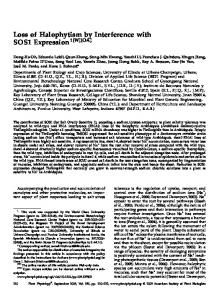
strain Stm-505 harbors, in addition to a transfer factor and a transfer-deficient Tc resistance plasmid, several other extrachromosomal elements that code for specific phage inhibition properties and are transfer deficient as well. Based on the results of these genetic studies, the number of plasmids was estimated to be at least four. Physical studies on the plasmids yielded more direct evidence on this point, as is shown below; these results will be presented first. Molecular properties of the plasmids in Stm-505. Agarose-gel electrophoresis of purified CCC DNA, isolated from strain Stm-505, indicated the presence of five different components (Fig. 1A). The molecular weights of four components were estimated from their relative mobilities to the standard reference plasmids and these were 5.8 x 10⁶, 2.9 x 10⁶, 2.5 x 10⁶, and 2.1 x 10⁶. No reliable molecular weight could be derived for the slowest migrating component, because the inversely proportional relationship between mobility and logarithm of the molecular weight does not hold for plasmids with a molecular weight of more than 25 x 106. These plasmids all tend to migrate at the same rate in 0.6% agarose gels (van Embden, unpublished data). A similar phenomenon has been observed with linear DNA molecules by Aay and Borst (1). Therefore, the molecular weight of the large circular component was derived from the sedimentation coefficient in sucrose gradients. CCC DNA from strain Stm-505 was sedimented in 5 to 20% sucrose gradients. Three distinct DNA species having S values of 72S, 46S, and 22-24S were present in this preparation (Fig. 2A). Prolonged centrifugation resolved the 22-24S DNA band into two components of 28S and 20-22S (Fig. 2B). Gamma ray
1419
irradiation with a dose of 920 rads resulted in a decrease of the 72S peak and a corresponding increase of the 46S peak (Fig. 2C). Apparently, the 72S material is composed of CCC DNA that is nicked to open circular DNA of 46S as a consequence of gamma ray irradiation. A value of 58 x 106 daltons was calculated for the 72S material. CCC DNA with S values of 28S and 20-22S correspond to molecular weights of 6.1 x 10⁶ and 2.8 x 10⁶ to 3.3 x 10⁶, respectively. These values are in agreement with those estimated from the mobility in agarose gels. Apparently the three smallest plasmids are not resolved in their separate components on the sucrose gradients. Confirmation for the CCC structure of the DNA bands in the agarose gels was obtained by gamma ray irradiation of the DNA preparation with a dose of 16,000 rads before gel electrophoresis. Again, all components migrated as discrete bands through the gels; however, the mobility of the four smaller components had decreased (Fig. 1B). These observations are consistent with the decrease in mobility to be expected when CCC DNA of a molecular weight ,pP!-20
*-- OC,
CCC PRI-20-
-.
OC PR!-23 -OC pRI-2.4 -OC DRP-25
CCC pRI-23CCC pRI-24CCC pRI-25-
FIG. 1. DNA bands in 0.6% agarose gels after electrophoresis in the presence of 5 pg of ethidium bromide per ml. Bands were made visible under ultraviolet light. (A) CCC DNA from Stm-505. (C) CCC DNA from the Tc-sensitive derivative S22. (B and D) Same as (A) and (C), respectively, but DNA preparations were gamma ray irradiated with a dose ofl 6,000 rads before electrophoresis. Plasmid pRI-19 is fully converted into its open circular (OC) form, whereas the smaller plasmids are partly nicked by irradiation with this dose.
into one of the five different phage patterns, B, C, D, E, or F. None of the transformants were able to transmit their acquired resistance to strain Stm-1N by conjugation, which is in agreement with the previous observation that none of the transconjugants with phage patterns identical to those of the transformants were transfer proficient (see Table 5). However, the resistance was easily mobilized to strain
Stm-1N by means of strain Stm-470, which harbors a transfer factor. CCC DNA isolated from Tc-resistant transformants with phage pattern 1, B, C, D, E, or F was analyzed by agarose-gel electrophoresis. It should be noted that strain Stm-1 carries a plasmid that has no function known to us. It migrated with the same mobility in the agarose gels as plasmid pRI-19 from strain Stm-505
VOL. 127, 1976
PHAGE TYPING AND PLASMIDS OF S. TYPHIMURIUM
E
I
E u
I
I
it
I
E
10 20 30 FIG. 2. Sucrose density gradient centrifugation of 3H-labeled CCC DNA isolated from Stm-505 (a). The procedures used are indicated in Materials and Methods. "4C-labeled bacteriophage lambda DNA, which had been heated for2 min at 70°C, was used as a reference marker (0). (A) Centrifugation for 90 min at 39,000 rpm; (B) centrifugation for 150 min at 39,000 rpm; (C) same as (A), but CCC DNA was gamma ray irradiated with 920 rads before centrifugation.
(Fig. 3). The Tc-resistant transformant S-1, with the phage pattern 1, carries only plasmid pRI-20 in addition to the cryptic plasmid (Fig. 3). This plasmid is also present in the trans-
1421
formants with phage patterns B, C, D, E, and F (Fig. 3; Table 6). The transformed strains S-2, S-3, and S-4, with phage patterns B, C, and D, respectively, were found to harbor, in addition to the cryptic plasmid and pRI-20, one more plasmid: pRI-25, pRI-23, or pRI-24, respectively (Fig. 3). Apparently, each of plasmids pRI-25, pRI-23, and pRI24 codes for specific Phi characteristics, and the phage patterns B, C, and D are the result of the introduction of the various plasmids into Stm-1. This is in contrast to plasmid pRI-20, which does not alter the phage sensitivity of strain Stm-1. The transformants S-5, S-6, and S-7, with phage patterns C, E, and F, respectively, were shown to carry more than one of the small Phi plasmids: pRI-23 + pRI-25, pRI-24 + pRI-25, and pRI-23 + pRI-24 + pRI-25, respectively (Fig. 3). CCC DNA derived from transformants S-1 through S-7 was used for transformation of competent Stm-1 cells, and Tc-resistant transformants were phage typed. The results (Table 6) are in agreement with the view that transformants with phage pattern 1 acquired only the Tc resistance plasmid pRI-20 and those with phage pattern B, C, D, E, or F received, in addition to pRI-20, one or more of the Phi plasmids pRI-23, pRI-24, and pRI-25. The transformation data suggest that the small Phi plasmids are co-transferred with the Tc resistance plasmid with high frequency in the transformation procedure. The experiments do not exclude the possibility that the co-transfer is due to the occurrence of recombinant molecules, between pRI-20 and the Phi plasmids, that are present in the DNA preparations and might not have been detected in the agarose gels. After introduction into strain Stm-1, these recombinant plasmids could then dissociate into their original components, i.e., pRI-20, pRI-23, pRI-24, and pRI-25. To learn whether the high rate of co-transfer could be explained in this way, strain Stm-1 was transformed with a mixture of CCC DNA isolated from strain S-1 (harboring pRI-20) and from the Tc-sensitive strain S-22 (carrying the plasmids pRI-19, pRI23, pRI-24, and pRI-25). Co-transfer of the Phi plasmids occurs with essentially the same frequency as in the transformation with CCC DNA isolated from type strain Stm-505 (Table 6). Using E. coli K-12 as a recipient, Cohen et al. (9) also observed the phenomenon of cotransformation, although the incidence was lower. It is concluded that the introduction of two or more independent plasmids into competent S. typhimurium cells can occur with a relatively high frequency, and this is not necessarily due to the presence of recombinant plasmid molecules.
1422
VAN EMBDEN, VAN LEEUWEN, AND GUINIE
J. BACTERIOL.
TABLE 6. Phage patterns of Tc-resistant Stm-1 transformants using CCC DNA from different donor strains and plasmid composition of donor strains Donor strain
505 S-i S-2 S-3 S-4 S-5 S-6 S-7 S-22 S-22 + S-1b
Phage No. of transformants with phage patterns: pattern
Presence of plasmids in donor Total no. strains as detected by electrophore-
of donor strain
1
B
C
analyzed D
E
F
505 A B C D C E F 505
46 35 16 18 23 7 30 20
6
4
12
14
21
13
103 35 30 30 44 20 50 60
5
20
14 12
21
5 2
8
1
2
5 11
1
3
2
13 13
-a
9
pRI-20
pRI-23
+ + + + +
+ + +
pRI-24
pRI-25
+
+
-
-
+ + + + + +
+
+ + +
+
+ + +
a -, No Tc-resistant transformants were obtained. ° Mixture of CCC DNA from both strains.
Function of plasmid pRI-19. As was shown above, none of the plasmids pRI-20, pRI-23, pRI-24, and pRI-25 are able to promote conjugal transfer. Therefore, the only plasmid considered for the transfer function would be pRI-19, which would be in agreement with the minimal size (about 20 x 106) of conjugative plasmids (8). Because of the presence in Stm-1 of the cryptic plasmid, which comigrates with pRI-19 in agarose gels, a transfer-proficient E. coli transconjugant was chosen for analysis of the plasmid content. Agarose-gel electrophoresis of CCC DNA isolated from strain E-3 (see Table 2) revealed the presence of four components with mobilities identical to pRI-19, pRI-20, pRI-23, and pRI-24. Sucrose density gradient centrifugation of tritium-labeled CCC DNA showed the presence of 72S, 28S, and 22S species (Fig. 4A and B). The 72S component was converted into 46S material upon gamma ray irradiation with a dose of 920 rads (Fig. 4C), and this species seems therefore equal in size with plasmid pRI19. It is concluded that pRI-19 carries the genes for conjugal transfer, because the other circular DNA components present in strain E-3, pRI-18, pRI-23, and pRI-24, have previously been shown to be non-self-transmissible. Plasmid properties of transconjugants. Conjugal transfer of the Tc resistance determinant of strain Stm-505 or mobilization of the streptomycin resistance plasmid of strain M74 by strain Stm-505 resulted in many different phage patterns of the recipient strain Stm-1 (Tables 4 and 5). The transfer-deficient transconjugants were found to have phage patterns 1, C, D, E, and F. This suggests that the nonself-transmissible plasmids pRI-23, pRI-24, and pRI-25 are frequently co-transferred with the drug resistance plasmid. Transfer-proficient transconjugants had
phage pattern 221, 222, or 335 (Table 5). The available evidence (Table 5) indicates that plasmid pRI-23 is present in strains with phage pattern 222, and the two plasmids pRI-23 and pRI-24 are present in strains with phage pattern 335. Agarose-gel electrophoresis with CCC DNA from strains with these phage patterns confirmed this hypothesis. Agarose-gel electrophoresis showed that strain S-14, with the phage pattern 221, was devoid of the small plasmids pRI-23, pRI-24, and pRI-25. This is consistent with the finding that none of the transconjugants of the mating S-14 x Stm-1R had phage pattern B, C, or D, which are characteristic for the small Phi plasmids. It is concluded that introduction of the transfer factor pRI-19 in Stm-1 gives rise to phage pattern 221 and that phage patterns 222 and 335 are produced when one or more of the Phi plasmids are co-transferred. Introduction of the lysogenic phage Sf-1 in plasmid-carrying strains. The influence on the phage pattern of all possible combinations of Phi elements of Stm-505 was studied by introducing them in Stm-1. Thus, six transformants carrying different combinations of the small Phi plasmids were lysogenized with prophage Sf-1. Transfer factor pRI-19 was introduced by mating with S-14 into the same six transformants, as well as into their Sf-1 lysogenic derivatives. No attempt was made to study the influence of prophage Sf-2 on the phage patterns. The presence of this lysogenic phage in Stm-1 inhibits the propagation of only a few typing phages, and therefore the influence of Sf-2 on the phage pattern of strains carrying most combinations of the other Phi elements would be masked. The resulting phage patterns of strains carrying the different combinations of Phi ele-
VOL. 127, 1976
PHAGE TYPING AND PLASMIDS OF S. TYPHIMURIUM
1423
SI S2 S3 S4 S5 S6 S7 Stm-l
cryptic plasmid -
pRI-20 -
pRI-23 pRI-24 pRI-25-
FIG. 3. Electrophoresis of CCC DNA isolated from strain Stm-1 and Tc-resistant transformants S-1 through S-7. Conditions of electrophoresis are those described in the legend to Fig. 1. The position of CCC components only is indicated. The other visible bands represent open circular DNA.
ments are shown in Table 7. Some strains carrying different combinations of plasmids had the same phage pattern. When all of the five Phi elements were introduced into strain Stm-1, the phage pattern characteristic for phage type 505 was recovered. Interestingly, Stm-1 carrying several combinations of these elements yielded phage patterns 501, 502, and 504, which are also found frequently in natural S. typhimurium isolates (30). Plasmids and lysogenic phages from natural isolates. To investigate whether the prophage and plasmid constitution of Stm-505 is representative for other isolates with the phage
pattern 505, we repeated all the genetic and molecular experiments described above, except the elimination of the Tc resistance. For this purpose the natural isolate 73-307 was used. The results of these experiments were essentially the same; two prophages and five different plasmids were found in this strain. All these elements had the same properties with regard to Phi character, transfer, drug resistance, and molecular size as those of Stm-505. From 11 other Tc-resistant natural isolates, CCC DNA was isolated and transformation to strain Stm-1 was done, which yielded again phage types 1, B, C, D, E, and F. Conjugal
1424
VAN EMBDEN, VAN LEEUWEN, AND GUINIE
J. BACTERIOL.
strains with phage type 505 were found not to carry the plasmid pRI-20, and they behaved identically to the Tc-cured strain S-22 with respect to their extrachromosomal properties. Each of 40 iiatural phage type 505 isolates tested produced two lysogenic phages with properties identical to Sf-1 and Sf-2. None of the strains produced colicin.
E
I
E
it CL
I
u
E
I"~
10
30 20 10 FIG. 4. Sucrose density centrifugation of CCC DNA isolated from strain E-3 (-), as described in the legend to Fig. 1. (A) Centrifugation for 90 min at 39,000 rpm; (B) centrifugation for 150 min at 39,000 rpm; (C) centrifugation as in (A), but CCC DNA was gamma ray irradiated with a dose of 920 rads before centrifugation.
transfer of the transfer factor led to phage pattern 221. Phage patterns 222 and 335 were found as well, indicating co-transfer of other Phi plasmids. Six Tc-sensitive S. typhimurium
DISCUSSION This study has shown that the two prophages and four of five plasmids carried by Stm-505 influence sensitivity towards the typing phages when these elements are introduced into the host Stm-1. Characteristic phage patterns arose when each of these Phi elements was transferred separately into strain Stm-1. The only plasmid that did not affect the phage susceptibility is the non-self-transmissible Tc resistance plasmid pRI-20. This plasmid, which has a molecular weight of 5.8 x 10⁶, resembles in size other non-selftransmissible drug resistance plasmids (13, 21, 28) and, because of its coexistence with the transfer-proficient plasmid pRI-19 in type strain 505, this plasmid complex is considered an "R-factor aggregate" (8) or a "class 2 Rfactor" (4). The transformation technique was used to introduce the plasmids into strain Stm-1, and it proved to be a powerful tool in establishing the function of the three small Phi plasmids, which have molecular weights between 2 and 3 million. We never observed the co-transformation of plasmid pRI-19, which might be caused by several factors such as competition with the small Phi plasmids, decreased permeability of competent Stm-1 cells for large DNA structures, or incompatibility with the resident cryptic plasmid in Stm-1. The phage patterns of Stm-1 derivatives carrying two or more of the Phi elements are consistent with the idea that the phage pattern is the result of superimposing the separate Phi properties of each element. Obviously, no synergism or antagonism between the nonchromosomal elements with respect to the interference with phage propagation was observed. If this would hold also for other natural Phi elements, this would greatly facilitate establishing the relationship between phage types of natural isolates and the carrying of plasmids and/or prophages. Phage patterns 221, 222, 335, 501, 502, 504, and 505, which were found after introduction of different combinations of the Phi elements in Stm-1, are identical to those of natural isolates with the corresponding phage types. One might
PHAGE TYPING AND PLASMIDS OF S. TYPHIMURIUM
VOL. 127, 1976
1425
TABLE 7. Phage pattern of strains carrying different combinations ofPhi elements Properties of recipient strains used
Phage pattern after introduction of: Phi plasmids present
Strain
S-i S-2 S-3 S-4 S-6 S-7
Phage pattern
pRI23
pRI-24
pRI-25
pRI-19
Prophage Sf1
1 B C D E F
+ +
+ + +
+ + +
221 221 222 220 220 335
A-1 A-1 A-2 A-4 A-4 A-5
anticipate the possibility that these natural isolates carry combinations of plasmids and prophages identical to those in the constructed strains. Preliminary results suggest that this is indeed true for phage types 501, 502, and 504. The genetic and molecular data obtained in this study of natural isolates of phage type 505, other than the type strain, suggest that this phage pattern is indicative of the presence of a particular set of Phi plasmids and prophages. The studies on strains carrying different combinations of plasmids and prophages indicated that some Phi elements can mask the Phi characteristics of other Phi elements (see Table 7). Although we believe that the prophages and plasmids from strains of phage type 505 play an important role in sensitivity to the typing phages, chromosomal determinants, which interfere with propagation of phages (11, 12), might be involved as well. Elimination of the Phi elements in strain Stm-505 would clarify the situation. However, the only element we were able to eliminate from strain Stm-505 was plasmid pRI-20, which does not interfere with phage propagation. The strains with phage type 505 predominate among natural isolates of S. typhimurium in The Netherlands and its adjacent countries (29). Studies of natural isolates with this phage type indicate that the described plasmids are very common in S. typhimurium strains isolated in this area. Experiments to establish whether these plasmids might have some function in the survival of salmonellae are in progress. ACKNOWLEDGMENTS We thank I. G. Andringa, N. W. Nagtegaal, M. M. M. van Nesselrooy, and D. Pruys for technical assistance and G. H. Hofmeester for carrying out the gamma-ray irradiation of DNA samples. LITERATURE CITED 1. Aay, C., and P. Borst. 1972. The gel electrophoresis of DNA Biochim. Biophys. Acta 269:197-200. 2. Anderson, E. S. 1966. Influence of the A transfer factor
3.
4.
5.
6. 7.
8. 9.
10.
11.
12. 13.
14.
15.
16. 17.
18.
pRI-19 + pro-
phage Sf-i 501 501 502 504 504 505
on the phage sensitivity of Salmonellae. Nature (London) 212:795-797. Anderson, E. S., and M. J. Lewis. 1965. Characterization of a transfer factor azsociated with drug resistance in Salmonella typhimurium. Nature (London) 208:843-849. Anderson, E. S., and E. Natkin. 1972. Transduction of resistance determinants and R factors of the transfer system by phage Plkc. Mol. Gen. Genet. 114:261-265. Bazaral, M., and D. R. Helinski. 1968. Characterization of multiple circular DNA forms of colicinogenic factor El from Proteus mirabilis. Biochemistry 10:3513-3519. Boyd, J. S. K. 1956. Immunity of lysogenic bacteria. Nature (London) 178:141. Boyd, J. S. K., and D. E. Bidwell. 1957. The type A phages of Salmonella typhimurium: identification by a standardized cross-immunity test. J. Gen. Microbiol. 16:217-228. Clowes, R. C. 1972. Molecular structure of bacterial plasmids. Bacteriol. Rev. 36:361-405. Cohen, S. N., A. C. Y. Chang, H. Boyer, and R. B. Helling. 1973. Construction of biologically functional bacterial plasmids in vitro. Proc. Natl. Acad. Sci. U.S.A. 70:3240-3244. Cohen, S. N., A. C. Y. Chang, and L. Hsu. 1972. Nonchromosomal antibiotic resistance in bacteria. VII. Genetic transformation of Escherichia coli by R-factor DNA. Proc. Natl. Acad. Sci. U.S.A. 69:2110-2114. Colson, C. A. M., and A. van Pel. 1970. Chromosomal location of host specificity in Salmonella typhimurium. J. Gen. Microbiol. 60:265-271. Colson, R. A., C. A. M. Colson, and A. van Pel. 1969. Host controlled restriction mutants of Salmonella typhimurium. J. Gen. Microbiol. 58:57-64. Falkow, S., J. D. A. van Embden, and P. Guerry. 1974. Molecular nature of two nonconjugative plasmids carrying drug resistance genes. J. Bacteriol. 117:619630. Guinke, P. A. M., W. H. Jansen, A. van Schuylenburg, and W. J. van Leeuwen. 1973. Bacteriophage typing of Salmonella typhimurium by use of a mechanized technique. Appl. Microbiol. 26:474-477. Guin&e, P. A. M., and J. Valkenburg. 1975. Salmonella isolations in The Netherlands, 1966-1973. Zentralbl. Bakteriol. Parasitenkd. Infektionskr. Hyg. Abt. 1 Orig. Reihe A 231:97-107. Guinee, P. A. M., W. J. van Leeuwen, and W. H. Jansen. 1972. New technique for biotyping. Appl. Microbiol. 23:1172-1174. Guinee, P. A. M., W. J. van Leeuwen, and D. Pruys. 1974. Phage typing of S. typhimurium in The Netherlands. 1. Phage typing system. Zentralbl. Bakteriol. Parasitenkd. Infektionskr. Hyg. Abt. 1 Orig. Reihe A 226:194-200. Korn, D., and A. Weisbach. 1962. Thymineless induc-
1426
19. 20. 21.
22.
23.
24. 25.
VAN EMBDEN, VAN LEEUWEN, AND
GUIN1gE
tion in Escherichia coli. Biochim. Biophys. Acta 61:775-790. Lennox, E. S. 1955. Transduction of linked genetic characters of the host by bacteriophage P1. Virology 1:190-206. Mandel, M., and A. Higa. 1970. Calcium dependent bacteriophage DNA infection. J. Mol. Biol. 53:159163. Milliken, C. E., and R. C. Clowes. 1973. Molecular structure plasmids of an R factor, its component drugresistance determinants and transfer factor. J. Bacteriol. 113:1026-1033. Novick, R. P., R. C. Clowes, S. N. Cohen, R. Curtiss III, N. Datta, and S. Falkow. 1976. Uniform nomenclature for bacterial plasmids: a proposal. Bacteriol. Rev. 40:168-189. Radloff, R. W., and J. Vinograd. 1967. A dye-bouyant density method for the detection and isolation of closed circular duplex DNA: the closed circular DNA in Hela cells. Proc. Natl. Acad. Sci. U.S.A. 57:15141520. Scholtens, R. T. 1950. Phage typing of Salmonella typhi in The Netherlands. Antonie van Leeuwenhoek J. Microbiol. Serol. 16:245-255. Scholtens, R. T., and J. A. Rost. 1972. Subdivision of Salmonella typhimurium into phage types and biotypes. Characterization of transferable factors by means of phage typing. The increase of drug-resist-
J. BACTERIOL.
ance in Salmonellas. Ann. Sclavo 14:309-344. 26. Sedlak, J., and H. Riswhe. 1968. Enterobacteriaceae Infektionen. Epidemiologie und Laboratoriumsdiagnostik. Georg Thieme, Leipzig. 27. Tsch&pe, H., A.-Z. Dragvs, H. Riache, and H. Kahn. 1974. Conversion of phage types of S. typhimurium due to different R-plasmids. Zentralbl. Bakteriol. Parasitenkd. Infektionskr. Hyg. Abt. 1 Orig. Reihe A 226:184-193. van Embden, J. D. A., and S. N. Cohen. 1973. Molecular 28. and genetic studies of an R-factor system consisting of independent transfer and drug-resistant plasmids. J. Bacteriol. 116:669-709. 29. van Leeuwen, W. J., and P. A. M. Guin6e. 1975. Frequency distribution of S. typhimurium phage types in various countries. Zentralbl. Bakteriol. Parasitenkd. Infektionskr. Hyg. Abt. 1 Orig. Reihe A 230:320-335. 30. van Leeuwen, W. J., D. Pruys, and P. A. M. Guin6e. 1974. Phage typing of S. typhimurium in The Netherlands. 2. Frequency distribution of S. typhimurium phage types in The Netherlands during 1971 and 1972. Zentralbl. Bakteriol. Parasitenkd. Infektionskr. Hyg. Abt. 1 Orig. Reihe A 226:201-206. 31. Watanabe, T., and T. Fukasawa. 1961. Episome-mev diated transfer of drug resistance in Enterobacteriaceae. II. Elimination of resistance factors with acridine dyes. J. Bacteriol. 81:679-683.