during REM sleep which is similar to the disconnection syndrome seen after corpus callosotomy. Other research suggests that an increase in interhemispheric.
BRAIN AND COGNITION
..
14, 113-125 (\990)
Interhemispheric EEG Coherence during Sleep and Wakefulness in Left- and Right-Handed Subjects
, ToRE NIELSEN, ALAIN ABEL, DoMINIQUE LORRAIN. AND JACQUES MONTPLAISIR
Centre D'etude du Sommeil, Hopital du Sacre-Coeur, and Universite de Montreal, Quebec, Canada
T.. 0
REM sleep is associated with the production of complex imagery sequences. Yet research is divided as to whether different brain regions are more or less coordinated in their functioning at this time. Some research suggests that there may occur a functional disconnection of the left and right cerebral hemispheres during REM sleep which is similar to the disconnection syndrome seen after corpus callosotomy. Other research suggeststhat an increase in interhemispheric coordination occurs. On the assumption that hemispheric coordination is reflected in the EEG coherence measure, we explored differences in interhemispheric coherence recorded in six left- and six right-handed normal subjects during periods of wakefulness. stage REM, stage 2, and stage 3/4 sleep. Strong evidence was found that mean EEG coherence values are larger during sleep than during waking and that they are approximately equal for the different stages of sleep. Frontal electrode placements demonstrated a slightly different pattern of coherence than central, parietal, or occipital placements. Furthermore, coherence values were larger for left-handed subjects over the occipital region during wakefulness, stage 2, and stage REM sleep, but not during stage 3/4 sleep. Coherence was not different for male and female subjects. These findings oppose the interpretation that a functional disconnection of hemispheres occurs during REM sleep and favor the interpretation that sleep in general is a state of heightened cortical coordination. Moreover, greater interhemispheric coherence over occipital brain regions in left-handed subjects suggests possible differences in the
cognitiveprocessesof thesesubjectsduringwakingand dreamingstates.
CI 1990
AcademicPress.Inc.
1/
We gratefully acknowledge the help of Guy Lapierre. who was central to the development of instrumentation for sleep stage and EEG coherence analyses. and Mereille Charron, who assisted in preparation of the manuscript. This research was supported by grants from the Medical ResearchCouncil of Canada and "Fonds de la Recherche en Sante du Quebec". Reprint requests should be addressed to Jacques Montplaisir. Centre d'etude du sommeil, Hopital du Sacre-Coeur. 5400 boul. Gouin Ouest. Montreal, Quebec H4J IC5. Canada. 113
0278-2626190$3.00
Copyrightc-. 1990by Academic Press, Inc. AU rights or reproduction in any rarm reserved.
114
NIELSEN ET AL.
INTRODUCTION Functional hemispheric disconnection in REM sleep. One of the earliest significant discoveries about REM sleep was its regular association with dream imagery (e.g., Dement & Kleitman. 1957). This association suggested that neural activity of the brain-and in particular of the left and right hemispheres of the brain-might be as active and coordinated during REM sleep as during wakefulness. Yet the available research is still inconclusive on this point. Two perspectives on hemispheric connection are frequently cited in the literature; these differ on the role they ascribe to the interhemispheric transfer of information during REM sleep. On the one hand, what may be referred to as the functional disconnection perspective suggests that information transfer is diminished during REM sleep. One component of this point of view is that the similarities between dream mentation and mentation associated with right hemisphere activity in waking states suggest that dream production might result primarily from right hemisphere activity (Bakan, 1978; Broughton, 1975; Galin, 1974). Several early studies of human subjects with right-sided lesions support this possibility (Cathala, Laffont, Gilbert, Esnault, Siksou, & Ming, 1982; Epstein, 1979; Humphrey & Zangwill, 1951; Kerr & Foulkes, 1981),and some neurophysiological studies also show a greater activation of the right hemisphere during REM sleep (Goldstein, Stoltzfus, & Gardocki, 1972;Hirshkovitz, 1979;Rosekind, Coates, & Zarcone, 1979),when vivid dreaming is more likely to occur. If such visuospatial functions as dream imagery production are, indeed, lateralized to the right hemisphere during REM sleep they may become functionally disconnected from left hemisphere linguistic functions at this time. This notion reflects the common observation that dream mentation is frequently difficult to recalI and verbalIy report and the experimental finding in cats that transfer across neural pathways in the corpus callosum is attenuated during REM sleep (Berlucchi, 1%5). The notion is also consistent with the finding that dream recall is poor after surgical disruption of the corpus callosum. SpecificalIy, dream recall in epileptic patients is reduced folIowing either complete callosotomy (Bogen, 1%9) or partial anterior or posterior calIosotomy (Montplaisir, Cote, Laverdiere, & St. Hilaire, 1985). Reduced dream recall folIowing anatomical disconnection of the hemispheres is thus analogous to what may be a less extreme form of "functional callosotomy" occurring during normal REM sleep. On the other hand, what may be referred to as the functional connection perspective proposes that interhemispheric transfer during REM sleep is augmented, not diminished. There is, in fact, research that challenges both the notion of right hemisphere specialization (e.g., Antrobus,
INTERHEMISPHERIC
EEG COHERENCE
115
1987;Greenberg & Farah. 1986;Kerr & Foulkes. 1981;Mum. Stefanini. Bonanni, Cei, Navona. & Denoth, 1984b)and the notion of hemispheric disconnection during sleep (e.g., Greenwood, Wilson, & Gazzaniga. 1977).Of particular relevance to this approach are studies using measures of EEG coherence that suggest that interhemispheric coordination is enhanced in REM sleep relative to NREM sleep. Studies on human infants indicate that during active sleep measures of interhemispheric EEG coherence are higher than in quiet sleep (e.g., Kuks, Vos, & O'Brien, 1988; Willekens, Dumermuth, Due, & Mieth, 1984). In adults. increases in interhemispheric coherence during REM sleep have been reported for some EEG frequency bands (Dumermuth, Lange, Lehmann. Meier, Dinklemann, & Molinari, 1983; Dumermuth & Lehmann, 1981), although there is some doubt as to the consistency of such findings (Banquet, 1983; Dumermuth & Lehmann, 1981). The prior studies are noteworthy becausethere is preliminary evidence that EEG coherence is a valid measure of interhemispheric connectivity (Montplaisir, Nielsen, COte, Boivin, Rouleau, & Lapierre, 1990). The latter research showed that EEG coherence is significantly reduced after surgical section of the corpus callosum, especially over those portions of the two hemispheres specifically innervated by fibers sectioned in the partial callosotomy procedure. Taken together, this research suggests that hemispheric connectivity-as measured by EEG coherence-may increase during certain stages of sleep relative to wakefulness. To further explore these two perspectives on hemispheric connectivity during sleep-specifically, to determine whether interhemispheric cortical processesbecome more or less connected during REM sleep relative to other states of the sleep/waking cycle-we sampled and compared mean EEG coherence values obtained from stage REM, stage 2, and stage 3/4 periods of sleep and from a period of relaxed, eyes-closed wakefulness. Hemispheric dominance and sleep. The differential activation of left and right hemispheres during sleep may be mediated by the relative hemispheric dominance or "handedness" of the individual (Mum et aI., 1984b). Some studies are consistent with the position that the nondominant hemisphere is relatively more active in REM sleep than in either NREM sleep or the waking state. Analyses of relative EEG power during various stages of sleep and wakefulness (Mum et aI., 1984b)indicate a relative increase in activity in the nondominant (right) hemisphere for right-handed subjects, and a relative decrease for left-handed subjects. Finally, a study of the waking EEG (Shaw, O'Connor, & Ongley, 1977) demonstrated that on verbal or spatial tasks a band coherence between Fz-P3 and Fz-P4 leads increased in most right-handed subjects and decreased in most left-handed subjects. To further examine this relationship between hemispheric dominance
116
NIELSEN ET AL.
and sleep, and, in particular, to examine whether EEG coherence across the sleep/wakefulness cycle differs for left- and right-handed subjects, we selected samples of both left- and right-handed subjects for all-night laboratory recording of the EEG. Gender differences and sleep. Brain activity during sleep may also be mediated by subject gender, an hypothesis suggested by McGlone's (1980) review of sex differences in brain asymmetry. She concluded tentatively that male brains are more functionally asymmetric than female brains; for example, clinical studies suggest that there is greater dependence on the left hemisphere for verbal functions in men than in women. Men and women may thus demonstrate different patterns of left-right hemispheric connectivity across the sleep/wakefulness cycle.
METHOD Six left-handed (mean age = 26 :!: 4.0 years) and six right-handed (mean age = 27 :!: 5.7 years) university students reponing no history of sleep disorders. no neurological conditions, and no use of psychotropic medications were selected to panicipate in this research. Subjects were solicited by advenisements posted on a local university campus; one subject was contacted in a local shop specializing in products for left-handed persons. Hand preference was determined using the test of manual dominance of Crovitz and Zener (1962)and by the requirement that left-handed subjects repon having at least one immediate
family member(Le.. parents.siblings)who is also left-handed. I Each of the two manual dominance subgroups was composed of three males and three females matched for age. Sleep was recorded and scored according to the standard method of Rechtschaffen and Kales (1968). EEG data was obtained using five electrodes placed symmetrically over each hemisphere according to the 10-20 system (Jasper, 1958). EEG data were analyzed from this system according to the parasaggital montage, which reflects conical activity in each of the adjacent electrode pairs along the anterior-posterior axis (e.g., left side: Fpl-F3, F3-C3, C3-P3, P3-0J). EEG channels were amplified and low-pass filtered at 50 Hz with Grass Model 7P511 amplifiers, and sampled at a rate of 200 Hz and stored on digital tape by a Compaq 386 personal computer. Power spectral analysis and smoothing of each channel was performed for each of four frequency bands with a commercially available software package for EEG spectral analysis (RHYTHM. 1989). Frequency bands were
definedas follows: B (0.5-4.0 Hz). 8 (4.0-8.0 Hz). a (8.0-13.0Hz). and13 (13.0-22.0Hz). Autospectra functions for each channel were calculated on 20 nonconsecutive 2.56-sec epochs (total time = 60 see) and weighted by a Hamming Window. Samples were recorded for the waking state (eyes closed), and for epochs of sleep stages 2. 3/4, and REM, all of which had been manually selected to be free from eye-movement and other muscle I
Subjects were also tested for handednessusing a visual gaze task and a dichotic listening
task (Bryden & Sprott. 1981). but these tasks did not produce a significantly different grouping of subjects. Specifically. of the six subjects classified as left-handed on the manual dominance task. five were also classified as left-handed on the visual gaze task. and four were classified as left-handed on the dichotic listening task. Of the six subjects classified as right-handed on the manual dominance task. one did not complete the other dominance tasks. four were classified as right-handed on the visual gaze task. and four were classified as right-handed on the dichotic listening task. These groupings did not produce a pattern of results in the data significantly different from those reported below for the grouping based on the manual dominance task.
-
INTERHEMISPHERICEEG COHERENCE
117
60 57" II ~
545" 48'
~ CI If!
45'
i
39'
42'
~ 3633' 30 Awake
*A
Stage 2
Stage 4
Stage REM
"'_--'
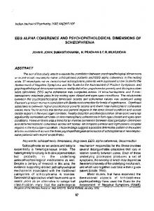
FtG. 1. Interhemispheric EEG coherence by sleep stage. all other means; p < .001.
'.
Awake mean differs from
anifact using the screen display and data selection features of the RHYTHM software. Interhemispheric coherence functions were calculated for each frequency band for each pair of homologous brain locations. A measure of mean EEG coherence was then calculated as the mean coherence of the four frequency bands (Le.. Mean EEG Coherence = (8 + 8 + a + {3)/4) for each stage for each subject. Statistical differences in Mean EEG Coherence were assessedusing MANDV A designs (BMDP4V) with two between-groups factors: Hand Preference (left. right) x Gender (female. male). and two repeated measures factors: Sleep Stage (wakefulness. stage 2. stage 3/4. stage REM) x Electrode Placement (frontal. central. parietal. occipital). Because of the small size of the subject sample. results from the Hand Preference x Gender interactions were not considered in any analyses. MANDV As were calculated using Mean EEG Coherence as the dependent measure. Differences and interactions of interest among repeated measures factors were subsequently determined using BMDP4V planned univariate contrasts. Also. the small sample size necessitated that interactions among the two repeated measures be assessedusing a pooled univariate estimate of F; df for these tests were thus reduced by the BMDP Geisser-Greenhouse Imhof formula to correct for the liberal estimate of F provided by the pooling procedure.
RESULTS Interhemisphericcoherenceas a function of sleep stage. Mean EEG coherencewas found to vary as a function of Sleep Stage(F(3, 6) 12.28,p = .0057;seeFig. I). This large effect was due to the fact that Mean EEG Coherenceduring wakefulnesswas significantlylower than during stage2 sleep(F(l, 8) = 41.84,P = .0002),stage3/4 sleep(F(l,
-
30.74. P = .0005), and stage REM sleep (F(l, 8) = 44.83, P 8) .0002). However. this Sleep Stage difference was qualified by a significant
118
NIELSEN ET AL. 50'
CJ_. °---
CJ_2
57' 54~
51-
1
T
48~ CJ
45-
.
If!
I
3633" 30 frontal
-A
.A
central
,,
pafletal
OCcIPItal
""
,--
FIG. 2. Interhemispheric EEG coherence by sleep stage and electrode placement. ", Awake mean differs from stage REM mean only; p < .05. *, Awake means differ from all other means for that placement; p < .05.
interaction with Electrode Placement(F(3, 27) = 4.29, p = .0109)2 indicating that the difference varied from anterior to posterior regions of the cortex. Specifically,univariatecontrastsrevealedthat MeanEEG Coherencefor wakefulnesswas significantlylower than it was for stages 2, 3/4, and REM sleepfor eachof the central (p = .0012,.0001,.0002, respectively),parietal(p = .0688,.0061,.0005),and occipital (p = .0020, .0001,.0002)placements,but wasonly significantlylower than coherence for stageREM sleepfor the frontal placements(p = .2425,.3163,.0430; see Fig. 2). Hemisphericdominanceand sleep.A main effect for Hand Preference was not observedfor MeanEEG Coherence(F(1, 8) = 1.00,p = .3466). However, a significantinteraction betweenHand Preferenceand Electrode Placement(F(3, 6) = 5.07,p = .0439)indicatedthat Mean EEG Coherencevaried by cortical region differentially for left- and righthandedsubjects.Specifically, valuesfor left-handedsubjectswere significantly higher than thosefor right-handedsubjectsfor occipital placements (F(1, 8) = 7.74,p = .0238),but not for frontal (F(1, 8) = 0.00, p = .9973),central (F(I, 8) = 0.05,p = .8300),or parietal (F(I, 8) = 0.03, p = .8759)placements(see Fig. 3). Moreover, this differencein occipital coherenceobtained for wakefulness(F(1, 8) = 11.44,p = .0096),stage2 sleep(F(I, 8) = 6.08,p = .0392),and stageREM sleep 2
Degreesoffreedom for the Sleep Stage x Electrode Placement interactions are adjusted
by the Geisser-Greenhouse
Imhof correction
factor.
~
~
INTERHEMISPHERIC
EEG COHERENCE
119
60
57 54
.. -
Left-handed
'"
Right-handed
T
Q)
~
51
~ 0 CJ w w c to
48
~
45 42 39 36 33 30 frontal * C1