BIOCELL 2015, 39(1): 1-7
ISSN 1667-5746 ELECTRONIC
Intermediate filament distribution patterns in maturing mouse oocytes and cumulus cells Maya Dyankova MARKOVA1, Venera Pantaleeva NIKOLOVA1, Irina Valcheva CHAKAROVA1, Ralitsa Stefanova ZHIVKOVA1, Rumen Kirilov DIMITROV2, Stefka Metodieva DELIMITREVA1 1 2
Department of Biology, Medical Faculty, Medical University of Sofia, Bulgaria Institute of Biology and Immunology of Reproduction, Bulgarian Academy of Sciences, Sofia, Bulgaria
Key words: meiosis; cytokeratin; vimentin; oogenesis; immunocytochemistry ABSTRACT: Successful oogenesis requires coordinated cytoskeletal rearrangements. While microfilaments and microtubules in mammalian oocytes are well studied, data about intermediate filaments are still scarce and controversial. We investigated intracellular distribution of vimentin and a set of cytokeratins in mouse oocytes and cumulus-oocyte complexes at different maturation stages using immunofluorescence and laser scanning confocal microscopy. Our results showed that conventional cytokeratins and vimentin are present and co-localize in mouse oocytes in the cortex as well as associated with the nucleus, meiotic spindle and meiotic chromosomes. During oogenesis, these intermediate filaments reorganize, moving from the cell periphery to the metaphase plate. Their distribution overlaps with that of fibrillar actin, except that actin does not associate with metaphase chromosomes. The same cytoskeletal proteins are present in cumulus cells and particularly in their trans-zonal projections. In foot processes, they localize in the periphery. The observed intermediate filament distribution patterns suggest transition to a more dynamic state of the cytoskeleton and a possible role in chromatin reorganization after germinal vesicle breakdown. The peripheral position in foot processes presumably allows oocyte-cumulus communications while providing adequate mechanical support.
Introduction Oocyte meiotic maturation depends on a series of chromatin rearrangements. The transition between germinal vesicle (GV) and germinal vesicle breakdown (GVBD) stages includes not only chromosome condensation but also shifting of heterochromatin from the nuclear periphery to the vicinity of nucleolus, leading to the formation of a dense perinucleolar rim called karyosphere (De La Fuente et al. 2004). Later stages include metaphase I (MI), completion of the 1st meiotic division and progression to metaphase II (MII), followed by arrest until fertilization. Chromatin rearrangements are coordinated to cytoskeletal changes. While much research has been devoted to tubulin and actin in mammalian oocytes, few studies have focused on intermediate filaments (IF). According to *Address correspondence to: Maya Markova,
[email protected] Received: July 16, 2014. Revised version received: November 4, 2014. Accepted: December 4, 2014.
some reports, cytoplasmic IF are not found in mammalian oocytes (Czernobilsky et al. 1985; Wendl et al. 2012). However, other studies have detected intermediate filament proteins, mostly cytokeratins, in oocyte cytoplasm. Lehtonen et al. (1983) have reported diffuse staining for cytokeratins in mouse oocytes, Gall et al. (1992) have observed cortical labeling in sheep oocytes, and Santini et al. (1993) have immunolocalized keratins 8, 18 and 19 in perinuclear position in human oocytes. Cytoskeletal sheets of 10-11 nm filaments positive for keratins 5, 6, 8, 16 and type Z as well as actin have been described in eggs of several mammalian species (Gallicano et al. 1994). The oocyte keratin cytoskeleton has been best described in the hamster. Immunocytochemical studies have found in prophase I-arrested hamster oocytes several large cortical keratin-containing aggregates dispersing to multiple smaller granular patches at MI and then to homogenously distributed small spots at MII (Plancha, 1996; Kabashima et al. 2010). Besides the cortical aggregates, the latter study described also a fine keratin filament network in the GV pe-
2
riphery. The authors hypothesized that the observed keratin redistribution reflected increasing complexity and a possible role to maintain cell integrity under physical stress after ovulation. Similar results have recently been obtained with mouse oocytes (Wei et al. 2013). For another IF protein, vimentin, negative reaction has been reported in mouse (Lehtonen et al. 1983) and human oocytes (Santini et al. 1993). A later study has detected vimentin in bovine and rhesus mature oocytes and zygotes by Western blotting (Payne et al. 2003). The authors observed vimentin filaments around zygote pronuclei and found that anti-vimentin antibody blocked pronuclear migration and fusion. The oocyte differentiates surrounded by cumulus cells with which it forms cumulus-oocyte complex (Alvarez et al. 2009). The cytoskeleton of cumulus cells is important for oogenesis because it provides mechanical basis for their interaction with the oocyte. They have been reported to contain both cytokeratins and vimentin, a phenomenon often observed in cultured cells but uncommon in tissues (Czernobilsky et al. 1985; Gall et al. 1992). Both these IF proteins, together with microtubules and microfilaments, are especially prominent in cytoplasmic projections of cumulus cells crossing zona pellucida and making contacts with the oocyte (Sutovský et al. 1994). However, the structural organization of IF in projections has not yet been characterized despite their role in oocyte differentiation.
MAYA DYANKOVA MARKOVA et al.
The insufficient and often contradictory data about cytoplasmic IF in mammalian oocytes and cumulus cells indicate the need of more research. Therefore, we performed a study by immunofluorescence and laser scanning confocal microscopy to detect cytokeratins and vimentin and to compare their distribution to that of fibrillar actin in mouse oocytes and cumulus-oocyte complexes at different maturation stages.
Materials and Methods The study was carried out in accordance with Bulgarian and EU legislation concerning animal experiments, and with permission of the Ethical Committee of the Medical University of Sofia. A total of 40 prepubertal (4-5 week old) BALB/c female mice were subjected to ovarian hyperstimulation with 5 IU of the follicle-stimulating hormone Menogon (Ferring, Kiel, Germany) and, 48 hours later, with 5 IU of the luteinizing hormone Novarel (Ferring). They were euthanized 16-18 hours after the first (16 animals) or second hormonal stimulation (24 animals). In the first case, ovaries were dissected and washed in physiologic saline solution. Oocytes at GV stage were obtained by puncture of antral follicles. In the second case, ovaries were dissected and washed together with oviducts. Mature (MII) oocytes were retrieved from
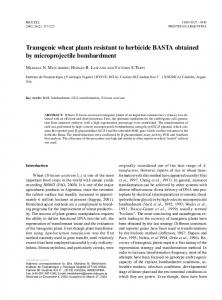
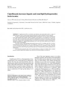
FIGURE 1. Oocytes stained for cytokeratins, vimentin and chromatin. A. GV stage, epifluorescence. Reaction for both types of IF is positive in the cortical layer and around the nucleus. B. Metaphase I, confocal microscopy. Staining is observed in the cortical layer, especially over the spindle, and the metaphase plate. C. Metaphase II, confocal microscopy. The oocyte cortex, spindle and chromosomes are labeled, as well as the fragmented polar body (left). Bars = 20 μm.
INTERMEDIATE FILAMENTS IN MOUSE OOCYTES
the ampulae of oviducts, while immature oocytes at GV, GVBD and MI stages were obtained by puncturing unovulated follicles. Oviducts and follicles were manipulated at 37 °C on a hot plate (Labotech, Ohlsbach, Germany) in Leibowitz medium (Sigma-Aldrich, Munich, Germany) supplemented with 0.3% bovine serum albumin (Sigma-Aldrich). After puncture, cells were washed in clean Leibowitz and briefly treated with 0.5 mg/ml hyaluronidase (Sigma-Aldrich) in the same medium to remove outer layers of cumulus. For half of the immature oocytes (without polar bodies), zona-attached cumulus cells were removed mechanically by pipetting through a fine glass pipette. The rest were investigated together with the cumulus cells connected to zona pellucida. For mature oocytes, all cumulus cells were lost spontaneously during hyaluronidase treatment. Immunofluorescence was carried out as previously described (Delimitreva et al. 2006). Oocytes were washed from the medium in washing buffer (phosphate-buffered saline, pH 7.2, with 0.3% bovine serum albumin). Then, they were fixed in prewarmed (37 °C) 2% paraformaldehyde in phosphate-buffered saline with 0.02% Triton X-100 (Sigma-Aldrich) for 45 min. After that, cells were washed twice in washing buffer for 10 min and once with washing buffer + 0.02% sodium azide. In this solution, they were stored overnight at 4 °C. For labeling of cytokeratins and vimentin, specific mouse monoclonal antibodies (Sigma-Aldrich) were used: clone PCK-26, recognizing cytokeratins 1, 5, 6, and 8, diluted 1:300 in washing buffer + 1% sodium azide; clone Vim-13.2, diluted 1:200; and affinity isolated anti-vimentin antibody produced in rabbit (Sigma-Aldrich), diluted 1:200, respectively. Cells were incubated in solution of anti-cytokeratin, anti-vimentin or anti-cytokeratin + rabbit anti-vi-
3
mentin antibody for 45 min at 37 °C, followed by washing twice in washing buffer + 1 μl/ml Tween-20 (Sigma-Aldrich) for 10 min at 37 °С. In negative controls, primary antibody was omitted. Then, FITC-conjugated secondary antibody (Sigma-Aldrich) for mouse primary antibodies and/ or TRITC-conjugated secondary antibody (Sigma-Aldrich) for the rabbit primary antibody, diluted 1:300 in washing buffer + 1% sodium azide, was applied for 45 min at 37°C in the dark. The solution contained also 10 μg/ml Hoechst 33258 (Sigma-Aldrich) for chromatin staining. When TRITC-conjugated secondary antibody was not used, 1 μg/ ml TRITC-conjugated phalloidin (Sigma-Aldrich) was added to the same solution for detection of fibrillar actin. The oocytes were subsequently washed four times in washing buffer + 1 μl/ml Tween 20 at 37oC for 10 min, except for the last wash which lasted 20-30 min. Thereafter, the samples were impregnated in increasing concentrations (5, 10, 30 and 50%) of polyvinyl alcohol (Fluka, Buchs, Switzerland) in washing buffer + 0.02% sodium azide. Each step was for 10 min at 37 °C. The oocytes were then placed in drops of 100% polyvinyl alcohol on a clean glass slide and covered with a coverslip. They were stored in the dark at 4 °C until observation. Samples were observed with an epifluorescence microscope Axioskop 20 (Zeiss, Goettingen, Germany). Some slides were also subjected to laser scanning confocal microscopy using Leica TSC SPE (Leica Microsystems, Wetzlar, Germany) microscope.
Results All three primary antibodies against IF proteins produced identical staining patterns. Initially, mouse anti-cytokeratin
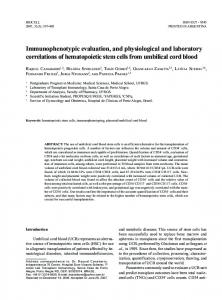
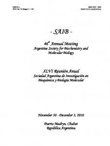
FIGURE 2. Immature prophase oocytes stained for IF, actin and chromatin, confocal microscopy. A. GV stage showing reaction for IF and actin in the cortical layer and the nuclear compartment, especially the karyosphere. Bar = 20 μm. B. GVBD stage. Condensing chromosomes are attached to the karyosphere. Diffuse reaction for IF and actin is observed in the chromatin region while a bright reaction is overlaying the karyosphere. A group of cumulus cells are also seen (bottom right). Bar = 10 μm.
4
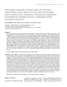
and rabbit anti-vimentin antibodies were used simultaneously, followed by FITC-labeled anti-mouse and TRITClabeled anti-rabbit secondary antibodies. A total of 36 oocytes were used, with 16, 3, 7 and 10 cells at stages GV, GVBD, MI and MII, respectively. The results showed full colocalization of cytokeratins and vimentin (Fig. 1). In the second series of experiments, anti-mouse antibodies against either cytokeratins or vimentin (collectively referred to as IF proteins below) were used with FITC-labeled secondary antibody, and TRITC-labeled phalloidin was applied for detection of actin. A total of 152 oocytes were used, with 56, 7, 41 and 48 cells at stages GV, GVBD, MI and MII, respectively. In immature prophase-arrested (GV) oocytes, positive reaction for IF and fibrillar actin was observed in the cortical layer underlying the oolemma and in perinuclear position (Fig. 1A shows this reaction for IF). After the karyosphere was formed and heterochromatin moved towards it, IF and actin were detected surrounding this structure (Fig. 2A). Both IF and microfilaments co-localized with the karyosphere heterochromatin. At GVBD, this bright reaction in the karyosphere remained while the inside of the nucleolus stayed dark and free from the investigated molecules (Fig. 2B). In MI and MII oocytes, IF were again localized in the cell cortex, forming a well visible cap corresponding to the actin cap. They were also observed in the vicinity of chromatin structures, overlying both the meiotic spindle and the chromosomes themselves (Fig. 1B, C, Fig. 3). Compared to GV and GVBD oocytes, cortical staining was decreased and less bright than the fluorescent reaction surrounding the metaphase plate. The described pattern was uniform, except for some individual variability in the intensity of cortical versus spin-
MAYA DYANKOVA MARKOVA et al.
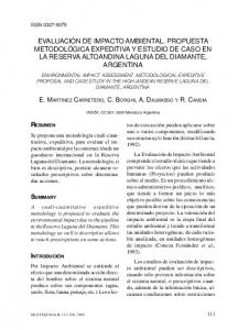
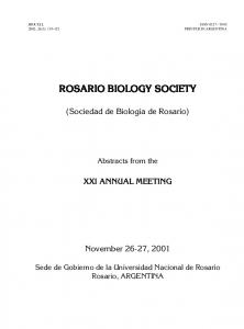
dle/chromosome-associated staining for IF in metaphase oocytes. Fibrillar actin mostly co-localized with IF but, unlike them, did not cluster around the chromosomes after GVBD. In metaphase oocytes, both IF and actin were detected around the meiotic spindle and the cap region but only IF were associated with the chromosomes. No differences were observed between MI and mature MII oocytes with regard to either IF or fibrillar actin. Positive reaction for the studied cytoskeletal proteins was observed not only in oocytes but also in cumulus cells surrounding the GV oocytes. The cytoplasm of cumulus cells stained positive for both IF and actin, with reaction for IF slightly brighter in the cell’s interior and for actin at the periphery. The staining for both groups of cytoskeletal proteins was most intensive in cytoplasmic projections through zona pellucida, visualized by confocal microscopy (Fig. 4). They could often be seen even when cumulus cells themselves had been detached during the preparation of the complex for microscopy; in this case, they were associated with smaller or larger cytoplasmic remnants. At the level of passing through the zona, projections were visualized as bright filaments. At their bulbous tips (foot processes), they produced fine ring-like structures in cross section. After MI assembly, few if any cumulus cells had remained attached to the oocyte, and we could rarely detect projections passing through zona pellucida.
Discussion The present immunocytochemical study of mouse oocytes and cumulus-oocyte complexes for vimentin and a set of keratins revealed not only presence of both types of IF pro-
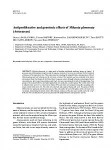
FIGURE 3. Metaphase oocytes stained for IF, actin and chromatin, confocal microscopy. A. MI. The cap is stained for IF and actin while only the IF show association with the chromosomes. The rest of the cortex only shows a weak reaction in this cell. B. MII. Meiotic spindle and polar body (bottom left) react for both IF and actin, and the MII plate for IF alone. Bars = 20 μm.
INTERMEDIATE FILAMENTS IN MOUSE OOCYTES
teins but also their full co-localization. Though unusual, such finding has been described in some cultured epithelial cells where newly synthesized vimentin distributes following the preexisting cytokeratin network (Pagan et al. 1996). The present data suggest that a similar situation may exist in mouse oocytes and cumulus cells from GV to MII stages. The double labeling co-localization of IF and fibrillar actin produced interesting results. In most tissues IF and microfilaments have little overlap (e.g. Parikh et al., 2008), with IF concentrating in the central part of the cell and actin in the periphery as we observed in cumulus cells. However, we found almost complete co-localization of the two cytoskeletal system in the oocyte, suggesting their close interaction during oogenesis. It should be mentioned that our results concerning distribution of fibrillar actin at different oocyte maturation stages were in agreement with reports of other authors (Azoury et al. 2008; Brunet and Verlhac, 2011). Biochemical studies have also revealed association of oocyte IF with components of actin cytoskeleton (Kadam et al. 2006). In the present work, localization of IF proteins in the oocyte cortex throughout all studied oogenesis stages was in agreement with earlier reports (Plancha, 1996; Kabashima et al. 2010; Wei et al. 2013), although differences were found in the intracellular IF distribution. In particular, while changes of keratin filament network from GV to MII
5
stages have been described by Kabashima et al. (2010) as extension and increasing complexity, our study found reduction of the peripheral layer of IF. We suppose that the role of this cortical IF network is to provide mechanical stability of the cell periphery, as in epithelial layers. We also hypothesize that, similarly to epithelial layers, this involves contacts with neighboring cells (i.e. the surrounding cumulus cells). In this respect, decreasing immunofluorescent reaction for IF in the cortex at later oogenesis stages could be expected with regard to the parallelly occurring gradual cumulus-oocyte disconnection. This reduction, similarly to the rearrangements of microtubules and microfilaments during oocyte maturation, could be a prerequisite for making the oocyte cytoskeleton more dynamic in order to allow polar body extrusion and zygote cleavage. Reaction for IF proteins in our study was observed also in the GV region of prophase-arrested oocytes and associated with the meiotic spindle and the metaphase plate of MI and MII oocytes. It is known that in a number of somatic cell types, cytoplasmic IF also tend to form a perinuclear network (Dupin et al. 2011) and to associate with the mitotic spindle (Chou et al. 2003) and chromosomes (Li et al. 2003). We observed increasing association of cytokeratins and vimentin with chromosomes and meiotic spindle during oocyte maturation parallelly to the relaxation of their connection to the cell periphery. This suggests a possible role
FIGURE 4. GV cumulus-oocyte complex stained for IF (A, B) and actin (A’, B’), confocal microscopy. A, A’. Projections of detached cumulus cells can be seen crossing zona pellucida (arrowheads). B, B’. Another focus of the same field. Foot processes in cross section show ringlike reaction (arrowheads). Bar = 5 μm.
6
of cytoplasmic IF for chromatin reorganization after GV breakdown. The only visible difference between IF and fibrillar actin distribution was the metaphase plate at MI and MII. While cytokeratin and vimentin co-localized with metaphase chromosomes, microfilaments showed no such association, suggesting that the two groups of cytoskeletal proteins have different roles in meiotic chromatin reorganization. According to studies on living mouse oocytes, the main function of fibrillar actin is to keep chromosomes in a limited area during spindle assembly and to transfer the spindle from the center of the oocyte to its periphery (Azoury et al. 2008). This is likely to require assembly around the entire set of chromosomes, while IF may be engaged in interaction with individual chromosomes. With regard to cumulus cells, the most interesting results of our study concerned the cytoplasmic projections documented by laser scanning confocal microscopy. In many GV oocytes, trans-zonal projections retained in place even after detachment of the respective cumulus cells. This indicates that the mechanical association of projections with oocyte was stronger than the cross-links between cytoskeletal components within cumulus cell cytoplasm. Such a result could be expected, given the importance of cumulus-oocyte contacts for normal oogenesis and particularly for meiosis arrest, resumption and progression (Barrett and Albertini, 2010; Solc et al. 2010). In agreement with earlier reports (Gall et al. 1992; Sutovský et al. 1994), IF and actin were stained more intensely in transzonal projections than in cytoplasm of cumulus cells, indicating concentration of cytoskeletal structures in projections to provide high mechanical strength despite the small cross section. At the level of foot processes, confocal microscopy showed ring-like reaction for both types of cytoskeletal proteins – a detail which, to our knowledge, has not been described before. This allocation of IF and microfilaments to the periphery presumably serves to guarantee sufficient mechanical stability while reserving the central part of the process for unhindered transport of substances to and from the oocyte. In conclusion, our study showed that conventional cytokeratins and vimentin are present in mouse oocytes in the cortex as well as associated with the nucleus, meiotic spindle and meiotic chromosomes. During oogenesis, these IF undergo reorganization, relaxing their connection to the cell periphery and concentrating around the metaphase plate, which suggests a possible role in chromatin reorganization after GV breakdown. The same IF proteins are present in cumulus cells and particularly in their trans-zonal projections, organized in a way to provide adequate mechanical support and at the same time to allow the oocyte-cumulus communications vital for successful oogenesis.
MAYA DYANKOVA MARKOVA et al.
Acknowledgements This study was funded by Medical University of Sofia grant No. 36/2012.
References Alvarez GM, Dalvit GC, Achi MV, Miguez MS, Cetica PD (2009). Immature oocyte quality and maturational competence of porcine cumulus-oocyte complexes subpopulations. Biocell 33: 167-177. Azoury J, Lee KW, Georget V, Rassinier P, Leader B, Verlhac MH (2008). Spindle positioning in mouse oocytes relies on a dynamic meshwork of actin filaments. Current Biology 18: 1514-1519. Barrett SL, Albertini DF (2010). Cumulus cell contact during oocyte maturation in mice regulates meiotic spindle positioning and enhances developmental competence. Journal of Assisted Reproduction and Genetics 27: 29-39. Brunet S, Verlhac MH (2011). Positioning to get out of meiosis: the asymmetry of division. Human Reproduction Update 17: 68-75. Chou YH, Khuon S, Herrmann H, Goldman RD (2003). Nestin promotes the phosphorylation-dependent disassembly of vimentin intermediate filaments during mitosis. Molecular Biology of the Cell 14: 1468-1478. Czernobilsky B, Moll R, Levy R, Franke WW (1985). Co-expression of cytokeratin and vimentin filaments in mesothelial, granulosa and rete ovarii cells of the human ovary. European Journal of Cell Biology 37: 175-190. De La Fuente R, Viveiros MM, Burns KH, Adashi EY, Matzuk MM, Eppig JJ (2004). Major chromatin remodeling in the germinal vesicle (GV) of mammalian oocytes is dispensable for global transcriptional silencing but required for centromeric heterochromatin function. Developmental Biology 275: 447-458. Delimitreva S, Zhivkova R, Isachenko E, Umland N, Nayudu PL (2006). Meiotic abnormalities in in vitro-matured marmoset monkey (Callithrix jacchus) oocytes: development of a non-human primate model to investigate causal factors. Human Reproduction 21: 240-247. Dupin I, Sakamoto Y, Etienne-Manneville S (2011). Cytoplasmic intermediate filaments mediate actin-driven positioning of the nucleus. Journal of Cell Science 124: 865-872. Gall L, De Smedt V, Ruffini S (1992). Co-expression of cytokeratins and vimentin in sheep cumulus-oocyte complexes. Alteration of intermediate filament distribution by acrylamide. Development Growth and Differentiation 34: 579-587. Gallicano GI, Larabell CA, McGaughey RW, Capco DC (1994). Novel cytoskeletal elements in mammalian eggs are composed of a unique arrangement of intermediate filaments. Mechanisms of Development 45: 211-226. Kabashima K, Matsuzaki M, Suzuki H (2010). Intermediate filament keratin dynamics during oocyte maturation requires maturation/ M-phase promoting factor and mitogen-activated protein kinase activities in the hamster. Reproduction in Domestic Animals 45: e184-e188. Kadam KM, D’Souza SJ, Bandivdekar AH, Natraj U (2006). Identification and characterization of oviductal glycoprotein-binding protein partner on gametes: epitopic similarity to non-muscle myosin IIA, MYH 9. Molecular Human Reproduction 12: 275-282. Lehtonen E, Lehto VP, Vartio T, Badley RA, Virtanen I (1983). Expression of cytokeratin polypeptides in mouse oocytes and preimplantation embryos. Developmental Biology 100: 158-165.
INTERMEDIATE FILAMENTS IN MOUSE OOCYTES
Li G, Tolstonog GV, Sabasch M, Traub P (2003). Type III intermediate filament proteins interact with four-way junction DNA and facilitate its cleavage by the junction-resolving enzyme T7 endonuclease I. DNA and Cell Biology 22: 261-291. Pagan R, Martín I, Alonso A, Llobera M, Vilaró S (1996). Vimentin filaments follow the preexisting cytokeratin network during epithelial-mesenchymal transition of cultured neonatal rat hepatocytes. Experimental Cell Research 222: 333-344. Parikh N, Nagarajan P, Sei-ichi M, Sinha S, Garrett-Sinha LA (2008). Isolation and characterization of an immortalized oral keratinocyte cell line of mouse origin. Archives of Oral Biology 53: 1091-1100. Payne C, Rawe V, Ramalho-Santos J, Simerly C, Schatten G (2003). Preferentially localized dynein and perinuclear dynactin associate with nuclear pore complex proteins to mediate genomic union during mammalian fertilization. Journal of Cell Science 116: 4727-4738. Plancha CE (1996). Cytokeratin dynamics during oocyte maturation in the hamster requires reaching of metaphase I. Differentiation 60: 87-98.
7
Santini D, Ceccarelli C, Mazzoleni G, Pasquinelli G, Jasonni VM, Martinelli GN (1993). Demonstration of cytokeratin intermediate filaments in oocytes of the developing and adult human ovary. Histochemistry 99: 311-319. Solc P, Schultz RM, Motlik J (2010). Prophase I arrest and progression to metaphase I in mouse oocytes: comparison of resumption of meiosis and recovery from G2-arrest in somatic cells. Molecular Human Reproduction 16: 654-664. Sutovský P, Fléchon JE, Pavlok A (1994). Microfilaments, microtubules and intermediate filaments fulfil differential roles during gonadotropin-induced expansion of bovine cumulus oophorus. Reproduction Nutrition Development 34: 415-425. Wei X, Xiangwei F, Guangbin Z, Jing X, Liang W, Ming D, Dianshuai Y, Mingxing Y, Jianhui T, Shien Z (2013). Cytokeratin distribution and expression during the maturation of mouse germinal vesicle oocytes after vitrification. Cryobiology 66: 261-266. Wendl J, Ebach K, Rodler D, Kenngott RA (2012). Immunocytochemical localization of cytoplasmic and nuclear intermediate filaments in the bovine ovary during folliculogenesis. Anatomia, Histologia, Embryologia 41: 190-201.