Intertester Reliability and Validity of Motion Assessments During Lumbar Spine Accessory Motion Testing Rob Landel, Kornelia Kulig, Michael Fredericson, Bernard Li and Christopher M Powers PHYS THER. Published online November 20, 2007 doi: 10.2522/ptj.20060179
The online version of this article, along with updated information and services, can be found online at: http://ptjournal.apta.org/content/early/2007/11/20/ptj.20060179 Collections
This article, along with others on similar topics, appears in the following collection(s): Injuries and Conditions: Spine Tests and Measurements
E-mail alerts
Sign up here to receive free e-mail alerts
Online First articles are published online before they appear in a regular issue of Physical Therapy (PTJ). PTJ publishes 2 types of Online First articles: Author manuscripts: PDF versions of manuscripts that have been peer-reviewed and accepted for publication but have not yet been copyedited or typeset. This allows PTJ readers almost immediate access to accepted papers. Page proofs: edited and typeset versions of articles that incorporate any author corrections and replace the original author manuscript.
Downloaded from http://ptjournal.apta.org/ by guest on June 1, 2013
Research Report
Intertester Reliability and Validity of Motion Assessments During Lumbar Spine Accessory Motion Testing Rob Landel, Kornelia Kulig, Michael Fredericson, Bernard Li, Christopher M Powers
Background and Purpose Posterior-anterior (PA) assessment of the lumbar spine correlates with radiographic signs of instability and can guide treatment choices, yet studies of the validity of lumbar PA assessments have not been conducted in vivo. The purposes of this study were to determine the intertester reliability of the PA examination in assessing intersegmental lumbar spine motion and to evaluate the validity of this procedure in vivo with dynamic magnetic resonance imaging (MRI).
Subjects Twenty-nine subjects with central lumbar pain participated in this study.
Methods Two physical therapists independently identified each subject’s most and least mobile lumbar segments using the PA procedure. Midsagittal lumbar images were obtained simultaneously during one examiner’s assessment. Lumbar segmental mobility was quantified from magnetic resonance images as the change in the intervertebral angle between the resting position and the end range of the PA force application. For each vertebral level tested, maximal sagittal-plane segmental motion was determined.
Results The intertester reliability for identifying the least mobile segment was good (agreement⫽82.8%, kappa⫽.71, 95% confidence interval [CI]⫽.48 to .94), but it was poor for identifying the most mobile segment (kappa⫽.29, 95% CI⫽⫺.13 to .71), despite good agreement (79.3%). The level of agreement between the PA assessments and intervertebral motion measured by MRI was poor (kappa⫽.04, 95% CI⫽⫺.16 to .24, and kappa⫽.00, 95% CI⫽⫺.09 to .08, for the least and most mobile segments, respectively).
Discussion and Conclusion Despite good intertester reliability for identifying the least mobile segment, PA assessments of lumbar segmental mobility did not agree with sagittal-plane motion measured by dynamic MRI. This finding calls into question the validity of the PA procedure for assessing intervertebral lumbar spine motion.
R Landel, PT, DPT, OCS, CSCS, is Associate Professor of Clinical Physical Therapy, Division of Biokinesiology and Physical Therapy at the School of Dentistry, University of Southern California, 1540 E Alcazar St, CHP 155, Los Angeles, CA 90033 (USA). Address all correspondence to Dr Landel at:
[email protected]. K Kulig, PT, PhD, is Associate Professor of Clinical Physical Therapy, Division of Biokinesiology and Physical Therapy at the School of Dentistry, University of Southern California. M Fredericson, MD, is Associate Professor, Department of Functional Restoration, Division of Sports Medicine, Stanford University, Palo Alto, Calif., and Director of Stanford Physical Medicine and Rehabilitation Clinics. B Li, PT, DPT, is Resident, USC Orthopedic Residency, Division of Biokinesiology and Physical Therapy at the School of Dentistry, University of Southern California. CM Powers, PT, PhD, is Associate Professor, Division of Biokinesiology and Physical Therapy at the School of Dentistry, University of Southern California. [Landel R, Kulig K, Fredericson M, et al. Intertester reliability and validity of motion assessments during lumbar spine accessory motion testing. Phys Ther. 2008; 88:xxx–xxx.] © 2007 American Physical Therapy Association Post a Rapid Response or find The Bottom Line: www.ptjournal.org
January 2008
Volume 88 Number 1 Downloaded from http://ptjournal.apta.org/ by guest on June 1, 2013
Physical Therapy f
1
Intertester Reliability and Validity of Motion Assessments
D
ecisions regarding the choice of interventions for low back pain are dependent on information obtained from accurate assessments. Accordingly, clinical assessments are used to identify impairments (eg, altered spinal mobility) that suggest specific treatment choices. It has been shown that the identification of hypermobility and hypomobility in the lumbar spine by use of a PA assessment procedure correlates with radiographic evidence of instability.1 In addition, the presence or absence of hypomobility and hypermobility has been shown to be useful in determining effective treatment choices.2– 4 Spinal mobility can be measured as total motion (ie, lumbar flexion or extension) or as segmental motion (ie, the motion between 2 vertebrae). In the clinical setting, segmental spinal mobility is assessed by applying a posterior-anterior (PA) force to a single vertebral spinous process with the individual in the prone position. This procedure purports to evaluate passive segmental lumbar mobility and is typically graded with a 3-point scale (hypermobile, normal, and hypomobile).5,6
For any clinical test to be useful, it must yield reliable and valid data. In addition, studies of the procedure must remain true to its clinical implementation and interpretation. Previous studies of lumbar segmental PA mobility and stiffness assessments demonstrated limited reliability.7,8 Binkley et al7 reported poor reliability for PA lumbar accessory mobility testing with a 9-point scale that ranged from 1 (excessive motion) to 9 (no motion), with the central point of 5 on the scale representing “normal” motion. Similarly, Maher and Adams8 reported poor reliability with an 11-point scale of stiffness rather than motion. The scale used by these authors ranged from ⫺5 (decreased stiffness) to 5 (increased 2
f
Physical Therapy
stiffness), with 0 representing “normal” stiffness. A limitation of the above-noted studies is that examiners were required to compare the mobility or stiffness of a specific lumber segment with “normal.” In order to achieve acceptable interrater reliability, a common definition of “normal” must exist between raters. This is problematic because the frame of reference used for comparisons is the qualitative aggregate of prior experience distilled into an expected amount of motion for a given segment. Because no 2 testers will have the same experiences, it follows that testers will not share a common reference standard for an expected amount of motion. Differences in examiners’ experience with and expectations for the amount of motion likely contributed to the poor intertester reliability in those investigations. Another methodological limitation of the studies of Binkley et al7 and Maher and Adams8 is that 9- and 11-point scales were used in those studies, respectively. These scales required examiners to make distinctions among small increments of motion, stiffness, or both. It is possible that clinicians may be able to agree on less precise assessments of segmental motion with the PA assessment procedure. We propose that evaluating segmental mobility with a within-subject dichotomous scale (identifying the most mobile and least mobile segments) may be better suited for an assessment of reliability by reducing the need to differentiate among potentially small amounts of motion and by eliminating comparisons with a preconceived notion of “normal.” The reliability of a clinical test has no meaning unless it has been shown to be valid. To date, studies of the validity of lumbar PA assessments have
not been conducted in vivo. Given that intersegmental motion of the lumbar spine can be evaluated during PA mobility assessments with dynamic magnetic resonance imaging (MRI),9 –11 validity testing can be conducted with a high degree of biofidelity. Recognizing the limitations of previous work in this area, our aims in the present study were to determine the intertester reliability of lumbar PA mobility assessments with a dichotomous within-subject scale and to assess the validity of the PA assessment procedure with dynamic MRI.
Method Subjects Twenty-nine subjects (13 men and 16 women) between the ages of 18 and 45 years (X⫽31.3 years) and with a diagnosis of nonspecific low back pain participated in this study. A physician screened all subjects for participation in the study through a careful history, self-administered Oswestry Disability Questionnaire and Medical Outcomes Study 36-Item Health Survey Questionnaire, physical examination, and standard anterior-posterior and lateral radiographs (oblique radiographs also were obtained when spondylolisthesis was suspected). Only subjects who reported a recent onset of centralized low back pain (within the last 3 months) at or above the level of the waist and increased pain with lumbar extension in standing were admitted to the study. Subjects over the age of 45 years were excluded to control for the possible confounding effects of osteoarthritis of the spine. Subjects were excluded from participation in the study if they had any of the following: disk pathology demonstrated during MRI, spinal malignancy, cardiovascular disease, evidence of spinal cord compression, aortic aneurysm, hiatal hernia, uncontrolled hypertension, spinal infection, severe respiratory disease,
Volume 88 Number 1 Downloaded from http://ptjournal.apta.org/ by guest on June 1, 2013
January 2008
Intertester Reliability and Validity of Motion Assessments pregnancy, abdominal hernia, prior low back surgery, gross spinal deformity, spondylolisthesis, known rheumatic joint disease, implanted biological devices that could interact with the magnetic field (eg, pacemakers, cochlear implants, ferromagnetic cerebral aneurysm clips), or signs or symptoms of neural compromise related to lumbar disk pathology (eg, pain radiating below the level of the buttocks, sensation changes in the lower extremities, diminished reflexes, lower-extremity weakness, urinary or fecal incontinence, increased peripheral pain with repeated lumbar extension). Instrumentation Dynamic imaging of the lumbar spine was performed with a vertically opened 0.5-T MRI system (Signa SP)* with a 56-cm opening that allowed the examiner access to the subject during testing. This system was equipped with a pulse sequence programming environment and realtime interactive MRI capability. Dynamic MRI was used instead of video fluoroscopy or radiography in order to eliminate exposure of the subject and the examiner to radiation. Previous studies9,11 demonstrated the feasibility of MRI for quantifying lumbar segmental motion, and the same procedure was used in the present study. Midsagittal-plane imaging of the lumbar spine was performed with a receive-only surface coil and an ultrafast spoiled GRASS (gradient-recalled acquisition in the steady state) pulse sequence. Images were obtained at a rate of one per second with the following parameters: repetition time⫽ 200 milliseconds, echo time⫽18 milliseconds, number of excitations⫽1.0, matrix⫽256⫻256, field of view⫽28⫻21 cm, and a 7-mm section thickness with an interslice * General Electric Medical Systems, 4855 W Electric Ave, Milwaukee, WI 53219-1628.
January 2008
spacing of 1 mm. The surface coil was flexible and was designed to expose the low back such that the examiner had direct access to palpate the lumbar spinous processes. Procedure Prior to participation, all subjects signed an informed consent form approved by the institutional review boards of the University of Southern California and Stanford University. Participants underwent 2 separate assessments of PA mobility of the lumbar spine: within the MRI environment and outside the MRI environment. Two different examiners performed the 2 assessments. Prior to data collection, the position of the subject and the assessment procedure were agreed upon and practiced by both examiners. For MRI, subjects were placed in the prone position, such that the spine and torso were positioned within the opening between the vertical magnets. Following subject positioning, a series of sagittal-plane localizing scans were obtained to ensure that the image plane captured the vertebral bodies and spinous processes of all lumbar vertebrae. Once the image plane was determined, continuous imaging (at a frequency of 1 Hz) commenced, starting with a static sagittal view of the lumbar spine in the resting position and continuing while the PA motion assessment was performed. All PA assessments within the MRI system were performed by a physical therapist with 15 years of manual therapy experience (KK). With the examiner standing, manual contact was made on the spinous processes with the hypothenar eminence just distal to the pisiform. A PA force, directed anteriorly in a direction perpendicular to the tangent of the arc of the lumbar lordosis, was applied to each vertebra, starting caudally at L5 and moving cranially to L1. Suffi-
cient force was applied to induce movement to the end of the available range of segmental motion and was comparable in magnitude to that of grade IV as defined by Maitland et al.12 The force was applied slowly (over approximately 1–2 seconds) and held at end range for at least 5 seconds in order to obtain a clear end-range image. Release of the force also occurred slowly (1–2 seconds), and then force was applied to the next vertebral level. Once the examiner released the force on L1 and a clear resting-position image was obtained, imaging was terminated. Following this procedure, the examiner recorded the lumbar segment deemed to be most mobile and that deemed to be least mobile. Following the PA motion assessment within the MRI system, the subject was moved to a room where a physical therapist with 16 years of manual therapy experience (RL) performed an identical PA motion assessment. The second therapist independently recorded the lumbar segment perceived to have the most movement and that perceived to have the least movement. The 2 examiners were unaware of the results of each other’s testing. Data Analysis Prior to analysis, all images were transferred from the MRI system console to a Macintosh G3 computer.† For the purposes of this study, only the images obtained at rest and at the end range of segmental motion were analyzed. A third investigator, who was unaware of all PA test results, made all measurements with National Institutes of Health software.‡ Lumbar segmental motion in the sagittal plane was quantified by measur† Apple Computer Inc, 1 Infinite Loop, Cupertino, CA 95014. ‡ National Institutes of Health, 9000 Rockville Pike, Bethesda, MD 20892.
Volume 88 Number 1 Downloaded from http://ptjournal.apta.org/ by guest on June 1, 2013
Physical Therapy f
3
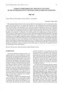
Intertester Reliability and Validity of Motion Assessments Table 1. Intertester Reliability for Least Mobile Segmenta
Tester 2
Tester 1 Level
5
4
5
0 (0)
3
2
1
Total 0
4
1 (0.1)
3
1
2
2 (0)
2
2
7 (3.0)
1
1
14 (8.8)
15
8
17
29
Total
0
1
3
3
10
a
Level indicates the lumbar spinal vertebra to which the posterior-anterior force was applied. Values in parentheses indicate the calculated frequencies of agreements expected by chance. Agreement⫽82.8%, kappa⫽.71, 95% confidence interval⫽.48 to .94.
To establish the intratester reliability of angular measurements, dynamic magnetic resonance images were obtained for 5 volunteers who were healthy on 2 separate occasions. Intraclass correlation coefficients were found to be excellent, ranging from .95 to .99 for all subjects. The standard error of measurement ranged from .40 to .66 degrees.
ing the intervertebral angle, which was defined as the angle formed by lines delineating adjacent vertebral end plates. This procedure has been described previously.9 –11 Lumbar segmental motion was defined as the difference between the intervertebral angles measured from the resting and the end-range images. An increase in intervertebral motion from the resting to the end-range positions was indicative of spinal extension. Conversely, a decrease in intervertebral motion was indicative of spinal flexion. The superior vertebra was used to define the target segment. For example, a PA force applied to L4 identified the target segment of L4 –L5. For each subject, the segments with the largest amount of motion and the smallest amount of motion were identified.
The kappa statistic was used to determine the level of agreement between the 2 examiners for identifying the most and least mobile segments with manual PA assessments (intertester reliability). In addition, the kappa statistic was used to determine the level of agreement between the measurements obtained by the examiner performing the PA assessment during MRI and the mea-
surements determined from images for identifying the most and least mobile segments (validity). The kappa results were interpreted as follows: excellent (ⱖ.75), fair to good (.40 – .74), and poor (ⱕ.40).13
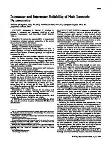
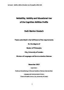
Results Intertester reliability for identifying the least mobile segment was good (kappa⫽.71, 95% confidence interval [CI]⫽.48 –.94, agreement⫽82.8%; Tab. 1). Intertester reliability for identifying the most mobile segment was poor (kappa⫽.29, 95% CI⫽⫺.13– .71, agreement⫽79.3%; Tab. 2). The validity of the PA assessments for identifying the least and most mobile segments also was poor (kappa⫽.04, 95% CI⫽⫺.16–.24, agreement⫽24.1% [Tab. 3], and kappa⫽.00, 95% CI⫽
Table 2. Intertester Reliability for Most Mobile Segmenta Tester 1 Level Tester 2
5 4
5 21 (19.9) 2
4
2
3
4 0 (0)
1
0 0 (0)
1 0 (0)
24
5
Total 24
1 Total
1
2 (0.7)
3 2
3
0
0
0
0 29
a Level indicates the lumbar spinal vertebra to which the posterior-anterior force was applied. Values in parentheses indicate the calculated frequencies of agreements expected by chance. Agreement⫽79.3%, kappa⫽.29, 95% confidence interval⫽⫺.13 to .71.
4
f
Physical Therapy
Volume 88 Number 1 Downloaded from http://ptjournal.apta.org/ by guest on June 1, 2013
January 2008
Intertester Reliability and Validity of Motion Assessments Table 3. Validity for Least Mobile Segmenta
MRI
Tester Level
5
4
5
0 (0)
4
3
2
2 (0.6)
1
Total
3
1
4
4
2
8
3
1 (0.3)
1
3
5
2
1
0 (2.0)
4
5
3
4 (3.25)
7
1 Total
0
2
2
11
14
29
a
Level indicates the lumbar spinal vertebra to which the posterior-anterior force was applied. Values in parentheses indicate the calculated frequencies of agreements expected by chance. Agreement⫽24.1%, kappa⫽.04, 95% confidence interval⫽⫺.16 to .24.
⫺.09–.08, agreement⫽27.6% [Tab. 4], respectively).
Discussion Intertester Reliability On the basis of the kappa statistic, the PA motion assessments of the lumbar spine performed by the 2 physical therapists were found to be reliable for assessing the segment that the therapists perceived as being least mobile. However, they were not reliable for assessing the segment that was perceived as being most mobile. Despite the large differences in the kappa statistics for determining the least and most mobile lumbar segments, the percentages of agreement between the 2 examiners were high and similar for both assessments.
Although the within-subject dichotomous scale used in this study would increase the odds of chance agreements among testers compared with the use of multiple-point scales, the kappa statistic accounts for the proportion of agreement expected by probability. In the least mobile segment assessment, for example, the sum of the frequencies of agreement expected by chance was 11.9 (Tab. 1). Thus, although the 2 examiners agreed on the least mobile segment 24 out of 29 times (82.8% agreement), nearly 12 of those agreements (41.4%) could be expected by chance alone. However, when the effects of chance were taken into consideration in computing the kappa statistic, the level of agree-
ment was still found to be high (kappa⫽.71). Because the kappa coefficient is not only a measure of agreement but also a measure of the variability in agreement, caution should be exercised when interpreting reliability indexes in the presence of homogeneous data. With respect to the most mobile segment assessment, the 2 examiners agreed nearly 80% of the time (23 of 29 observations); nonetheless, the kappa statistic was only .29. However, inspection of the data in Table 2 shows that nearly all of the observations were made at L5. This clustering of data resulted in a high frequency of expected chance agreement at this level (19.9) and, there-
Table 4. Validity for Most Mobile Segmenta Tester
MRI
Level
5
4
3
2
1
Total
5
8 (7.4)
1
9
4
4
0 (0.6)
4
3
4
1
2
3
1
5
1
24
4
1 Total
0 (0)
1
6
0 (0.4)
4 0 (0)
0
1
0
6 29
a Level indicates the lumbar spinal vertebra to which the posterior-anterior force was applied. Values in parentheses indicate the calculated frequencies of agreements expected by chance. Agreement⫽27.6%, kappa⫽.00, 95% confidence interval⫽⫺.09 to .08.
January 2008
Volume 88 Number 1 Downloaded from http://ptjournal.apta.org/ by guest on June 1, 2013
Physical Therapy f
5
Intertester Reliability and Validity of Motion Assessments fore, resulted in a lower kappa statistic. Despite the varied kappa statistic results for the assessment of the most and least mobile lumbar segments, our findings represent an improvement in intertester reliability for PA mobility testing over that reported in previous investigations.7,8 As stated above, we believe that previous investigations were limited by the fact that clinicians were required to compare the motion of a target segment with a poorly defined “normal” amount of motion and were asked to distinguish among small increments of perceived motion. Although the within-subject dichotomous scale used in the present study appeared to improve intertester agreement, further work is needed to better define the circumstances under which manual assessments should be evaluated. It could be argued that the first tester may have altered the mobility of the spine in performing the assessment, affecting the second tester’s findings. For example, Lee et al14 found that cervical lordosis increased with repeated PA loading. One would expect that this process would have preferentially altered the least mobile segments, rendering them more mobile. If this were the case, then the reliability of identifying the least mobile segments would have been adversely affected, whereas the most mobile segments would have been rendered even more mobile, thereby improving the examiner’s reliability. The fact that the opposite findings occurred suggests that there was no change in mobility as a result of the first examiner’s PA assessment. Validity As illustrated by the low kappa values and percentages of agreement, manual assessment of lumbar segmental mobility with the PA procedure did not agree with the measure6
f
Physical Therapy
ment of sagittal-plane segmental motion by dynamic MRI. This finding was consistent for the assessments of the least and most mobile segments. This finding challenges the validity of the PA assessment procedure and calls into question what clinicians perceive during this procedure. Previous work9,15 showed that applying a PA force at one spinous process causes motion at the target vertebra and that this motion will be propagated caudally and cranially. Therefore, motion measured at a single segment likely underrepresented the amount of excursion felt by the examiner performing the PA assessment. To test this hypothesis, the sum of the angular measurements obtained for each lumbar segment during the application of a PA force to that segment was calculated, providing the total lumbar spine sagittal plane motion that was induced. However, comparing total lumbar intervertebral angle measurements with the examiners’ identification of the most and least mobile segments did not improve the validity of the test. Dynamic MRI was chosen to validate the PA procedure on the basis of the assumption that the determination of least and most mobile lumbar segments was based on the examiners’ perception of intervertebral motion. However, it is likely that clinicians also take into consideration the resistance to motion in making their determination of hypomobility, hypermobility, or both.16 The appropriate term for describing resistance to motion from an applied force is stiffness.17–20 The possibility that the examiners’ judgments were influenced by perceived stiffness instead of motion could explain the poor agreement between the manual assessments and the MRI measurements. Maher and Adams21 reported that the PA technique could be used to reliably judge the stiffness of different
metal springs but not the PA stiffness of human spines.17,21 The authors concluded that there are dimensions other than mechanical stiffness in the PA examination that confound the reliability of the assessment. The MRI findings of the present study suggest that motion is not one of those dimensions. Although the present study represents one of several attempts to validate motion testing during the application of a PA maneuver,9,22–24 our findings contribute to the mounting evidence illustrating the difficulty of assessing joint mobility with manual techniques.8,17,20,24 –29 With the increasing accessibility of dynamic MRI and nonferromagnetic force sensors, future studies should focus on measuring the amount of force produced by the clinician and correlating it with the amount of motion measured on MRI in order to estimate the actual stiffness of the spine. There are several limitations of the present investigation. First, the examiners in the present study were making judgments on relatively young subjects with a diagnosis of nonspecific low back pain. Future studies should consider the assessment of a broader age group. Second, during the application of the PA force at the target vertebral level, linear displacement in the form of PA translation of the lumbar vertebra also occurs.22 A clinician could erroneously interpret this translatory motion as being intersegmental motion. Measurement of this translation requires a stable point of reference within each MRI image from which the distance to the vertebra can be measured and translation can be calculated. Without a reference point that is consistent from image to image, it is impossible to measure the amount of linear translation. No attempt was made to obtain this measurement in the present study; this topic could be a focus for future re-
Volume 88 Number 1 Downloaded from http://ptjournal.apta.org/ by guest on June 1, 2013
January 2008
Intertester Reliability and Validity of Motion Assessments search. Finally, given the extensive exclusion criteria used, many types of patients who might commonly be tested clinically were eliminated from the present study.
Conclusion The results of this study revealed that 2 examiners applying a PA force to a lumbar spinous process could agree on the lumbar segment that they perceived to be the least mobile segment, but they were less reliable in judging the segment deemed to be the most mobile segment. Manual assessment of lumbar segmental mobility did not agree with sagittal-plane motion measured by dynamic MRI. This finding calls into question the validity of the PA procedure as a method for assessing intervertebral motion of the lumbar spine. It is possible that clinicians are basing their manual PA assessments on perceived stiffness instead of intervertebral motion; however, further research is needed to test this hypothesis. Dr Landel and Dr Powers provided concept/ idea/research design. Dr Landel, Dr Kulig, Dr Li, and Dr Powers provided writing. Dr Landel, Dr Kulig, Dr Fredericson, and Dr Powers provided data collection. Dr Landel, Dr Li, and Dr Powers provided data analysis. Dr Powers provided project management, fund procurement, and institutional liaisons. Dr Fredericson provided subjects. Dr Kulig, Dr Fredericson, and Dr Li provided consultation (including review of manuscript before submission). The study protocol was reviewed and approved by the institutional review boards of the University of Southern California and Stanford University. The results of this study, in part, were presented at the Combined Sections Meeting of the American Physical Therapy Association; February 20 –24, 2002; Boston, Mass. This article constituted partial fulfillment of Dr Li’s graduation requirements for the USC Orthopedic Physical Therapy Residency Program. This research was supported by a grant from the Foundation for Physical Therapy.
January 2008
This article was received June 22, 2006, and was accepted July 26, 2007. DOI: 10.2522/ptj.20060179
References 1 Fritz JM, Piva SR, Childs JD. Accuracy of the clinical examination to predict radiographic instability of the lumbar spine. Eur Spine J. 2005;14:743–750. 2 Fritz JM, Whitman JM, Childs JD. Lumbar spine segmental mobility assessment: an examination of validity for determining intervention strategies in patients with low back pain. Arch Phys Med Rehabil. 2005;86:1745–1752. 3 Childs JD, Fritz JM, Flynn TW, et al. A clinical prediction rule to identify patients with low back pain most likely to benefit from spinal manipulation: a validation study. Ann Intern Med. 2004;141: 920 –928. 4 Fritz JM, Whitman JM, Flynn TW, et al. Factors related to the inability of individuals with low back pain to improve with a spinal manipulation. Phys Ther. 2004;84: 173–190. 5 Grieve G. Mobilisation of the Spine: A Primary Handbook of Clinical Method. Edinburgh, United Kingdom: Churchill Livingstone; 1991:164. 6 Maitland GD, Hengeveld E, Banks K, English K. Vertebral Manipulation. 6th ed. Oxford, United Kingdom: ButterworthHeinemann; 2001:152. 7 Binkley J, Stratford PW, Gill C. Interrater reliability of lumbar accessory motion mobility testing. Phys Ther. 1995;75: 786 –792; discussion 793–795. 8 Maher C, Adams R. Reliability of pain and stiffness assessments in clinical manual lumbar spine examination. Phys Ther. 1994;74:801– 809; discussion 809 – 811. 9 Kulig K, Landel R, Powers CM. Assessment of lumbar spine kinematics using dynamic MRI: a proposed mechanism of sagittal plane motion induced by manual posterior-to-anterior mobilization. J Orthop Sports Phys Ther. 2004;34:57– 64. 10 Beneck GJ, Kulig K, Landel RF, Powers CM. The relationship between lumbar segmental motion and pain response produced by a posterior-to-anterior force in persons with nonspecific low back pain. J Orthop Sports Phys Ther. 2005;35: 203–209. 11 Powers CM, Kulig K, Harrison J, Bergman G. Segmental mobility of the lumbar spine during a posterior to anterior mobilization: assessment using dynamic MRI. Clin Biomech. 2003;18:80 – 83. 12 Maitland GD, Hengeveld E, Banks K, English K. Vertebral Manipulation. 6th ed. Oxford, United Kingdom: ButterworthHeinemann; 2001. 13 Fleiss JL. Statistical Methods for Rates and Proportions. 2nd ed. New York, NY: Wiley-Interscience; 1981. 14 Lee YW, McGregor AH, Bull AMJ, Wragg P. Dynamic response of the cervical spine to posteroanterior mobilisation. Clin Biomech. 2004;20:228 –231.
15 Lee YW, Tsung BY, Tong P, Evans J. Bending stiffness of the lumbar spine subjected to posteroanterior manipulative force. J Rehabil Res Dev. 2005;42:167–174. 16 Petty NJ, Maher C, Latimer J, Lee M. Manual examination of accessory movements: seeking R1. Man Ther. 2002;7:39 – 43. 17 Maher CG, Latimer J, Adams R. An investigation of the reliability and validity of posteroanterior spinal stiffness judgments made using a reference-based protocol. Phys Ther. 1998;78:829 – 837. 18 Bjornsdottir SV, Kumar S. Posteroanterior spinal mobilization: state of the art review and discussion. Disabil Rehabil. 1997; 19:39 – 46. 19 Latimer J, Lee M, Adams RD. The effects of high and low loading forces on measured values of lumbar stiffness. J Manipulative Physiol Ther. 1998;21:157–163. 20 Maher CG, Simmonds M, Adams R. Therapists’ conceptualization and characterization of the clinical concept of spinal stiffness. Phys Ther. 1998;78:289 –300. 21 Maher CG, Adams R. Is the clinical concept of spinal stiffness multidimensional? Phys Ther. 1995;75:854 – 860; discussion 861– 864. 22 Lee R, Evans J. An in vivo study of the intervertebral movements produced by posteroanterior mobilization. Clin Biomech (Bristol, Avon). 1997;12:400 – 408. 23 McGregor AH, Wragg P, Gedroyc WM. Can interventional MRI provide an insight into the mechanics of a posterior-anterior mobilisation? Clin Biomech (Bristol, Avon). 2001;16:926 –929. 24 Simmonds MJ, Kumar S, Lechelt E. Use of a spinal model to quantify the forces and motion that occur during therapists’ tests of spinal motion. Phys Ther. 1995;75: 212–222. 25 Mootz RD, Keating JC Jr, Kontz HP, et al. Intra- and interobserver reliability of passive motion palpation of the lumbar spine. J Manipulative Physiol Ther. 1989;12: 440 – 445. 26 Riddle DL. Measurement of accessory motion: critical issues and related concepts. Phys Ther. 1992;72:865– 874. 27 Smedmark V, Wallin M, Arvidsson I. Interexaminer reliability in assessing passive intervertebral motion of the cervical spine. Man Ther. 2000;5:97–101. 28 Chiradejnant A, Maher CG, Latimer J. Objective manual assessment of lumbar posteroanterior stiffness is now possible. J Manipulative Physiol Ther. 2003;26: 34 –39. 29 Panzer DM. The reliability of lumbar motion palpation. J Manipulative Physiol Ther. 1992;15:518 –524.
Volume 88 Number 1 Downloaded from http://ptjournal.apta.org/ by guest on June 1, 2013
Physical Therapy f
7
Intertester Reliability and Validity of Motion Assessments During Lumbar Spine Accessory Motion Testing Rob Landel, Kornelia Kulig, Michael Fredericson, Bernard Li and Christopher M Powers PHYS THER. Published online November 20, 2007 doi: 10.2522/ptj.20060179
http://ptjournal.apta.org/subscriptions/
Subscription Information
Permissions and Reprints http://ptjournal.apta.org/site/misc/terms.xhtml Information for Authors
http://ptjournal.apta.org/site/misc/ifora.xhtml
Downloaded from http://ptjournal.apta.org/ by guest on June 1, 2013