Pathology - Research and Practice xxx (xxxx) xxx–xxx
Contents lists available at ScienceDirect
Pathology - Research and Practice journal homepage: www.elsevier.com/locate/prp
Intrahepatic cholangiocarcinoma after Fontan procedure in an adult with visceral heterotaxy ⁎
Dehua Wanga, , Darren Marshallb, Gruschen Veldtmanb, Anita Guptaa, Andrew T. Troutc, Juan Villafaned, Kevin Bovea a
Division of Pathology and Laboratory Medicine, Cincinnati Children’s Hospital Medical Center, Cincinnati, OH, 45229, United States Cincinnati Adult Congenital Heart Disease Program, Cincinnati Children’s Hospital Medical Center, Cincinnati, OH, 45229, United States c Department of Radiology, Cincinnati Children’s Hospital Medical Center, Cincinnati, OH, 45229, United States d Department of Pediatrics, University of Kentucky, Louisville, KY 40202, United States b
A R T I C LE I N FO
A B S T R A C T
Keywords: Cholangiocarcinoma Fontan palliation Heterotaxy Cholangiociliopathy Atypical ductular proliferation MYST3 mutation
Hepatic dysfunction, including development of hepatocellular carcinoma and other liver lesions has been increasingly reported following Fontan procedure for congenital heart disease. We report a unique case of intrahepatic cholangiocarcinoma 28 years after a Fontan procedure in a 31year old female with heterotaxy syndrome. The subcapsular mass-forming tumor was composed of poorly differentiated tumor cells arranged in small vague glandular or slit-lumen nests, and focally fused or anastomosing large trabecular patterns within the prominent fibrotic stroma. The tumor cells with immunoreactivity to CK7, CK19, Cam5.2, COX2, EMA, BCL-2, MOC-31 and AE1/AE3, supported a diagnosis of intrahepatic cholangiocarcinoma. Focal atypical ductular proliferation within the background liver may represent a precursor lesion to this tumor. Dysmorphic cilia observed by electron microscopy examination in the background liver may suggest cholangiociliopathy in heterotaxy. MYST3 mutation at Q1388H detected in intrahepatic cholangiocarcinoma is reported for the first time.
1. Introduction The Fontan procedure, designed for staged surgical palliation of tricuspid atresia, has since been expanded to palliate all forms of single ventricle physiology. As surgical techniques and postoperative survival have improved, the management of these patients has progressively incorporated screening for, and management of chronic complications such as progressive hepatic fibrosis and hepatic neoplasms that develop in the context of congestive hepatopathy. Hepatocellular carcinoma (HCC) is the most commonly described hepatic malignancy in patients after Fontan palliation [1–3]. In this report, we describe a patient who developed peripheral intrahepatic cholangiocarcinoma 28 years following Fontan procedure. 2. Case report 2.1. Clinical history A 31 year old female had complex congenital heart disease characterized by heterotaxy syndrome with left atrial isomerism,
⁎
polysplenia and dextrocardia with surgical palliation including modified left Blalock–Taussig shunt on day 17 of life, followed by a Fontan procedure at 4 years of age. She subsequently required epicardial pacing with a dual chamber pacemaker for complete heart block. At age 32, she presented with shoulder pain, and non-specific right epigastric discomfort. Cardiac catheterization demonstrated good Fontan hemodynamics with no evidence of pathway obstruction and no abnormalities of pulmonary vascular resistance or ventricular function. Computed tomography (CT) of the abdomen showed an enlarged liver with subtle surface nodularity and a 5 cm, subcapsular mass in the right lobe (Fig. 1, anatomic left lobe in heterotaxy syndrome) that was hypoenhancing in both the arterial and portal venous phases. Initial alpha fetoprotein (AFP) was mildly elevated to 39 ng/ml (reference range: ≤8 ng/ml). The gallbladder was normal in appearance without wall thickening, cholelithiasis or pericholecystic fluid. Intrahepatic and extrahepatic bile ducts were not dilated. The kidneys, pancreas and spleen were normal. Clinical manifestations for primary ciliary dyskinesia (PCD) were absent. No history of alcohol intake, viral hepatitis, obesity or diabetes was recorded in the clinical notes. An ultrasound guided liver biopsy was performed, and the initial pathological findings were
Corresponding author. E-mail address:
[email protected] (D. Wang).
https://doi.org/10.1016/j.prp.2018.03.016 Received 10 December 2017; Received in revised form 28 February 2018; Accepted 13 March 2018 0344-0338/ © 2018 Elsevier GmbH. All rights reserved.
Please cite this article as: Wang, D., Pathology - Research and Practice (2018), https://doi.org/10.1016/j.prp.2018.03.016
Pathology - Research and Practice xxx (xxxx) xxx–xxx
D. Wang et al.
HepPar-1(Fig. 2F) and AFP excluded typical HCC. Compelling morphological evidence for combined hepatocellular-cholangiocarcinoma was lacking. However, some morphological feature of small duct or ductular-like tumor nests in this tumor may mimic combined hepatocellular-cholangiocarcinoma with stem-cell features, intermediate-cell subtype or cholangiocellular type. With the absent expression of hepatic markers and hepatic progenitor markers such as arginase, HepPar-1, AFP, CD10, CEA, c-kit (CD117), CD34, CD56 (NCAM) and vimentin, combined hepatocellular-cholangiocarcinoma is unlikely. The tumor cells were also negative for mucin by mucicarmine, Alcian blue and PAS after diastase treatment. Overall, macroscopic, morphological and immunohistochemical findings were most consistent with poorly differentiated intrahepatic cholangiocarcinoma, mass-forming type. The features of tumor infiltrating the periphery, fibrotic stroma and excessive tumor vasculature (Fig. 3F) were suggestive of subclassification as peripheral small duct type or ductular/cholangiolar type ICC recently proposed [6]. Background liver showed incomplete bridging stage 3 fibrosis (Fig. 4A), sinusoidal fibrosis, variable sinusoidal dilation, and mild macro/microvesicular steatosis in Zone 2 of hepatocellular lobules (Fig. 4B). Within the non-neoplastic background liver, we also found a few scattered microscopic foci of atypical ductular proliferation unrelated to overt fibrosis or vascular invasion by the main tumor. These lesions had nuclear atypia, forming compact ductules which mimicked tumor but were negative for the tumor markers such as Glypican 3 (Fig. 4C–E). Electron microscopy (EM) examination of the background liver showed dysmorphic cilia in the small bile ducts (Fig. 4F). Cytogenetic study showed a normal female karyotype with 46, XX [19]. To identify cancer-related mutations for therapeutic purposes, targeted next generation sequencing was performed on the paraffin-embedded tumor tissue at FoundationOne (Cambridge, MA). This assay is designed to include 315 genes as well as introns of 28 genes involved in rearrangements, known to be somatically altered in human solid tumors that are validated targets for therapy. Of unknown significance, a single genomic alteration, MYST3 mutation at Q1388H (at mutant allele frequency > = 10%) was identified in this tumor.
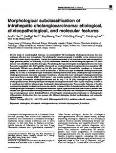
Fig. 1. Axial CT performed with intravenous contrast showed an irregular, hypoenhancing mass (arrow) in the right hepatic lobe (anatomic left lobe in heterotaxy syndrome) with overlying capsular retraction. The surface contour of the liver was nodular.
consistent with a poorly differentiated carcinoma favoring primary intrahepatic cholangiocarcinoma (ICC). Subsequent F-18 Fluorodeoxyglucose (FDG) PET/CT showed abnormal FDG uptake by the dominant liver mass (SUVmax = 4.8) as well as two smaller foci of abnormal increased metabolic activity adjacent to the dominant liver mass concerning for additional tumors. The patient underwent resection of the anatomic left lateral section of the liver. 2.2. Pathological findings and targeted next generation sequencing result The liver specimen was serially sectioned perpendicular to the resection margin to reveal an irregular 6.3 × 5.6 × 3.1 cm tumor mass with firm grey-whitish cut surface (Fig. 2A), abutting the liver capsule. A 2.4 × 2.0 × 1.2 cm small nodule with similar gross findings was 0.5 cm away from the dominant mass. The resection margin was negative for the tumor. The background liver had a non-uniform vague or incomplete nodular appearance. No additional masses or lesions were identified. The dominant tumor mass and the small nearby independent nodule were composed of poorly differentiated, small to medium-sized tumor cells which were occasionally pleomorphic. The tumor cells were arranged in small vague glandular or slit-lumen glandular nests, occasionally fused or anastomosed into large trabecular tumor nests within prominent fibrotic stroma (Fig. 2B–C). The infiltrating tumor cells entrapped adjacent hepatocytes (Fig. 2D, F) enveloping portal area structures such as small ducts/ductules (Fig. 2E) at the tumor periphery. The differential diagnoses included cholangiocarcinoma, hepatocellular carcinoma (HCC), combined hepatocellular-cholangiocarcinoma, metastatic adenocarcinoma and neuroendocrine tumor. The tumor cells were positive for CK7 (Fig. 2E), CK19 (Fig. 3A), Cam5.2 (Fig. 3B), COX2 (Fig. 3C), EMA, BCL-2, AE1/AE3 and MOC-31, an immunoprofile that supported a diagnosis of intrahepatic cholangiocarcinoma. These markers can also be positive in metastatic adenocarcinomas. Giving that the tumor cells were negative for CEA, CK20, CDX2, TTF1, ER, PR, GCDFP-15 and mammoglobulin, metastatic adenocarcinoma from GI, pancreas, breast, lung, kidney, ovary and thyroid were excluded. In the solid trabecular nests, the tumor cells were weakly positive for synaptophysin, a finding deemed insufficient for the diagnosis of neuroendocrine tumor, and interpreted as slight neuroendocrine differentiation in this tumor. Notably, immunoreactivity for synaptophysin has been reported in some cases of cholangiocarcinoma [4]. Although Glypican-3 immunoexpression within the tumor cells (Fig. 3D) raised the possibility of hepatocellular carcinoma or combined hepatocellular-cholangiocarcinoma, Glypican-3 has also been reported to be expressed in some cases of cholangiocarcinoma [5]. In addition, negativity for hepatic markers such as arginase (Fig. 3E),
3. Discussion Hepatic dysfunction after the Fontan operation is thought to be related to a combination of venous congestion of the liver and the relatively low cardiac output which chronically reduces hepatic arterial blood supply [2,7]. The creation of the Fontan circulation relieves the pulmonary overcirculation, systemic desaturation, and ventricular volume overload inherent to single ventricle physiology by redirecting returning systemic venous blood directly to the pulmonary circulation and allowing the single functional ventricle to solely pump oxygenated blood into systemic arterial circulation. The attainment of this physiologic relief comes with a trade-off as the achievement of a Fontan circulation produces novel pathophysiologic derangements, namely chronic elevation of central venous pressure (CVP) and a decrease in cardiac output; a dangerous combination that can portend end organ dysfunction [8]. The liver is unique in that it has a dual blood supply with about 20% of its blood supply coming from the systemic arterial system via the hepatic artery and the remaining 80% coming in the form of partially deoxygenated blood via the portal vein [9]. The elevated CVP seen in patients with a Fontan circulation is associated with a relative deprivation in portal venous inflow with a compensatory and relative increase in hepatic arterial blood supply to the liver, the socalled arterial buffer response. Hepatic arterial flow is vulnerable to the decreased cardiac output associated with the Fontan circulation limiting the extent to which the hepatic arterial buffer response can secure liver blood supply [10]. This combination of factors conspires to create a state of chronically decreased oxygen delivery to the liver which predisposes to organ damage. This hepatic injury can range from reversible fibrosis to irreversible cirrhosis and neoplasia. 2
Pathology - Research and Practice xxx (xxxx) xxx–xxx
D. Wang et al.
Fig. 2. Intrahepatic cholangiocarcinoma. Peripheral ICC demonstrated a mass-forming growth pattern with blurred border and an adjacent small nodule arising in a stage 3 fibrotic liver (A). The tumor cells were arranged in vague small glandular or slit-lumen nests, sometime fused or anastomosed within the prominent fibrotic stroma (B, x100; C, x400). The infiltrative tumor cells comingled with adjacent hepatocytes (D, x200), or portal tracts with small ducts or ductules in the tumor periphery (E, x100). CK7 highlighted tumor cells (week staining), and small ducts, ductules (strong staining) (E, x200). The tumor cells were negative for HepPar 1, in contrast, entrapped hepatocytes were positive by HepPar 1 (F, x200).
features. Liau JY et al. [13] showed that patients with cholangiolar type ICC had significant better prognosis than those with bile duct type ICC. However, the advanced peripheral small duct or ductular/cholangiolar type ICC has tendency for intrahepatic metastasis6. One small tumor nodule next to the dominant tumor in this patient may suggest intrahepatic metastasis. It has been hypothesized that peripheral small duct type ICCs are derived from small bile ducts originated from hepatic progenitor cells or transformation of ductules [6]. Mucin hypersecretion is rarely seen in peripheral small duct type ICCs. ICCs with coexpression of NCAM/BCL-2 frequently develop in viral hepatitis and develop into mass-forming peripheral type ICCs. ICCs with NCAM positivity and a background of viral hepatitis may be related to origin from hepatic progenitor cells. Our case revealed peripheral massforming tumor location and typical histological characteristics of small duct type or ductular/cholangiolar type ICC. However, the tumor cells
The timeline for the development of significant hepatic dysfunction following Fontan is not definitively known. Previous autopsy studies [2] have shown an incremental incidence of portal and sinusoidal fibrosis and cirrhosis with time after Fontan. Further, regenerative and hyperplastic nodules as well as hepatic adenomas and hepatocellular carcinoma are being increasingly reported after Fontan [7]. To date, there are numerous cases of HCC have been reported after Fontan procedure without other associations [1–3,11,12]. Risk for HCC appears after the first decade of Fontan physiology and rises thereafter to as high as 5% [12]. To our knowledge, ours is the first report of an adult with intrahepatic cholangiocarcinoma in the context of the Fontan procedure. ICCs are heterogeneous carcinomas arising from different anatomical sites of the liver. ICCs can be subclassified into bile duct type and small duct type or ductular/cholangiolar type according to histological
Fig. 3. The immunoprofile of ICC. The tumor cells were positive for CK19 (A, x200), Cam5.2 (B, x100), COX2 (C, x200), Glypican 3 (D, x200), and negative for arginase (E, x200). There was a prominent tumor microvasculature around tumor nests and within stroma highlighted by CD34 (F, x40).
3
Pathology - Research and Practice xxx (xxxx) xxx–xxx
D. Wang et al.
Fig. 4. The background liver. Stage 3 hepatic fibrosis with incomplete bridging was highlighted by Trichrome stain (A, x20). Macro/microvesicular steatosis was on the right and sinuses dilation was on the left (B, x200). Microscopic nests of periportal ductular proliferation with mild cytological atypia, forming compact ductules with small or indistinct lumens and scant stroma in the background (C, x200, inset x400). The proliferating ductules were strongly positive for CK7 in contrast to variably weekly positive biphenotypic hepatocytes in the peripheral (D, x200). That lesion was negative for Glypican 3 as demonstrated in red circle (E, x200). EM (F, x10 K; upper panel: 15 K; lower panel: 40 K) demonstrated dysmorphic cilia that were exceptionally long, undulating rather than straight and have disorganized microtubules.
Heterotaxy patients have a high prevalence of primary cilia dyskinesia (PCD) which is a autosomal recessive disorder of the motile cilia [17]. Diseases of the nonmotile cilia are commonly associated with a spectrum of fibrocystic changes in the kidneys and the liver, including polycystic kidney disease, nephronophthisis and renal cystic dysplasia, congenital hepatic fibrosis, Caroli's disease, and polycystic liver disease. Our patient had no clinical manifestations of PCD or nonmotile ciliopathy associations mentioned above, but EM study demonstrated dysmorphic cilia in small bile ducts of this patient’s background liver. Defects in the structure and/or function of cholangiocyte cilia lead to cholangiociliopathies that result in cholangiocyte hyperproliferation [18]. Noted microscopic nests of atypical bile ductular proliferation may be a consequence of cholangiociliopathy in this patient. Loss of cholangiocyte cilia (sensory and nonmotile) has been described in association with dysregulation of several molecular pathways resulting in cholangiocarcinoma development and progression in experimental studies18. Extrahepatic cholangiocarcinoma have been briefly reported in older adults with situs inversus independent of preceding Fontan operation [19]. It is unknown whether susceptibility to this malignancy is directly influenced by situs abnormalities or cholangiociliopathy. A recent study revealed a high prevalence of IDH1 or IDH2 mutations in peripheral cholangiolar type ICCs [13]. The tumor of this patient was not immunoreactive to IDH1, and no IDH1/IDH2 mutations were detected by targeted next generation sequencing. Instead, MYST3 mutation at Q1388H was detected in the tumor of this patient. MYST3 gene (KAT6) encodes histone lysine acetyltransferase protein. Somatic sequence alterations in MYST3, primarily missense mutations have been observed at low frequency in a range of solid tumors [20], but the functional impact of these alterations remains uncharacterized. MYST3Q1388H has been only reported in renal clear cell carcinoma sample in The Cancer Genome Atlas (TCGA) database but has not been reported in intrahepatic cholangiocarcinoma.
in this case only demonstrated weak expression of BCL-2 and no distinct expression of CD56 (NCAM), which may suggest alternative pathway of carcinogenesis associated with precirrhosis related to a Fontan procedure. Aishima et al. [6] proposed two models of ICCs progression, the bile duct type and peripheral small duct type ICCs. Some bile duct type ICCs seem to develop from premalignant lesions, i.e. biliary intraepithelial neoplasia (BilIN), and intraductal papillary neoplasms of bile duct (IPNB). Chronic hepatitis and cirrhosis are postulated to be part of the background of peripheral mass-forming type ICCs. The canals of Hering and smallest interlobular bile ducts/ductules are candidate cell origins of peripheral small duct type ICCs. Premalignant lesions such as BilIN in bile duct type ICCs have not been detected in small duct type ICCs, but the neoplastic epithelium might evolve from the small interlobular ducts/ductules or canals of Hering [6]. Bile ductules and canals of Hering are highly reactive and proliferative in livers with a wide variety of disorders [14]. A few reports [15] on ICC arising in bile duct adenoma or malignant transformation of bile duct adenoma to ICC have suggested the possibility of precursor lesions of peripheral ICC. We identified the microscopic nests of ductular proliferation with nuclear atypia within the background liver, mimicking bile duct adenoma or ductular/cholangiolar ICC, but not enough to support a diagnosis of metastatic ICC confirmed by immunohistochemical study. These foci of compact atypical ductules may represent a precursor to this tumor. The etiopathogenesis of either HCC or cholangiocarcinoma after Fontan procedure is unclear. The advanced liver disease included stage 3–4 hepatic fibrosis and macrovesicular/microvesicular steatosis in the non-neoplastic portion of this patient’s liver were evidence of chronic venous congestion and fatty liver disease of unknown etiology as well as possible intermittent hypoxic injury. The ductular type of intrahepatic cholangiocarcinoma is known to associate with cirrhosis [14]. Fontan physiology complicated by cardiac cirrhosis may contribute to occurrence of this tumor similar to the proposed mechanism of HCC development after Fontan procedure. ICC arising in the cirrhotic or precirrhotic liver shows higher density of tumor microvessels and arteries than that in ICC with background liver showing a normal histology or non-specific reactive changes [16]. This case also showed the feature of increased density of tumor microvessels and arteries (Fig. 3F).
4. Conclusion This is the first description of an adult with intrahepatic cholangiocarcinoma within a fibrotic liver 28 years after a Fontan procedure. The Fontan physiology complicated by cardiac precirrhosis/cirrhosis may contribute to the occurrence of the described ICC which is 4
Pathology - Research and Practice xxx (xxxx) xxx–xxx
D. Wang et al.
similar to the proposed mechanism in HCC. Unfortunately, the advanced peripheral small duct or ductular/cholangiolar type ICC has tendency for intrahepatic metastasis [6]. Clinical follow up is recommended in patients with Fontan procedure for fibrosis, cirrhosis and screening for liver malignancy, particularly in adults. Molecular study for liver malignancy associated with Fontan procedure is warranted to increase understanding of this carcinogenic pathway.
[5] D. Baumhoer, L. Tornillo, S. Stadlmann, M. Roncalli, E.K. Diamantis, L.M. Terracciano, Glypican 3 expression in human nonneoplastic, preneoplastic, and neoplastic tissues: a tissue microarray analysis of 4,387 tissue samples, Am. J. Clin. Pathol. 129 (2008) 899–906. [6] S. Aishima, Y. Oda, Pathogenesis and classification of intrahepatic cholangiocarcinoma: different characters of perihilar large duct type versus peripheral small duct type, J. Hepato. Pancreat. Sci. 22 (2015) 94–100. [7] J. Rychik, G. Veldtman, E. Rand, P. Russo, J.J. Rome, K. Krok, D.J. Goldberg, A.M. Cahill, R.G. Wells, The precarious state of the liver after a Fontan operation: summary of a multidisciplinary symposium, Pediatr. Cardiol. 33 (2012) 1001–1012. [8] M. Gewillig, The fontan circulation, Heart 91 (2005) 839–846. [9] Z. Kan, D.C. Madoff, Liver anatomy: microcirculation of the liver, Semin. Interv. Radiol. 25 (2008) 77–85. [10] C. Eipel, K. Abshagen, B. Vollmar, Regulation of hepatic blood flow: the hepatic arterial buffer response revisited, World J. Gastroenterol. 16 (2010) 6046–6057. [11] E. Martinez-Quintana, A. Monescillo, F. Rodriguez-Gonzalez, Hepatocellular carcinoma in a non-failing Fontan circulation, Rev. Esp. Enferm. Dig. 109 (2017) 375. [12] S.K. Asrani, C.A. Warnes, P.S. Kamath, Hepatocellular carcinoma after the Fontan procedure, N. Engl. J. Med. 368 (2013) 1756–1757. [13] J.Y. Liau, J.H. Tsai, R.H. Yuan, C.N. Chang, H.J. Lee, Y.M. Jeng, Morphological subclassification of intrahepatic cholangiocarcinoma: etiological, clinicopathological, and molecular features, Mod. Pathol. 27 (2014) 1163–1173. [14] Y. Nakanuma, M. Sasaki, H. Ikeda, Y. Sato, Y. Zen, K. Kosaka, K. Harada, Pathology of peripheral intrahepatic cholangiocarcinoma with reference to tumorigenesis, Hepatol. Res. 38 (2008) 325–334. [15] K.B. Lee, Histopathology of a benign bile duct lesion in the liver: morphologic mimicker or precursor of intrahepatic cholangiocarcinoma, Clin. Mol. Hepatol. 22 (2016) 400–405. [16] J. Xu, S. Igarashi, M. Sasaki, T. Matsubara, N. Yoneda, K. Kozaka, H. Ikeda, J. Kim, E. Yu, O. Matsui, Y. Nakanuma, Intrahepatic cholangiocarcinomas in cirrhosis are hypervascular in comparison with those in normal livers, Liver Int. 32 (2012) 1156–1164. [17] A.J. Shapiro, S. Tolleson-Rinehart, M.A. Zariwala, M.R. Knowles, M.W. Leigh, The prevalence of clinical features associated with primary ciliary dyskinesia in a heterotaxy population: results of a web-based survey, Cardiol. Young 25 (2015) 752–759. [18] A.P. Mansini, E. Peixoto, K.M. Thelen, C. Gaspari, S. Jin, S.A. Gradilone, The cholangiocyte primary cilium in health and disease, Biochim. Biophys. Acta (2017). [19] B.C. Organ, L.J. Skandalakis, S.W. Gray, J.E. Skandalakis, Cancer of bile duct with situs inversus, Arch. Surg. 126 (1991) 1150–1153. [20] J. Chen, F.H. Herlong, J.R. Stroehlein, L. Mishra, Mutations of chromatin structure regulating genes in human malignancies, Curr. Protein Pept. Sci. 17 (2016) 411–437.
Conflict of interest statement None of the authors have or had any relevant of relationship with any commercial interests, as pertaining to this manuscript. Funding disclosures None. Acknowledgement We would like to thank Christopher Woods for his excellent technique assistance. References [1] D. Josephus Jitta, L.J. Wagenaar, B.J. Mulder, M. Guichelaar, D. Bouman, J.P. van Melle, Three cases of hepatocellular carcinoma in Fontan patients: review of the literature and suggestions for hepatic screening, Int. J. Cardiol. 206 (2016) 21–26. [2] K. Pundi, K.N. Pundi, P.S. Kamath, F. Cetta, Z. Li, J.T. Poterucha, D.J. Driscoll, J.N. Johnson, Liver disease in patients after the fontan operation, Am. J. Cardiol. 117 (2016) 456–460. [3] A.A. Ghaferi, G.M. Hutchins, Progression of liver pathology in patients undergoing the Fontan procedure: chronic passive congestion, cardiac cirrhosis, hepatic adenoma, and hepatocellular carcinoma, J. Thorac. Cardiovasc. Surg. 129 (2005) 1348–1352. [4] Y. Nakanuma, Y. Sato, K. Harada, M. Sasaki, J. Xu, H. Ikeda, Pathological classification of intrahepatic cholangiocarcinoma based on a new concept, World J. Hepatol. 2 (2010) 419–427.
5