Clinical Anatomy 26:827–832 (2013)
ORIGINAL COMMUNICATION
Intraoperative Small Bowel Length Measurements and Analysis of Demographic Predictors of Increased Length EZRA N. TEITELBAUM,1 KHASHAYAR VAZIRI,1* SARA ZETTERVALL,1 RICHARD L. AMDUR,1 AND BRUCE A. ORKIN2 1
Department of Surgery, George Washington University, Washington, District of Columbia 2 Division of Colon and Rectal Surgery, Rush University Medical Center, Chicago, Illinois
Few studies have measured small bowel length (SBL) in live humans and many textbooks base their “normal” SBL values on cadaver data. Here, we present a series of intraoperative SBL measurements and analyze predictors of increased length. SBL from ligament of Treitz to ileocecal valve was measured in patients undergoing laparotomy for colorectal resection. Patients with Crohn’s disease and those who had undergone prior bowel resections were excluded. In the 240 patients studied, mean SBL was 506 6 105 (285–845) cm. Height was positively associated with increased SBL (P < 0.001) and men had longer SBL than women (533 vs. 482 cm, P < 0.001). A multivariate linear regression model using patient sex, age, height and weight was significant (P 5 0.001) and the predictors explained 8% of the variance in SBL. In this model, only height was independently predictive of increased SBL (P 5 0.03). Correlation results differed between sexes. In men, height correlated with increased SBL (r 5 0.20; P 5 0.03), whereas in women it did not. In men, age had a positive correlation with SBL at a trend level (r 5 0.17; P 5 0.08), whereas in women age had a negative correlation with SBL (r 5 20.18; P 5 0.04). The mean SBL was 506 cm in live patients, as compared with the 600–700 cm range derived from prior cadaver studies. Male sex and height had positive correlations with SBL. SBL may decrease with age in women but not in men. Clin. Anat. 26:827–832, 2013. VC 2013 Wiley Periodicals, Inc. Key words: gastric bypass; intestinal anatomy; small bowel length; short gut syndrome
INTRODUCTION Although seemingly a basic anatomic measurement, there exists considerable uncertainty regarding the normal length of the human small intestine. Anatomy, gastroenterology, and surgery textbooks provide normal values for small bowel length (SBL) ranging from 400 to 800 cm (Greenfield and Mulholland, 2001; Gray et al. 2005; Schwartz and Brunicardi, 2010), with many stating a norm of 600–700 cm (Rosse et al., 1997; Yamada and Alpers, 2009; Moore et al., 2010; Patton et al., 2010; Sleisenger et al., 2010). These measurements are largely derived from studies of human cadavers and may differ from actual
C V
2013 Wiley Periodicals, Inc.
physiologic length in vivo. Few data exist from measurements in live humans. The findings of prior studies using both live and cadaveric measurements (Treves, 1885; Underhill, 1955; Guzman et al., 1977; *Correspondence to: Khashayar Vaziri, George Washington University, Department of Surgery, 2150 Pennsylvania Ave., NW, Suite 6B, Washington, District of Columbia 20037. E-mail:
[email protected] Received 3 August 2012; Revised 28 January 2013; Accepted 30 January 2013 Published online 20 March 2013 in Wiley Online Library (wileyonlinelibrary.com). DOI 10.1002/ca.22238
828
Teitelbaum et al.
TABLE 1. Description and Distribution of Measurements Variable SBL (cm) Age Height (cm) Weight (kg)
Mean
Standard deviation
Range
Skewness
Kurtosis
506 55 169 77
105 15 10 19
285–845 20–86 138–196 41–175
0.52 20.09 0.13 0.95
0.17 20.52 20.31 2.56
Nordgren et al., 1997; Hounnou et. al., 2002; Hosseinpour and Behdad, 2008) provide conflicting correlations between patient sex, age, height, weight and SBL. Rather than simply an academic metric, SBL is of important clinical value in many situations. Surgeons must be careful to preserve intestinal length in patients undergoing small bowel resection for obstruction, ischemia or inflammatory bowel disease (IBD) to avoid postoperative short gut syndrome (Scolapio, 2004). Although the small bowel is often measured in these circumstances, it may be difficult to run the small bowel in its entirety to determine an accurate measure of remaining length when extensive adhesions, inflammation and distension are present. Additionally, during Roux-en-y gastric bypass and other anatomy-altering procedures of the fore and midgut, typically only the length of the “Roux limb” (i.e., the jejunal conduit to the stomach) is measured, leaving the length of the distal, and functionally absorptive, small bowel unknown. For these reasons, having an accurate estimation of normal SBL is clinically relevant. Here, we present a series of intraoperative SBL measurements taken in patients undergoing laparotomy. Specific attention is paid to analyzing potential patient-specific predictors of SBL.
METHODS SBL was measured in patients undergoing laparotomy for colorectal resection at a single institution. The small bowel was measured from the ligament of Treitz to the ileocecal valve (jejunum and ileum) along its anti-mesenteric border with a 25 cm umbilical tape immediately upon entering the peritoneal cavity. No traction was applied to the bowel during measurement and manipulation was kept to a minimum. Exclusion criteria included patients with Crohn’s disease, adhesions that precluded measurement of the entire bowel length, peritonitis, and those who had undergone prior bowel resections (either small intestine, colon, or rectum). Patients who had prior intra-abdominal surgery without bowel resection or and who had no significant adhesions (e.g., appendectomy or gynecologic procedures) were included. Demographic data including sex, age, height, and weight were recorded prospectively at the time of surgery. Data were analyzed from a database after removal of patient protected health information. Statistical analysis was performed using SPSS software (version 20, IBM, Armonk, NY) and SAS (version 9.2, Cary, NC). Associations between sex, age, height, weight and SBL were first examined individually using
univariate linear analyses. Separate univariate analyses of age, height, and weight with SBL were then performed within the male and female subgroups. Finally, two multivariate linear regression models were created to predict SBL: the first using sex, age, height and weight as predictors, and the second using the same predictors with additional interaction terms for sex 3 height and sex 3 age. Sensitivity analyses were performed: one to examine the effect of multicollinearity, the other to examine the effect of non-normality of data distributions. A two-tailed P-value < 0.05 was considered statistically significant in all cases. Values throughout are presented as: mean 6 standard deviation (minimum 2 maximum).
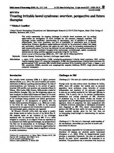
RESULTS From May of 1997 to December of 2008, SBL was measured intraoperatively in 240 patients. 127 patients were female (53%). The mean age was 55 6 15 (range 20–86) years, mean height was 169 6 10 (range 138–196) cm and mean weight was 77 6 19 (range 41–175) kg (Table 1). Mean SBL from the ligament of Treitz to ileocecal value was 506 6 105 (range 285–845) cm (Fig. 1). Skewness was between 21 and 1 for all continuous variables, so parametric analyses were conducted and a normal distribution (for SBL, age, weight and height) was assumed in order to perform multiple linear regression testing. To determine whether these distributional assumptions
Fig. 1. Histogram distribution.
of
SBL,
showing
a
normal
Small Bowel Length Measurement
829
TABLE 2. Univariate Correlation Between Demographics and SBL Variable
Pearson or Phi coefficient
P value
20.24 20.01 0.26 0.11