vaccines targeting the most frequent HPV types associated with cervical cancer (HPV-16 and HPV-18) alone or in combination with the most frequent types that ...
Current Research, Technology and Education Topics in Applied Microbiology and Microbial Biotechnology A. Méndez-Vilas (Ed.) _______________________________________________________________________________________
Introduction of PapilloCheck®, a microarray-based assay for the detection and genotyping of HPV, into the clinical laboratory D.A. Pilger 1, T.T. Moreira2, D.M. Leinberger3, and V.V. Cantarelli4,5 1
Faculdade de Farmácia, Universidade Federal do Rio Grande do Sul, Av. Ipiranga n. 2752 Sala 304 E, Porto Alegre, RS, 90.610-000. Brazil 2 Molecular Biology Section, Weinmann Laboratorio, Ramiro Barcelos 910/5, Porto Alegre – RS – 90035-001 – Brazil. 3 Greiner Bio-One GmBH, BioChip Group, Maybachstr. 2, 72636 Frickenhausen, Germany. 4 Department of Biomedicine, Institute of Health Sciences, Universidade Feevale, RS-239, 2755, Novo Hamburgo – RS – 93510-250, Brazil. 5 Laboratório Qualitá. Av. Dr. Mauricio Cardoso, 833, Hamburgo Velho, Novo Hamburgo – RS - Brazil Infection of the genital mucosa by certain types of human papillomavirus (HPV) may lead to warts or irregular cell growth. Persistence of HPV infection, particularly by those belonging to the high-risk types (HR-HPV), is associated with an increased risk for cervical cancer development. Low-risk HPV (LR-HPV) types are more often associated with benign warts. Microarray technology has been introduced into the clinical laboratory for HPV detection. One such method is the PapilloCheck® (Greiner Bio-One GmbH, Germany), which is able to detect and identify 24 HPV types. Here we describe the comparison of results for HPV detection in 176 genital samples analysed by PapilloCheck® and Hybrid Capture (HC2). The performance of PapilloCheck® was considered adequate and this methodology was introduced into the routine testing for HPV in our clinical laboratory. Keywords: Microarray; PapilloCheck®, DNA chip; HPV; HPV genotyping
1. Introduction Infection of the genital mucosa by certain types of human papillomaviruses (HPV) often causes irregular cell growth and warts. Although some HPV infections may regress spontaneously, persistence of HPV infection, particularly caused by those types identified as high-risk types (HR-HPV), is associated with an increased risk of developing cervical cancer [1,2]. Low-risk HPV types (LR-HPV) are more often associated with benign genital warts [3]. Recently, vaccines targeting the most frequent HPV types associated with cervical cancer (HPV-16 and HPV-18) alone or in combination with the most frequent types that cause genital warts (HPV-6 and HPV-11), have been developed [4]. Until now, several molecular methods have been used to detect HPV DNA in genital samples [5]. The Hybrid Capture 2 (HC2) HPV DNA assay (Qiagen) has been widely used to detect HPV types belonging to either the low or high-risk group. However, HC2 neither distinguishes between the several types that comprise each group nor is it able to determine the presence of multiple types in the sample. In this assay, potentially present HPV DNA hybridizes with an RNA probe cocktail and the RNA:DNA hybrids are then captured with antihybrid antibodies. The subsequent detection is based on a chemiluminescence reaction. The method is based on signal amplification and can detect HRHPV types 16, 18, 31, 33, 35, 39, 45, 51, 52, 56, 58, 59 and 68, as well as LR-HPV types 6, 11, 42, 43 and 44 using two separate reactions. However, despite being considered adequately sensitive for HPV DNA detection in clinical settings, the high-risk probe cocktail is known to cross-react with other related or non-related HPV types [6]. More recently, microarray technology has been incorporated into the clinical laboratory. For HPV detection, PapilloCheck® (Greiner Bio-One GmbH, Germany) is a microarray-based assay, which is able to simultaneously detect and identify 24 different HPV types [7]. This method has been compared with several other available methods, including HC2 [8,9,10] and the good clinical performance of PapilloCheck® has been assessed in two studies published recently [8,10]. The HPV types currently detected by PapilloCheck® are: High-Risk types: 16, 18, 31, 33, 35, 39, 45, 51, 52, 53, 56, 58, 59, 66, 68, 70, 73, and 82 Low-Risk types: 6, 11, 40, 42, 43 and 44 PapilloCheck® is a PCR-based assay that uses a new set of primers which targets the E1 region of the HPV genome (Figure 1). The fluorescently labeled PCR products are then hybridized to specific DNA probes fixed onto the surface of a DNA chip. In our lab, PapilloCheck® was introduced in 2008 and has been used as a diagnostic tool to detect and genotype HPV present in clinical samples. A representation of all steps involved in HPV testing by PapilloCheck® can be viewed in Figure 2.
©FORMATEX 2010
1437
Current Research, Technology and Education Topics in Applied Microbiology and Microbial Biotechnology A. Méndez-Vilas (Ed.) _______________________________________________________________________________________
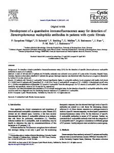
long control region
E6 E7
L1 E1
HPV16 7906 bp
PapilloCheck® E1 PCR product
L2 E5
E2 E4
Figure 1. PapilloCheck® uses a new set of primers which targets the E1 region of the HPV genome.
2. Methodology 2.1. Sample Collection According to the manufacturer (Greiner Bio-One), PapilloCheck® has been validated using DNA prepared from cervical smears collected in different transport media, e.g. PreservCyt®, Hologic (Bedford, MA USA), Surepath™, BD (Franklin Lakes, NJ USA), STM™ Qiagen (Gaithersburg Inc., Gaithersburg USA). In our initial studies, we used 176 genital samples collected using the digene specimen collection Kit (Qiagen), which were assayed by HC2 and then kept frozen (-20°C) until needed. Subsequently, 1.0 ml of the remaining genital sample was used for DNA extraction, as indicated below. More recently, Greiner Bio-One developed a dedicated PapilloCheck® Collection Kit for the collection of cervical samples consisting of a cervical brush and a vial containing a transport medium. We extended the usage of the cervical brush to obtain samples including endocervical, urethral, and other genital samples. Additionally, biopsies (e.g. from warts), preserved in transport medium were used for the analysis. 2.2. DNA Extraction Different manual and automated DNA extraction methods can be used to obtain purified nucleic acids from clinical samples in general and for the extraction of viral and human genomic DNA from human cervical samples in particular. In our case, frozen genital samples (see 2.1) were allowed to thaw and then 1.0 ml aliquot was used for DNA extraction using the High Pure PCR Template Preparation Kit (Roche) in combination with the MagNA Pure LC platform (Roche). After extraction and purification, samples were eluted with appropriated elution buffer to a final volume of 100µl. The QIAamp DNA Mini Kit (Qiagen) is now used in our laboratory. The volume of sample used is dependent on the extraction method and the manufacturer’s instruction must be consulted before establishing a certain method. In the meantime, Greiner Bio-One developed two dedicated manual PapilloCheck® DNA Extraction Kits either applying single column or 8 column DNA extraction. The isolated DNA must be subjected to PCR amplification prior to hybridization of the PCR products onto DNA probes fixed on the PapilloCheck® Chip.
1438
©FORMATEX 2010
Current Research, Technology and Education Topics in Applied Microbiology and Microbial Biotechnology A. Méndez-Vilas (Ed.) _______________________________________________________________________________________
1. Sample Collection Cervical smear
4. Hybridisation 15 mins at RT
2. DNA Extraction 60-90 mins
5. Washing 2 mins
3. PCR 150 mins
6. Scanning 10 mins
7. Data Analysis 5 mins
Figure 2: Schematic representation of all steps and the required time for HPV detection by PapilloCheck®
2.3. DNA Amplification After the extraction of viral and human genomic DNA from a cervical specimen a 350 bp fragment of the E1 gene is amplified in the presence of a set of HPV specific primers by polymerase chain reaction (PCR). In each run, a series of positive and negative controls are included to avoid false negative or false positive results, respectively. To avoid false negative results that might result from a deficient sample collection, a fragment of the human “house keeping gene” ADAT1 (Adenosine deaminase, tRNA-specific1) is amplified in the same reaction, which assures that an adequate amount of human DNA is present in each reaction. The correct performance of the PCR is monitored by parallel amplification of a DNA fragment of a control template present in the PapilloCheck® PCR-MasterMix. For each sample a PCR reaction mix is loaded into appropriate PCR tubes, and consists of 0.2 µl Taq DNA polymerase and 5 µl of previously extracted DNA added to 19.8 µl of PapilloCheck® PCR-MasterMix, which is then submitted to the following PCR cycling: 95°C for 15 min, followed by 40 cycles applying: 95°C for 30 sec, 55°C for 25 sec, and 72°C for 45 sec, and then 15 cycles of 95°C for 30 sec and 72°C for 45 sec. At the end the temperature is hold at 4-8°C. The manufacturer recommends the use of HotStarTaq DNA Polymerase (Qiagen). In our lab, Platinum® Taq DNA Polymerase (Invitrogen) was used for PCR reactions and showed equivalent performance. To prevent carry-over contaminations, PapilloCheck® PCR-MasterMix contains dUTP, and any PCR product originated from previous reactions can be eliminated via Uracil-DNA Glycosylase (UNG) treatment before PCR amplification (37°C for 20 min). It is not necessary to check for the presence of PCR products using agarose or polyacrylamide gel electrophoresis at the end of the PCR reaction. All the necessary steps to validate the reaction are incorporated in the DNA chip used subsequently. 2.4. DNA Hybridization After amplification, 5 µl of the amplification products (E1 PCR product and controls) are mixed with hybridization mix and then hybridised to the on-chip controls and HPV-specific DNA-probes attached to the DNA-chip surface. Hybridization is performed in a water vapour saturated atmosphere at room temperature (RT) for 15 min. After hybridization, any unbound DNA is removed by washing with specific washing buffers. The three rapid washing steps are performed with a dedicated wash solution for 10s at RT, 1 min at 50°C and again 10s at RT. After a drying step, the PapilloCheck® DNA-chip is scanned using the CheckScanner™, a two-colour laser microarray scanner with excitation
©FORMATEX 2010
1439
Current Research, Technology and Education Topics in Applied Microbiology and Microbial Biotechnology A. Méndez-Vilas (Ed.) _______________________________________________________________________________________
wavelengths of 532 nm and 635 nm. Every DNA-chip contains 12 DNA-arrays allowing the simultaneous analysis of 12 cervical samples. Each control and type specific HPV DNA probe is printed in five replicates (Figure 3).
Figure 3: Each Slide permits the simultaneous analysis of up to 12 samples. Each well contains type specific HPV DNA probes in replicates of 5 as well as several controls.
2.5. Data Analysis The final evaluation of the PapilloCheck® DNA-chip is carried out using the CheckScanner™ and CheckReport™ software, respectively. The signals created by the hybridized, fluorescently labelled PCR products are automatically read out and potentially present DNA of 24 HPV types is detected and identified. Additional features of the innovative software allow automatic sample tracking, report generation and data collection. Scanning up to 4 DNA-chips enables a flexible throughput of up to 48 analyses in parallel 2.6. Quality Controls A special feature of the PapilloCheck® assay is the presence of comprehensive on-chip controls, which render the analysis highly reliable. Several control systems monitor all critical steps of the assay and chip processing, e.g. specimen quality and DNA extraction (sample control), quality of the PCR reaction (PCR control) and the efficiency of the hybridization (hybridization control), as well as spot homogeneity and printing quality (orientation control and printing control). In addition to the presence or absence of HPV types the CheckReport™ software automatically shows both the corresponding values of the controls and the detected HPV types in a detailed report. For the read-out of the different controls both excitation wavelengths of the CheckScanner™ are used. Whereas for the control of the assay performance (sample and PCR control) the red channel is used (excitation wavelength of 635 nm), the quality of the hybridization and the chip is assessed in the green channel (excitation wavelength of 532 nm). Sample Control: PapilloCheck® tests for the quality of the specimen and/or the DNA extraction by amplifying a fragment of the housekeeping gene ADAT1 (see 2.3) finally leading to a signal on the sample control spots on the PapilloCheck® Chip if human DNA is present in the extracted DNA from the cervical specimen. If no or insufficient amplification of ADAT1 takes place the CheckReport™ software reports the signal as “sample control failed” and the analysis has to be repeated.
1440
©FORMATEX 2010
Current Research, Technology and Education Topics in Applied Microbiology and Microbial Biotechnology A. Méndez-Vilas (Ed.) _______________________________________________________________________________________
PCR control: PapilloCheck® also controls the quality of the PCR reaction. A fragment is amplified from a control template (see 2.3) present in the PapilloCheck® PCR-MasterMix resulting in a signal on the PCR control spots on the PapilloCheck® Chip. The quality of the amplification reaction is also automatically assessed by the CheckReport™ software. If the PCR performance decreases below a threshold the signal of this control is displayed as “PCR control failed”. 2.7. Results and discussion In our lab, we have compared the performance of PapilloCheck® with the HC2, the most widely used HPV test. Initially, we analyzed 176 genital samples by both methods. For PapilloCheck®, the DNA microarrays were automatically analyzed using the CheckScanner™ (Greiner Bio-One) and CheckReport™ software to identify HPV genotype(s) present in each sample. Since only high-risk HPV types (HR-HPV) were detected by HC2, only samples that were positive for HR-HPV by PapilloCheck® were included in the statistical analysis. Overall, the agreement was 84.7%, with 88.8% and 78.3% of positive and negative agreement, respectively. The Cohen’s kappa coefficient was 67.6%. Fifteen samples were positive by PapilloCheck® and negative by HC2. In all cases a high-risk HPV type was detected by PapilloCheck®. These samples were further confirmed to harbour HPV DNA by an alternative method [11] and thus considered false negative by HC2. On the other hand, twelve samples were positive by HC2 and negative by PapilloCheck®. In general, these samples tended to have low RLU values and were negative by the alternative PCR assay, and were considered false positive by HC2. PapilloCheck® allowed for rapid detection and identification of the HPV genotypes involved in these infections, a characteristic that may be useful for follow-up, since only long-term persistence of the same HPV genotype is associated with increased risk for the development of cervical cancer. Among our patients, several HPV types other than HPV-16 and HPV-18 were found in a relatively high proportion of samples. Interestingly, coinfection by two or more HPV types was observed in 42.5% of the HPV positive patients. Among these, 21.6% were coinfected by 2 HPV types, and 20.8% had 3 or more types involved. The following HPV types were more likely to be found in association (coinfection) with other types: 6, 16, 35, 39, 42, 44, 51, 52, 53, 56, 59, 66, 68, 73 and 82 (p