|
Received: 8 June 2017 Accepted: 13 June 2017 DOI: 10.1002/brb3.775
ORIGINAL RESEARCH
Hippocampal volume, social interactions, and the expression of the normal repertoire of resident–intruder behavior Eszter Kalman | Kevin A. Keay School of Medical Sciences (Anatomy & Histology), The University of Sydney, Sydney, NSW, Australia Correspondence Eszter Kalman, Laboratory of Neural Structure & Function, School of Medical Sciences (Anatomy & Histology), The University of Sydney, Sydney, NSW, Australia. Email:
[email protected] Funding information This study was supported by NHMRC (Australia) and NG Macintosh Memorial Fund
Abstract Introduction: Reduced hippocampal volumes are reported in individuals with disrupted emotional coping behaviors in both human clinical conditions and in experimental animal models of these populations. In a number of experimental animal models, it has been shown that social interactions can promote resilience and buffer the negative neural consequences of stimuli that disrupt effective coping. Methods: Hippocampal and dentate gyrus volumes were calculated in 54 male Sprague Dawley rats; (1) single housed (n = 12), (2) single housed and exposed to daily 6-min social interactions testing in a resident–intruder paradigm (n = 11); (3) group housed (n = 12); (4) single housed and sham injured (n = 12); (5) single housed, sham injured, and social interactions tested (n = 7). Results: We present data which shows that even a brief daily exposure to a conspecific in resident–intruder social interactions test is sufficient to prevent the reduction in hippocampal volume triggered by single housing. Conclusion: When considered with previously published data, these findings suggest that the expression of the full repertoire of social, nonsocial, dominance, and submissive behaviors in response to the physical presence of an intruder in the home cage plays a significant role in this maintenance of hippocampal volume. KEYWORDS
corticosterone, dominance behavior, hippocampus morphology, housing, nerve injury, rat, social buffering
1 | INTRODUCTION
our previously published data showed that following sciatic nerve
Reduced hippocampal volumes are reported in patients with post-
played by a rat in response to the presence of an intruder into the
traumatic stress disorder or major depressive disorder in whom
territory of their home cage is substantially changed. This effect on
emotional coping behaviors are disrupted (Gilbertson et al., 2002;
resident–intruder social interactions is characterized by significantly
O’Doherty, Chitty, Saddiqui, Bennett, & Lagopoulos, 2015; Saylam,
reduced dominance behaviors and more frequent approach–avoid
Üçerler, Kitiş, Ozand, & Gönül, 2006; Sheline, Gado, & Kraemer,
like behaviors (Kalman & Keay, 2014; Monassi, Bandler, & Keay,
2003; Sheline, Sanghavi, Mintun, & Gado, 1999). In a preclinical an-
2003). Rats with reduced dominance behaviors had significantly
imal model of nerve injury, similar findings have been reported in
smaller hippocampal volumes compared to both nerve injured rats
injured rats with comorbid alterations in emotional coping behav-
whose resident–intruder social interactions were unchanged, and
iors, an effect that is not seen in nerve-injured rats whose emotional
sham-injured rats with normal levels of dominance behavior (Kalman
coping behaviors are unchanged (Kalman & Keay, 2014). Specifically,
& Keay, 2014).
injury, in a subgroup of rats, the repertoire of behaviors usually dis-
This is an open access article under the terms of the Creative Commons Attribution License, which permits use, distribution and reproduction in any medium, provided the original work is properly cited. © 2017 The Authors. Brain and Behavior published by Wiley Periodicals, Inc. Brain and Behavior. 2017;e00775. https://doi.org/10.1002/brb3.775
wileyonlinelibrary.com/journal/brb3 | 1 of 7
|
KALMAN and KEAY
2 of 7
The coincident hippocampal volume changes and reduction of
Rats were single housed in a clear Perspex cage (40 × 36 × 24 cm)
dominance behavior in injured rats raise the question of whether it
to which they were habituated (14 days). The group housed rats lived
is the expression of the full repertoire of resident–intruder social be-
in polycarbonate cages with a wire lid (53 × 37 × 25 cm) in groups of
haviors that in some way protects the hippocampus from such shrink-
4–6. Food and water were available ad libitum. All rats were housed
age. In the visible burrow system paradigm (Blanchard et al., 1995),
and tested under a reversed dark–light cycle (12 h:12 h). Resident–in-
dominant rats are readily identified by their behaviors and their lev-
truder social interactions testing was conducted on groups 2 and 5 as
els of dominance are related to increased hippocampal neurogenesis.
described earlier. The resident–intruder testing procedure was mod-
Dominant rats have more cells in the dentate gyrus of the hippocam-
ified from that described by Koolhaas et al. (2013) and is described
pus than subordinate rats, an increase that is related primarily to the
in detail by Monassi et al. (2003). In brief, an age, weight, and sex-
survival of new neurons rather than increased levels of proliferation
matched intruder was introduced into the home cage of each singly
(Kozorovitskiy & Gould, 2004). It is often inferred, although not ex-
housed rat. The interactions of the two rats were recorded for 6 min
perimentally demonstrated, that changes in hippocampal volume are a
using an infrared camera (DCRA-C155; Sony). The behaviors of each
consequence of either altered neurogenesis or the altered survival of
resident rat were scored from the video record and classified into one
newly generated neurons. It is possible therefore that the relationship
of the four mutually exclusive categories. Dominance behavior: stand-
between the expression of dominance behaviors and enhanced sur-
ing on top of the supine intruder, biting, chasing, aggressive groom-
vival of hippocampal neurons relates to our observation that rats who
ing, boxing, and sideway lateral pushing. Social behavior: sniffing and
do not show the normal repertoire of resident–intruder social behav-
exploration of the intruder specifically focused around the anogeni-
iors have smaller hippocampal volumes.
tal region. Nonsocial: cage exploration and self-grooming. Submissive:
In the resident–intruder test, resident rats are singly housed to
defensive alerting, fleeing behavior, and supine postures.
enable the establishment of a territory in the home cage. It is import-
Rats were behaviorally tested for 11 days (i.e., 5 test days, a rest
ant to note that each rat is in visual, auditory, and olfactory contact
day, and then 6 further test days). The rats receiving a sham injury
with other identically housed individuals, however, they do not have
were tested for 5 days prior to the injury, testing was not conducted
physical contact. Social interactions with a conspecific intruder, intro-
on the day of surgery, then testing resumed for 6 days after the sur-
duced into the home cage are measured daily (Monassi et al., 2003).
gery. In the behavioral test, the same intruder rat never met a resident
In this study, we investigated whether the expression of the nor-
rat more than twice and never on consecutive days.
mal repertoire of resident–intruder social behaviors maintains hip-
Rats in groups 4 and 5 were anesthetized with isoflurane (2%–3%
pocampal volume. Specifically, we measured hippocampal volumes
adjusted) to perform the sham surgery. An incision was made in the
in: (1) rats able to express the normal repertoire of social behaviors
skin overlying the right thigh muscles. The muscles were parted gently
in the resident–intruder test; (2) rats similarly housed with no op-
by blunt dissected and the sciatic nerve revealed at its trifurcation.
portunity to express the normal repertoire of social behaviors; and
The skin was then sutured and the rat was allowed to recover under
(3) rats housed in a social group under standard laboratory housing
close observation. The period of anesthesia and surgical procedures
conditions.
lasted no longer than 20 min (for further detail, see Kalman & Keay,
In an earlier study looking at the impact of nerve injury on hip-
2014). On completion of resident–intruder testing, the brain of each
pocampal volume, we reported that nerve-injured, sham-injured, and
rat was removed following transcardial perfusion with saline followed
uninjured rats each of which showed the normal repertoire of social
by paraformaldehyde (4%). A one in five series of coronal 40 μmol/L
behaviors in the resident–intruder test had identical hippocampal vol-
sections were cut on a cryostat, mounted on slides, and Nissl stained.
umes. We followed up these observations in this study and compared
A photomicrograph (40× magnification) was taken of each section and
sham-injured rats that had undergone social interactions testing, with
image analysis software was used to calculate the surface area of the
sham-injured rats with no opportunity to express the normal reper-
hippocampus and dentate gyrus on every section (Schneider, Rasband
toire of social behaviors in the resident–intruder test. We hypothe-
& Eliceiri, 2012). Hippocampal and dentate gyrus boundaries were
sized a reduction in hippocampal volume in the group of rats with no
drawn onto each photomicrograph (cf. Kalman & Keay, 2014). Paxinos
opportunity to express the normal levels of dominance behavior.
and Watson’s stereotaxic atlas was used to define the anatomical boundaries of the hippocampus and the dentate gyrus (Paxinos &
2 | METHODS
Watson, 2007). The total volumes of the hippocampus and dentate gyrus were calculated using Cavalieri’s method (Rosen & Harry, 1990), in which the surface areas of each of these structures are calculated in
Fifty-four male Sprague Dawley rats were used in this experiment. The
equidistant serial coronal sections and then multiplied by the distance
University of Sydney, Animal Care and Ethics Committee approved all
between the adjacent sections. The surface areas of the hippocampus
procedures (#3920, #4852, and #776). The rats were randomly allo-
on serial coronal sections for its entire rostrocaudal extent are shown
cated into five groups: (1) single housed (n = 12); (2) single housed and
in Figure 1c. The total hippocampal volumes calculated from these
social interactions tested (n = 11); (3) group housed (n = 12); (4) single
sections are shown in Figure 1a.
housed and sham injured (n = 12); (5) single housed, sham injured, and social interactions tested (n = 7).
The dorsal hippocampus is defined anteriorly by the presence of CA3 cells at approximately the coronal level represented at −1.72 mm
|
3 of 7
KALMAN and KEAY
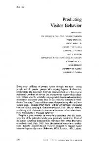
F I G U R E 1 Mean durations (±SEM) of dominance, nonsocial, social, and submissive behaviors expressed by (a) single-housed and social interactions tested rats and (b) single-housed, social interactions tested, and sham-injured rats on days 3–12 of resident–intruder social interactions testing
bregma in the atlas of Paxinos and Watson (2007) and posteriorly by
The volumes of hippocampus and dentate gyrus were compared
the transitional zone of the fimbria and its replacement by the oriens
between all groups using multivariate analysis of variance (MANOVA).
layer of the hippocampus. A distinction aided by the fact that the fi-
This analysis was conducted following preliminary assumption testing
bers of the fimbria are parallel to the plane of sectioning and the oriens
for linearity, normality, univariate, and multivariate outliers as well as
layer runs perpendicular to the plane of sectioning. The intermediate
homogeneity of variance. A MANOVA was performed for the follow-
hippocampus is defined anteriorly by the disappearance of the fimbria
ing datasets: (1) left dentate gyrus (dorsal, ventral, posterior), (2) right
and the appearance of the ventral subregions. Its posterior border is
dentate gyrus (dorsal, ventral, posterior), (3) left hippocampus (dorsal,
defined by the merging of the dorsal and ventral granular layers of
intermediate, posterior), and (4) right hippocampus (dorsal, interme-
the dentate gyrus. This border was selected in order to maintain con-
diate, posterior). Statistical significance was determined for p .44 is considered significant for
from bregma. In the region of CA1, we defined the border using a per-
N = 18; Figure 2b).
pendicular line drawn from the hippocampal fissure to the alveolus. The lateral border was established by the change in staining between oreins of the hippocampus and the alveolus. At the point where the
3 | RESULTS
CA2 region changes into the ventral subiculum a line was drawn between the end of the pyramidal layer and the hippocampal fissure. The
Social interactions in the resident–intruder test are stable over the
boundaries of the dentate gyrus were defined by the border of the
11-day testing procedure (see Figure 1a). When exposed to an in-
molecular and granular layers which are readily identified in thionin-
truder rat, the resident rats spend almost half of the 6-min test period
stained sections. The posterior dentate subregion was defined ante-
engaged in nonsocial behavior, which is predominantly self-grooming
riorly as the point at which the dorsal and ventral regions merge and
and exploring the cage. Dominance is the next most frequent behavior
posteriorly as the point at which the granular layer has disappeared.
expressed and comprises mainly of the resident standing on top of
Comparisons of sections from randomly selected rats, perfused on
the supine intruder, aggressive grooming of the intruder, and chasing
different days, were used to compare cortical thickness, the width of
and sideways lateral pushing. Negligible time is spent in submissive
the diencephalon, and the distance from the dorsal surface to superior
behaviors (