J_ID: PEPS Customer A_ID: PEPS22625 Cadmus Art: PEPS22625 Ed. Ref. No.: 2014-00055.R1 Date: 16-February-15
Stage:
Page: 1
Invited Review Targeting Protein2Protein Interfaces Using Macrocyclic Peptides AQ2
Meng Gao,1 Kui Cheng,1 Hang Yin1,2 1
Department of Chemistry, Center of Basic Molecular Science, Tsinghua University, Beijing, China 100082
2
Department of Chemistry and Biochemistry, the BioFrontiers Institute, University of Colorado Boulder, Boulder, CO 80309-0596 Received 15 December 2014; revised 29 January 2015; accepted 1 February 2015 Published online 00 Month 2015 in Wiley Online Library (wileyonlinelibrary.com). DOI 10.1002/bip.22625
INTRODUCTION ABSTRACT: Protein2protein interactions (PPIs) are critical in numerous biological processes including signaling transduction, function regulations, and disease development. To regulate PPIs has been thought to be challenging due to their highly dynamic and expansive interfacial areas. Nonetheless, successful examples have been reported of targeting PPI using small molecules, peptides, and proteins. Peptides, especially macrocyclic peptides have proven to be a particularly useful tool to inhibit PPIs for their exquisite potency, stability and selectivity. Herein we review the recent developments of this area of research, focusing on the macrocyclic peptides isolated from natural products, identified from library screening, and rationally designed based on strucC 2015 Wiley Periodicals, Inc. tures, as PPI regulators. V
Biopolymers (Pept Sci) 00: 000–000, 2015. Keywords: macrocyclic peptide; protein2protein interface; rational design; inhibitor
This article was originally published online as an accepted preprint. The “Published Online” date corresponds to the preprint version. You can request a copy of any preprints from the past two calendar years by emailing the Biopolymers editorial office at
[email protected]. Correspondence to: Hang Yin; e-mail:
[email protected] Contract grant sponsor: National Institutes of Health Contract grant numbers: R01GM101279, R01GM103843 Contract grant sponsor: Tsinghua University Meng Gao and Kui Cheng contributed equally to this work. C 2015 Wiley Periodicals, Inc. V
P
rotein2protein interactions (PPIs) regulate numerous essential processes of life, including replication and transcription of nucleic acids, protein biosynthesis, signal transduction, metabolic cycle, and also viral diffusion and virus survival in host cell.1–5 Hence, extensive researches focusing on regulation of PPI have been carried out in the last decade.6–8 Current PPI mediators are mainly divided into three groups: small molecules, linear/cyclic peptides, and proteins.9–12 In this review, we focus on the macrocyclic peptide PPI mediators as they have the following advantages. Firstly, peptides are large enough to render an extensive, continuous binding surface that can competitively inhibit PPIs.13,14 The polypeptide backbones usually make them more soluble in water than small molecule organic compounds. Secondly, when compared to proteins, peptides can be more readily synthesized, characterized and purified.14,15 Finally, compared to linear peptides, cyclic peptides are: (A) more rigid, therefore demand less entropy penalty upon association with their targets, which in turn elevates their binding affinities (Figure 1); and (B) more stable in complex biological environments due to enhanced resistance to proteolytic degradation.14–16 In term of their functions, macrocyclic peptide PPI regulators can be generally described as activators and inhibitors. An activator can facilitate protein2protein associations,17 thereby increasing the PPI-mediated cellular response. For example, peptide A (Figure 2) was found to activate human melanocortin 3 receptor (hMC3R), an inhibitory autoreceptor on the surface of proopiomelanocortin neurons.17–20 Meanwhile, the macrocyclic peptide inhibitors suppress the PPI-induced respnse by disrupting the protein2protein complex formation. For example, macrocyclic peptide mimetic B (Figure 2) showed significant inhibition of the interaction between mixed lineage leukemia 1 (MLL1) and its mediator menin with a Ki
PeptideScience Volume 00 / Number 00
ID: padmavathym Time: 21:15 I Path: N:/3b2/PEPS/Vol00000/150013/APPFile/JW-PEPS150013
1
F1
F2
J_ID: PEPS Customer A_ID: PEPS22625 Cadmus Art: PEPS22625 Ed. Ref. No.: 2014-00055.R1 Date: 16-February-15
2
Stage:
Page: 2
Gao et al. Table I Tbe sequences of macrocyclic peptides isolated from natural products Peptides
PPI Targets
l
(PAR-2)-KLK5
2
p53-MDM2
Peptide Sequences
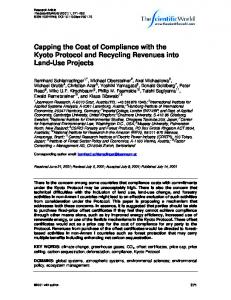
C O L O R FIGURE 1 Cyclic peptides are more rigid than their linear counterparts upon binding to proteins, leading to more potent inhibitors of PPIs.
of 4.7 nM.21 In this mini-review, representative macrocyclic peptide PPI regulators identified from natural products and peptide library screening, as well as developed base on known protein structures are discussed.
MACROCYCLIC PEPTIDES ISOLATED FROM NATURAL PRODUCTS Kallikrein kinin system regulates angiocarpy, urinary and nervous systems, and other physiological functions.22 Sunflower trypsin inhibitor-1 (SFTI-1) isolated from Escherichia coli has been shown to be an inhibitor of Kallikrein-5 (KLK5) by blocking its interaction with protease activated receptor-2 (PAR-2).23 Because of the difficulty of their purifiT1 cation, SFTI-1 analogues 1 (Table I) was prepared using
solid-phase peptide synthesis (SPPS) with an overall 25% isolated yield.23 The SFTI-1 analogue, macrocyclic peptide 1, was stabilized by the introduction of the disulfide linkage. This effective inhibition to KLK5 was monitored by the decreased Ca21 influx. And this stronger inhibitory activity implies the potential therapeutic application to atopic dermatitis.23 P53, “the guardian of the genome”, is well known for its role as an administrator of cell proliferation, growth and apoptosis.24 Over 50% of human tumors contain p53 mutations.24 Oncogene murine double minute-2 (MDM2) can tightly bind to the DNA-binding domain of p53, consequently regulating transcription of the genes and apoptosis.25 Duncan et al. found a macrocyclic peptide, chlorofusin (2) (Table I), derived from Microdochium caespitosum can inhibit the p53-MDM2 interaction by competing with p53 binding with MDM2.26 The titration of 2 in the dissociationenhanced lanthanide fluorescent immunoassay (DELFIA)-
FIGURE 2 The chemical structures of macrocyclic peptide agonist, A, and antagonist, B, of protein-protein interactions.
Biopolymers (Peptide Science)
ID: padmavathym Time: 21:15 I Path: N:/3b2/PEPS/Vol00000/150013/APPFile/JW-PEPS150013
J_ID: PEPS Customer A_ID: PEPS22625 Cadmus Art: PEPS22625 Ed. Ref. No.: 2014-00055.R1 Date: 16-February-15
AQ1
Stage:
Page: 3
Targeting Protein2Protein Interfaces Using Macrocyclic Peptides
3
Table II The sequences of macrocyclic peptides selected from library screening Pepticies
PPI Targets
3
IN-LEDGF/p75
4
CtBP1-CtBP2
5
TNFa-TKFRl
6
p53-HDM2
modified enzyme linked immunosorbent assay (ELISA) determined the inhibitory potency of 2 with an IC50 value of 4.6 lM.26 For the p53-MDM2 interaction, there have been a number of elegant studies on potent small molecule and peptidomimetic inhibitors which have been reviewed elsewhere.27,28
MACROCYCLIC PEPTIDES SELECTED FROM LIBRARY SCREENING Developing highly efficient anti-human immunodeficiency virus (HIV) drug remains a formidable challenge to scientists around the world. In recent years, inhibiting HIV type 1 (HIV1) integrase (IN) that inserts viral cDNA into the human genome has gained much interests.13 One of widely known IN-related proteins is its cellular cofactor, LEDGF/p75 that facilitates IN binding to viral cDNA and makes HIV-1 replication.29 By screening a cyclic peptide library derived from the T2 LEDGF p75 361–370 fragment, peptide 3 (Table II) was identified to be a potent IN inhibitor. Peptide 3 effectively binds to IN, inhibiting its catalytic activity in vitro with high stability (effective even after eight days).30 As a result to search for transcription factors PPI regulators, macrocyclic peptide 4 was selected from screening of a 64 million-membered combinatory cyclic peptides library. Peptide 4 disrupts the NADH-dependent C-terminal binding protein (CtBP) dimerization.28 It was shown that peptide 4 modulates the maintenance of mitotic fidelity in breast cancer
Peptide Sequences
cells (strongly dependent on glycolysis) by breaking the CtBP dimerization without affecting the mitotic fidelity of cells (independent of glycolysis).31 The effect of peptide 4 on disrupting of CtBP dimerization was confirmed using microscopy in COS-7 monkey kidney cells co-transfected YFP–CtBP and CFP–CtBP vectors.28 Tumor necrosis factor-alpha (TNFa) is a pleiotropic inflammatory cytokine related to many disease developments including tuberculosis, septic shock, and various chronic inflammatory disorders.32,33 TNFa-induced response starts with the binding of TNFa trimer to the extracellular domain of TNFa receptor 1 (TNFR1) followed by inhibitory protein release.33 Hence blocking the TNFa-TNFR1 interaction is a potential strategy to regulate the TNFa signaling. By building and screening the bicyclic peptide library, Lian et al. found peptide 5 (Table II) can strongly bind to TNFa and inhibit the TNFa-TNFR1 interaction, significantly extending cell life.34 Biotinylated TNFa was immobilized on the Neutravidin-coated microtiter plate before incubated with horseradish peroxidase (HRP)-conjugated TNFR1 with different concentrations of peptide 5. The amount of HRP2TNFR1 bound to each well was then quantitated by an ELISA assay. Peptide 5 was found to inhibit TNFa-TNFR1 with an IC50 value of 3.1 6 0.3 lM.34 As mentioned above, since its discovery, p53 has been an important target for anti-cancer drug development. As the negative regulator of p53, human double minute-2 (HDM2) compromises the cancer-preventing functions of
Biopolymers (Peptide Science)
ID: padmavathym Time: 21:15 I Path: N:/3b2/PEPS/Vol00000/150013/APPFile/JW-PEPS150013
J_ID: PEPS Customer A_ID: PEPS22625 Cadmus Art: PEPS22625 Ed. Ref. No.: 2014-00055.R1 Date: 16-February-15
4
Stage:
Page: 4
Gao et al.
Table III The sequences of macroeyelie peptides developed by structure-based rational design Peptides
PPI Targets
7
TLR4-MD2
8
TLR4-MD2
9
TLR4-MD2
20
CagL-avb3
11
coat protem-integrin
28
SPSB2-JNOS
p53. Intensive research efforts to develop disruptors of the association of p53 and HDM2 have been successfully carried out, including some macrocyclic peptides as inhibitors to the PPI between p53 and HMD2.9,35 A b-Hairpin peptidomimetic, peptide 6 (Table II), with 6-chloro group incorporated in its Aib-Pmp-Trp23-Glu indole side chain, was identified from screening a phage display peptide library.36 Using an ELISA assay, the authors found that peptide 6 competes with p53 by recognizing the same binding pocket on the surface of HDM2, consequently inhibits the p53-HDM2 association with IC50 value of 5 6 1 nM.36
MACROCYCLIC PEPTIDES DEVELOPED BY STRUCTURE-BASED RATIONAL DESIGN Toll-like receptors (TLRs) are a protein family and an integral part of innate and adaptive immune systems. All TLR response is activated by their respective signal molecules, pathogenassociated molecular patterns (PAMPs).37 It is known that deficiency or excess of TLR4-mediated immune response, dependent on the heterodimer of TLR4 and myeloid differentiation factor 2 (MD2) formation, both can result in pathological disorders.38,39 Recently, a number of reports have appeared on a variety of agonists and antagonists for regulation of the TLR4MD2 interaction, including synthetic molecule, linear peptides and cyclic peptides.40,41 Most recently, Gao et al. compared linear peptides with cyclic peptides in the regulation of TLR4 sigT3 naling and found that macrocyclic peptides 7 and 8 (Table III) cause synergistic activation with lipopolysaccharide (LPS) to turn on TLR4 signaling monitored by nitric oxide production.42 It is worth noting that these macrocyclic peptides were rationally designed by computer simulation. Using the Rosetta
Peptide Sequences
program, the authors designed de novo MD2 mimics that target the TLR4-MD2 protein-protein interface.42 In a separate study, macrocyclic peptides designed based on the natural sequence of MD2 were also found to inhibit the LPS-induced proinflammatory cytokine release.41 Experimental and computational simulation results demonstrated that the MD2 mimic macrocyclic peptide 9 (Table III) can occupy the MD2-binding region on the TLR4 surface, blocking the LPS-induced inflammation signal.41 Using direct biophysical binding assays, Liu et al. validated that the inhibitor peptide 9 is selective and specific for TLR4 in the presence of other TLRs.43 Peptide 7 and 8 both can synergistically enhance the LPS-induced TLR4 signal, while their corresponding linear peptides showed little effects. This observation suggests that the disulfide bond bridged macrocyclic peptide scaffold is essential for their agonistic activities. As a comparison, macrocyclic peptide 9, a truncated functional region of human MD2, can inhibit the TLR4-MD2 interaction, leading to specific inhibition of the TLR4 signaling. Exact mechanism of the different functions of peptide 7, 8 and 9 remains elusive at present. Nonetheless, structurally homologous ligands caused either agonistic or antagonistic effects to various TLRs have been observed with their native ligands as well,44 implying minimal conformational regulations of these innate immune receptors may result in opposite inflammatory response in cells. Helicobacter pylori is a widely studied gastric bacterium and carcinogen. H. pylori targets gastric cells using the RGD sequence (-Arg-Gly-Asp-) of the protein cytotoxin-associated gene L (CagL) that binds to integrins a5b1 and avb3 on the host cell surface.45 An approach for H. pylori related gastric diseases like chronic atrophic gastritis and peptic ulcers, is to obstruct the CagL-avb3 interaction. For example, a cyclic RGD derivative 10 (Table III) designed as CagL functional mimics Biopolymers (Peptide Science)
ID: padmavathym Time: 21:15 I Path: N:/3b2/PEPS/Vol00000/150013/APPFile/JW-PEPS150013
J_ID: PEPS Customer A_ID: PEPS22625 Cadmus Art: PEPS22625 Ed. Ref. No.: 2014-00055.R1 Date: 16-February-15
AQ1
Stage:
Page: 5
Targeting Protein2Protein Interfaces Using Macrocyclic Peptides
5
Table IV Tbe sequences of maeroeyelie peptides synthesized by different cyclization methods Peptides
PPI Targets
13
BCL9-(b-catenin)
14
RAKK-RANKL
15
ExoS-(14-3-3)
16
(BCL-2)-BAK
17
p53-MDM2
18(WP9QY)
Peptide Sequences
RAKK-RANKL
occupies the binding region of CagL-avb3 PPI and recognizes avb3.45 A cell adhesion assay showed that macrocyclic peptide 10 has higher binding affinity with integrins a5b1 and avb3 than CagL.45 Moreover, the NMR measurements and molecular dynamics (MD) calculations demonstrated that the inhibition of the CagL-avb3 interaction mainly relies on secondary structure elements, further identified the RGD sequence is favored.45 In addition, the cyclic peptide 11 (Table III) with RGD sequence was also used to phage delivery in tumor cells recently. Phage VIII, amplified by isopropyl b-D-1thiogalactopyranoside (IPTG) and decorated with peptides 11 on its major coat protein, can bind with integrins on tumor
cells surface and consequently internalized by host cells.46 The amount of 11 decorated can be modulated by IPTG levels and the internalized phage can be quantified by fluorescent microscopy imaging.46 Another example is the macrocyclic peptide inhibitor of the inducible nitric oxide synthase (iNOS). In immune system, nitric oxide controlled by iNOS is an essential molecule to host body for its role of safety alarm when meeting with exogenous threats. It is known that iNOS is regulated by splA kinase and ryanodine receptor (SPRY) domain-containing suppressor of cytokine signaling box protein2 (SPSB2).47 Inhibiting the association of protein SPSB2 and iNOS is a reasonable strategy.
Biopolymers (Peptide Science)
ID: padmavathym Time: 21:15 I Path: N:/3b2/PEPS/Vol00000/150013/APPFile/JW-PEPS150013
J_ID: PEPS Customer A_ID: PEPS22625 Cadmus Art: PEPS22625 Ed. Ref. No.: 2014-00055.R1 Date: 16-February-15
6
Stage:
Page: 6
Gao et al.
Macrocyclic peptide 12 (Table III) containing the specific binding sequence of iNOS, DINNN, can bind with SPSB2 instead of iNOS in bone marrow-derived macrophages (BMDM) lysates. It is developed to inhibit the interaction of SPSB2iNOS successfully with potential to be an anti-infective drug.47
COMMON STRATEGIES OF RING INTRODUCTION Finally, we will discuss a few commonly used methods employed in the cyclic peptides synthesis. In many naturally occurring cyclic peptides, the rings are formed through disulfide bonds between cysteine residues. However, disulfide bonds may not be maintained under physiological conditions. The “click chemistry”48 has been widely used as a synthetic method to generate macrocyclic peptides.49 A good example is that Kawamoto et al. used Huisgen1,3-dipolar cycloaddition reaction to synthesize triazole-stapled B-cell leukemia/lymphoma 9 T4 (BCL9) a-helical peptides 13 (Table IV), which can inhibit the interaction between BCL9 and b-catenin.50 In addition, utilizing “amine cross-linkages”51 (cyclization through a secondry amine) instead of disulfide bonds becomes an alternative method.51 For instance, the WP9QY (Table IV) derivative peptide 14 (Table IV), with amine cross-linkage replacement of disulfide bond, resulted in higher stability and less deactivaition in vitro. With the improvement in enzymatic stability, the WP9QY derivative 14 can be used to target the interaction of receptor activator of NF-jB (RANK) and receptor activator of NF-jB ligand (RANKL) by binding to RANKL and inhibit RANKL-induced abnormal immunization. Another choice is to replace disulfide bonds by “hydrophobic cross-links”52 (cyclization linked by aliphatic hydrocarbon) to facilitate peptide folding.52 Upon cross-linking with olefin metathesis catalyzed by ruthenium complex, the macrocyclic compound 15 (Table IV) turns into inhibitor of interaction between human adaptor protein 14-3-3 and virulence factor exoenzyme S (ExoS) with strongly enhanced binding affinity with 14-3-3.52 Finally and importantly, two classic examples using olefin metathesis to generate hydrocarbon-stapled peptides are presented here. Walensky et al. successfully synthesized “stapled” peptide 16 (Table IV) with stable a-helix conformation.53 This peptide was able to recognize the BH2-binding pocking on the surface of pro-survival oncogenic protein, B-cell CLL/lymphoma 2 (BCL-2), activating the apoptotic pathway to kill leukemia cells.53 Similarly, peptide 17 (Table IV) was designed by Beak et al. for inhibiting MDM2-p53 interaction with an allhydrocarbon staple that conformationally and proteolytically stabilized the short peptide.54 Overall, the cross-link strategy is becoming a useful tool to provide potent peptide PPI inhibitors.
OUTLOOK In this mini review, we briefly discuss macrocyclic peptides as PPI modulators, including representative macrocyclic peptides isolated from natural products, selected from library screening, and obtained through structure-based designs. New technologies such as computer simulation55 provide clear guidance for rational designs and preliminary structure–activity relationship information. We expect to see even more remarkable progress of this research area in the next a few years. With more information available for previously hard-to-get protein complex structures and more computing powers, rationally targeting protein2protein interfaces by macrocyclic peptide may prove to be a generally applicable method to generate new candidates for drug developments. Nonetheless, challenges remains: first, some macrocyclic peptides are hard to synthesize. The synthesis is particularly difficulty with those macrocyclic peptides with more than one ring in a single molecule. Furthermore, the stereochemistry and backbone substitution patterns of unnatural macrocyclic peptides may pose further challenge in their library preparation. Second, the complex crystal structure of macrocyclic peptide and protein can be difficult to get, restricting the further understanding of the mechanism between the targeted receptors and their macrocyclic peptide ligands. This is especially true for some protein groups such as transmembrane proteins, which compose a substantial portion of relevant drug targets. Third, average macrocyclic peptides are still larger than conventional drug-like small molecules. This is helpful when one targets PPIs as they can make more extensive contact with the protein target and can do so without excessive entropic penalty. However, their elevated molecular weights make bioavailability less optimal, which need to be taken into consideration during further therapeutic developments.
REFERENCES 1. McGhee, J. D.; Hippel, P. H. V. J Mol Biol 1974, 86, 469–489. 2. Zhang, A. X.; Wassarman, K. M.; Ortega, J.; Steven, A. C.; Storz, G. Mol Cell 2002, 9, 11–22. 3. Pawson, T.; Nash, P. Gene Dev 2000, 14, 1027–1047. 4. Lomberk, G.; Mathison, A. J.; Grzenda, A.; Seo, S.; Demars, C. J.; Rizvi, S.; Bonilla-Velez, J.; Calvo, E.; Fernandez-Zapico, M. E.; Iovanna, J.; Buttar, N. S.; Urrutia, R. J Biol Chem 2012, 287, 13026–13039. 5. He, Y. P.; Nakao, H. H.; Tan, S. L.; Polyak, P. J.; Neddermann, P.; Vijaysri, S.; Jacobs, B. L.; Katze, M. G. J Virol 2002, 76, 9207– 9217. 6. Cusick, M. E.; Klitgord, N.; Vidal, M.; Hill, D. E. Hum Mol Genet 2005, 14, R171–R181. 7. Vidal, M. Febs Lett 2009, 583, 3891–3894.
Biopolymers (Peptide Science)
ID: padmavathym Time: 21:15 I Path: N:/3b2/PEPS/Vol00000/150013/APPFile/JW-PEPS150013
J_ID: PEPS Customer A_ID: PEPS22625 Cadmus Art: PEPS22625 Ed. Ref. No.: 2014-00055.R1 Date: 16-February-15
AQ1
Stage:
Page: 7
Targeting Protein2Protein Interfaces Using Macrocyclic Peptides 8. White, T. C.; Berny, M. A.; Robinson, D. K.; Yin, H.; DeGrado, W. F.; Hanson, S. R.; McCarty, O. J. T. Febs J 2007, 274, 1481– 1491. 9. Fischer, P. M. Int J Pept Res Ther 2006, 12, 3–19. 10. Arkin, M. R.; Wells, J. A. Nat Rev Drug Discov 2004, 3, 301– 317. 11. Angeles Bonache, M.; Balsera, B.; Lopez-Mendez, B.; Millet, O.; Brancaccio, D.; Gomez-Monterrey, I.; Carotenuto, A.; Pavone, L. M.; Reille-Seroussi, M.; Gagey-Eilstein, N.; Vidal, M.; de la Torre-Martinez, R.; Fernandez-Carvajal, A.; Ferrer-Montiel, A.; Teresa Garcia-Lopez, M.; Martin-Martinez, M.; Jesus Perez de Vega, M.; Gonzalez-Muniz, R. ACS Comb Sci 2014, 16, 250– 258. 12. Granier, S.; Terrillon, S.; Pascal, R.; Demene, H.; Bouvier, M.; Guillon, G.; Mendre, C. J Biol Chem 2004, 279, 50904–50914. 13. Maes, M.; Loyter, A.; Friedler, A. Febs J 2012, 279, 2795–2809. 14. Benyamini, H.; Friedler, A. Future Med Chem 2010, 2, 989– 1003. 15. Katz, C.; Levy-Beladev, L.; Rotem-Bamberger, S.; Rito, T.; Rudiger, S. G. D.; Friedler, A. Chem Soc Rev 2011, 40, 2131– 2145. 16. Gavenonis, J.; Sheneman, B. A.; Siegert, T. R.; Eshelman, M. R.; Kritzer, J. A. Nat Chem Biol 2014, 10, 716–722. 17. Mayorov, A. V.; Cai, M.; Palmer, E. S.; Liu, Z.; Cain, J. P.; Vagner, J.; Trivedi, D.; Hruby, V. J. Peptides 2010, 31, 1894– 1905. 18. Butler, A. A. Peptides 2006, 27, 281–290. 19. Cone, R. D. Endocr Rev 2006, 27, 736–749. 20. Mayorov, A. V.; Cai, M. Y.; Chandler, K. B.; Petrov, R. R.; Van Scoy, A. R.; Yu, Z. R.; Tanaka, D. K.; Trivedi, D.; Hruby, V. J. J Med Chem 2006, 49, 1946–1952. 21. Zhou, H.; Liu, L.; Huang, J.; Bernard, D.; Karatas, H.; Navarro, A.; Lei, M.; Wang, S. J Med Chem 2013, 56, 1113–1123. 22. Swedberg, J. E.; Nigon, L. V.; Reid, J. C.; de Veer, S. J.; Walpole, C. M.; Stephens, C. R.; Walsh, T. P.; Takayama, T. K.; Hooper, J. D.; Clements, J. A.; Buckle, A. M.; Harris, J. M. Chem Biol 2009, 16, 633–643. 23. Shariff, L.; Zhu, Y.; Cowper, B.; Di, W.-L.; Macmillan, D. Tetrahedron 2014, 70, 7675–7680. 24. Levine, A. J.; Momand, J.; Finlay, C. A. Nature 1991, 351, 453– 456. 25. Oliner, J. D.; Pietenpol, J. A.; Thiagalingam, S.; Gvuris, J.; Kinzler, K. W.; Vogelstein, B. Nature 1993, 362, 857–860. 26. Duncan, S. J.; Gruschow, S.; Williams, D. H.; McNicholas, C.; Purewal, R.; Hajek, M.; Gerlitz, M.; Martin, S.; Wrigley, S. K.; Moore, M. J Am Chem Soc 2001, 123, 554–560. 27. Wang, W.; Hu, Y. Med Res Rev 2012, 32, 1159–1196. 28. Liu, M.; Pazgier, M.; Li, C.; Yuan, W.; Li, C.; Lu, W. Angew Chem Int Edit 2010, 49, 3649–3652. 29. Cherepanov, P.; Maertens, G.; Proost, P.; Devreese, B.; Van Beeumen, J.; Engelborghs, Y.; De Clercq, E.; Debyser, Z. J Biol Chem 2003, 278, 372–381. 30. Hayouka, Z.; Hurevich, M.; Levin, A.; Benyamini, H.; Iosub, A.; Maes, M.; Shalev, D. E.; Loyter, A.; Gilon, C.; Friedler, A. Bioorgan Med Chem 2010, 18, 8388–8395.
7
31. Birts, C. N.; Nijjar, S. K.; Mardle, C. A.; Hoakwie, F.; Duriez, P. J.; Blaydes, J. P.; Tavassoli, A. Chem Sci 2013, 4, 3046–3057. 32. Kawakami, M.; Cerami, A. J Exp Med 1981, 154, 631–639. 33. Chen, G. Q.; Goeddel, D. V. Science 2002, 296, 1634–1635. 34. Lian, W.; Upadhyaya, P.; Rhodes, C. A.; Liu, Y.; Pei, D. J Am Chem Soc 2013, 135, 11990–11995. 35. Bautista, A. D.; Appelbaum, J. S.; Craig, C. J.; Michel, J.; Schepartz, A. J Am Chem Soc 2010, 132, 2904–2096. 36. Garcia-Echeverria, C.; Chene, P.; Blommers, M. J. J.; Furet, P. J Med Chem 2000, 43, 3205–3208. 37. Beutler, B. Nature 2004, 430, 257–263. 38. Akira, S.; Uematsu, S.; Takeuchi, O. Cell 2006, 124, 783–801. 39. Due, M. R.; Piekarz, A. D.; Wilson, N.; Feldman, P.; Ripsch, M. S.; Chavez, S.; Yin, H.; Khanna, R.; White, F. A. J Neuroinflamm 2012, 9, 200. 40. Cheng, K.; Wang, X.; Zhang, S.; Yin, H. Angew Chem Int Edit 2012, 51, 12246–12249. 41. Slivka, P. F.; Shridhar, M.; Lee, G.-i.; Sammond, D. W.; Hutchinson, M. R.; Martinko, A. J.; Buchanan, M. M.; Sholar, P. W.; Kearney, J. J.; Harrison, J. A.; Watkins, L. R.; Yin, H. ChemBioChem 2009, 10, 645–649. 42. Gao, M.; London, N.; Cheng, K.; Tamura, R.; Jin, J.; SchuelerFurman, O.; Yin, H. Tetrahedron 2014, 70, 7664–7668. 43. Liu, L.; Ghosh, N.; Slivka, P. F.; Fiorini, Z.; Hutchinson, M. R.; Watkins, L. R.; Yin, H. ChemBioChem 2011, 12, 1827–1831. 44. Park, B. S.; Song, D. H.; Kim, H. M.; Choi, B.-S.; Lee, H.; Lee, J.-O. Nature 2009, 458, 1191–1195. 45. Conradi, J.; Huber, S.; Gaus, K.; Mertink, F.; Royo Gracia, S.; Strijowski, U.; Backert, S.; Sewald, N. Amino Acids 2012, 43, 219–232. 46. Choi, D. S.; Jin, H.-E.; Yoo, S. Y.; Lee, S.-W. Bioconjugate Chem 2014, 25, 216–223. 47. Yap, B. K.; Leung, E. W. W.; Yagi, H.; Galea, C. A.; Chhabra, S.; Chalmers, D. K.; Nicholson, S. E.; Thompson, P. E.; Norton, R. S. J Med Chem 2014, 57, 7006–7015. 48. Kolb, H. C.; Finn, M. G.; Sharpless, K. B. Angew Chem Int Edit 2001, 40, 2004–2021. 49. Saludes, J. P.; Morton, L. A.; Ghosh, N.; Beninson, L. A.; Chapman, E. R.; Fleshner, M.; Yin, H. ACS Chem Biol 2012, 7, 1629–1635. 50. Kawamoto, S. A.; Coleska, A.; Ran, X.; Yi, H.; Yang, C.-Y.; Wang, S. J Med Chem 2012, 55, 1137–1146. 51. Aoki, K.; Maeda, M.; Nakae, T.; Okada, Y.; Ohya, K.; Chiba, K. Tetrahedron 2014, 70, 7774–7779. 52. Glas, A.; Bier, D.; Hahne, G.; Rademacher, C.; Ottmann, C.; Grossmann, T. N. Angew Chem Int Edit 2014, 53, 2489–2493. 53. Walensky, L. D.; Kung, A. L.; Escher, I.; Malia, T. J.; Barbuto, S.; Wright, R. D.; Wagner, G.; Verdine, G. L.; Korsmeyer, S. J. Science 2004, 305, 1466–1470. 54. Baek, S.; Kutchukian, P. S.; Verdine, G. L.; Huber, R.; Holak, T. A.; Lee, K. W.; Popowicz, G. M. J Am Chem Soc 2011, 134, 103– 106. 55. Joce, C.; Stahl, J. A.; Shridhar, M.; Hutchinson, M. R.; Watkins, L. R.; Fedichev, P. O.; Yin, H. Bioorg Med Chem Lett 2010, 20, 5411–5413.
Biopolymers (Peptide Science)
ID: padmavathym Time: 21:15 I Path: N:/3b2/PEPS/Vol00000/150013/APPFile/JW-PEPS150013