Am J Physiol Heart Circ Physiol 293: H3448–H3455, 2007. First published October 5, 2007; doi:10.1152/ajpheart.00764.2007.
Involvement of COX-1 in A3 adenosine receptor-mediated contraction through endothelium in mice aorta Habib R. Ansari,1 Ahmed Nadeem,1 Stephen L. Tilley,2 and S. Jamal Mustafa1 1
Department of Physiology and Pharmacology, Center for Interdisciplinary Research in Cardiovascular Sciences, Robert C. Byrd Health Sciences Center, West Virginia University, Morgantown, West Virginia; 2Department of Medicine, Division of Pulmonary and Critical Care Medicine, University of North Carolina at Chapel Hill, Chapel Hill, North Carolina Submitted 3 July 2007; accepted in final form 2 October 2007
Ansari HR, Nadeem A, Tilley SL, Mustafa SJ. Involvement of COX-1 in A3 adenosine receptor-mediated contraction through endothelium in mice aorta. Am J Physiol Heart Circ Physiol 293: H3448–H3455, 2007. First published October 5, 2007; doi:10.1152/ajpheart.00764.2007.—We investigated whether A3 adenosine receptor (A3AR) is involved in endothelium-mediated contraction through cyclooxygenases (COXs) with the use of wild-type (WT) and A3 knockout (A3KO) mice aorta. A3ARselective agonist, Cl-IBMECA, produced a concentration-dependent contraction (EC50: 2.9 ⫾ 0.2 ⫻ 10⫺9 M) in WT mouse aorta with intact endothelium (⫹E) and negligible effects in A3KO ⫹E aorta. At 10⫺7 M, contractions produced by Cl-IBMECA were 29% in WT ⫹E, while being insignificant in A3KO ⫹E aorta. Cl-IBMECA-induced responses were abolished in endothelium-denuded tissues (⫺E), in both WT and A3KO aorta. A3AR gene and protein expression were reduced by 74 and 72% (P ⬍ 0.05), respectively, in WT ⫺E compared with WT ⫹E aorta, while being undetected in A3KO ⫹E/⫺E aorta. Indomethacin (nonspecific COXs blocker, 10⫺5 M), SC-560 (specific COX-1 blocker, 10⫺8 M), SQ 29549 (thromboxane prostanoid receptor antagonist, 10⫺6 M), and furegrelate (thromboxane synthase inhibitor, 10⫺5 M) inhibited Cl-IBMECA-induced contraction significantly. Cl-IBMECA-induced thromboxane B2 production was also attenuated significantly by indomethacin, SC-560, and furegrelate in WT ⫹E aorta, while having negligible effects in A3KO ⫹E aorta. NS-398 (specific COX-2 blocker) produced negligible inhibition of Cl-IBMECA-induced contraction in both WT ⫹E and A3KO ⫹E aorta. Cl-IBMECA-induced increase in COX-1 and thromboxane prostanoid receptor expression were significantly inhibited by MRS1523, a specific A3AR antagonist in WT ⫹E aorta. Expression of both A3AR and COX-1 was located mostly on endothelium of WT and A3KO ⫹E aorta. These results demonstrate for the first time the involvement of COX-1 pathway in A3ARmediated contraction via endothelium. endothelium; A3 knockout; cyclooxygenases; thromboxane A2 pathway; thromboxane prostanoid receptors; contraction ADENOSINE IS A UBIQUITOUS endogenous substance, which is involved directly or indirectly in regulating the vascular tone (35). The physiological effects of adenosine are mediated by activation of cell surface adenosine receptors (ARs). Four subtypes of receptors, namely A1, A2A, A2B, and A3ARs, are known so far whose vascular effects vary with the receptor subtype. The A3AR is the most recently identified subtype and is negatively coupled to adenylate cyclase, similar to A1AR (16). The role of ARs in aortic contraction and relaxation has been studied in several species earlier. A2BAR and A2AAR have
Address for reprint requests and other correspondence: S. J. Mustafa, Dept. of Physiology & Pharmacology, Center for Interdisciplinary Research in Cardiovascular Sciences, Robert C. Byrd Health Science Center, West Virginia Univ., Morgantown, WV 26505 (e-mail:
[email protected]). H3448
been shown to have vasorelaxant, whereas A1AR has been shown to have vasoconstrictive properties in mice and rats (1, 18, 36, 37). However, the role of A3AR has not been studied in mice aorta, although its protective role in ischemia-reperfusion injury in the heart has been defined earlier (15). A3AR activation in one study has been shown to cause vasoconstriction in hamster arterioles (30); however, it was not explored whether this response was endothelium dependent or independent. Endothelial-derived mediators play an important role in modulating vascular tone. In vascular diseases, such as hypertension and diabetes, the endothelial cells become dysfunctional, which is characterized by reduced release of endothelial-derived relaxing factors (13, 39) or increased release of contractile mediators (17, 29). Endothelium-dependent contractions are mediated by products of cyclooxygenases (COXs), because blockers of these enzymes, such as indomethacin, have been shown to inhibit contractile response in vessels (40). COXs modulate biological and pathological processes primarily through their active metabolites, namely prostaglandin (PG) E2, PGI2, thromboxane (Tx) A2, PGF2␣, and PGD2 (5, 32). These prostanoids diffuse to the smooth muscle and activate Tx prostanoid (TP) receptors, finally causing contraction in a variety of tissues (8, 41). However, the pathway accounting for endothelium-dependent contraction in relation to A3AR is not well understood. COXs catalyze the synthesis of PGs from arachidonic acid, and two isozymes encoded by different genes, COX-1 and COX-2, mediate this process (14, 32). Accumulating evidence indicates that COX-1 and COX-2 activities differentially influence the renal and cardiovascular functions (6, 26, 34, 42). It has been shown in various studies that COX-1, being a constitutive enzyme, is primarily responsible for basal prostanoid production in the aorta (27), whereas COX-2, being an inducible enzyme, may be more important under inflammatory/ disease conditions (7). The present study is a further step in exploring the contribution of each of these COX isoforms in mice aorta and their relationship with A3AR. Therefore, we have used A3AR agonists/antagonists and COX blockers, such as indomethacin (a nonspecific COX blocker), SC-560 (specific COX-1 blocker), NS-398 (specific COX-2 blocker), SQ 29548 (TP receptor antagonist), and furegrelate [Tx synthase (TS) inhibitor] to elucidate the role of COX-1/COX-2 in A3AR-mediated aortic contraction. Our results indicate that both A3AR and COX-1 are mostly expressed on endothelium, where the activation of A3AR leads to endothelium-dependent The costs of publication of this article were defrayed in part by the payment of page charges. The article must therefore be hereby marked “advertisement” in accordance with 18 U.S.C. Section 1734 solely to indicate this fact.
0363-6135/07 $8.00 Copyright © 2007 the American Physiological Society
http://www.ajpheart.org
A3AR-MEDIATED ENDOTHELIUM-DEPENDENT VASOCONSTRICTION
contraction and TxB2 production, mainly via COX-1 in wildtype (WT) mice, while being absent in A3 knockout (KO) mice aorta. MATERIALS AND METHODS
All of the experimental protocols were performed according to the guidelines and approval of the Animal Care and Use Committee at West Virginia University. A3KO mice were generated, as previously described (28) and backcrossed 12 generations to the Balb/C background. The corresponding WT (Balb/C) mice were purchased from Harlan Sprague-Dawley (Indianapolis, IN). Preparation of isolated mouse aorta. A3KO and their corresponding WT mice (10 –12 wk old) were used in this study. The mice were killed by deep anesthesia with pentobarbital sodium (100 mg/kg ip) followed by thoracotomy. Then the aorta was gently removed and cleaned of fat and connective tissue. The aorta was cut transversely into four rings that measured ⬃3– 4 mm in length. Extreme care was taken not to damage the endothelium in the rings. In some rings, the endothelium was removed mechanically by placing a piece of thin wire in the lumen and rubbing the aortic ring. The rings were mounted vertically between two wire hooks. Rings were then suspended in 10-ml organ baths containing Krebs-Henseleit buffer. The KrebsHenseleit buffer (pH 7.4), containing 118 mM NaCl, 4.8 mM KCl, 1.2 mM MgSO4, 1.2 mM KH2PO4, 25 mM NaHCO3, 11 mM glucose, and 2.5 mM CaCl2, was maintained at 37°C with continuous bubbling of 95% O2 and 5% CO2. The aortic rings were equilibrated for 90 min with a resting force of 1 g, with changes of the bathing solution at 15-min interval. Isometric force measurement. For measurement of isometric force response, the tissues were allowed to equilibrate for 90 min. The resting force of 1 g has been used in our laboratory earlier in mouse thoracic aorta (1, 36). At the end of the equilibration period, the tissues were contracted with 50 mM KCl to check the viability of individual aortic rings. The rings were washed with Krebs-Hanseleit buffer and were constricted with phenylephrine (PE, 10⫺7 M) for pharmacological studies. The changes in tension were monitored continuously with the fixed range precision force transducer connected to the differential amplifier (BIOPAC system). The data were recorded on a digital acquisition system and analyzed using Acknowledge 3.5.7 software (BIOPAC system). Contraction experiments. The tissues were contracted with PE (10⫺7 M) to produce consistent submaximal (⬃90%) response in our experiments. The integrity of the vascular endothelium was assessed pharmacologically by acetylcholine (10⫺7 M) to produce relaxation of PE precontracted rings. The tissues that did not elicit reproducible and stable contraction with PE (10⫺7 M) and relax ⬍50% with acetylcholine (10⫺7 M) were discarded from the study. The endotheliumdenuded aortic rings showed no relaxation response to acetylcholine. The aortic rings were then washed several times with Krebs-Hanseleit buffer and allowed to equilibrate for 30 min before the experimental protocol began. In experiments where the involvement of endothelium was investigated, endothelium was removed using a thin stainless steel wire. Experimental protocol. The concentration-response curves (CRCs) for agonists, Cl-IBMECA and CCPA, were run in parallel in aortic rings from both WT and A3KO mice. In all of the cases, agonists were added in a cumulative fashion to yield the CRCs. In the experiments where the effects of antagonist were measured, it was added 30 min before contraction of the tissue with PE and was present throughout the experiment. Contractile responses were expressed as a percent increase in the contraction with respect to PE in response to each concentration of agonist used. Real-time PCR: A3AR and COX-1 genes expression. The isolation of total RNA from the mouse aortic tissue with and without endothelium was performed as described previously by our laboratory (1). Briefly, the aortic tissues were homogenized using the TRIzol reagent AJP-Heart Circ Physiol • VOL
H3449
followed by DNase treatment. This was followed by conversion of 0.5 g of total RNA into cDNA using high-capacity cDNA archive kit (Applied Biosystems) in a total volume of 100 l. The real-time PCR was performed using ABI PRISM 7300 Detection System (Applied Biosystems) using Taqman Universal Mastermix (Applied Biosystems, Branchburg, NJ). The reaction volume (25 l) included 12.5 l of 2⫻ Taqman Universal Mastermix, 1 l of cDNA, and 1.25 l of 20⫻ FAM-labeled Taqman gene expression assay master mix solution. For the real-time PCR of the A3AR and COX-1 genes, the Taqman inventoried gene expression products were purchased from Applied Biosystems (Foster City, CA). The 18S rRNA (ribosomal RNA) was used as an endogenous control. The fold difference in expression of target cDNA was determined using the comparative threshold cycle (CT) method. The ⌬CT value was determined in each experiment by subtracting the average 18S CT value from the corresponding average CT for A3AR and COX-1 in aorta, with and without endothelium. The fold difference in gene expression of the target was calculated as described earlier (22). Measurement of TxB2 levels. TxB2 levels were measured according to the method of Thakali et al. (38), with little modification using an enzyme immunoassay (EIA) kit (Cayman Chemicals, Ann Arbor, MI). In brief, cleaned mice aorta were incubated at 37°C for 90 min in 1 ml of Krebs-Henseleit buffer, as described for organ bath studies. After incubation, tissues were treated with agonist/anatagonist, and incubation continued as indicated. The incubations were stopped by placing the aortic tissues in microfuge tubes on dry ice. TxB2 (a stable TxA2 metabolite) was measured in the aortic tissues using the kit. The tubes containing aortic tissues were thawed and homogenized in EIA buffer, followed by centrifugation for 30 min at 14,000 rpm. Supernatant (50 l) was added to a secondary antibody-coated 96well plate to determine the amount of TxB2 present in each sample. A standard TxB2 curve was also performed (described under Data analysis). The total protein was determined by Bradford protein assay. TxB2 levels were normalized to total protein in each sample and expressed as picograms per milligram protein. Western blotting: A3AR, COX-1, and TP receptor proteins expression. The aortic tissues [intact endothelium (⫹E)] from both WT and A3KO mice were homogenized in the lysis buffer (pH 8) containing 50 mM Tris 䡠 HCl, 40 mM Na4P2O7, 50 mM NaF, 5 mM MgCl2, 0.1 mM Na3VO4, 10 mM EGTA, 2 mM PMSF, 1% Triton X-100, aprotinin (10 g/ml), and leupeptin (10 g/ml). In the experiments where A3AR-mediated modulation in COX-1 and TP receptor expression was to be assessed, the aortic tissues (⫹E) from both WT and A3KO mice were processed similar to the organ bath experiments and treated with and without A3AR agonist (Cl-IBMECA, 10⫺7 M). For measurements of COX-1 and TP receptor expressions by Western blotting, anti-COX-1 and -TP receptor polyclonal antibodies from Cayman Chemicals (Ann Arbor, MI) were used, as described previously (25, 45). For A3AR expression, rabbit polyclonal A3 antiserum was used as described earlier by Feoktistov et al. (12). Aliquots of the aortic tissues (40 g protein/well) were separated on 10% SDS-PAGE. Prestained low-range marker (Bio Rad cat. no. 161-0305) was run parallel. Proteins were transferred onto nitrocellulose membrane and probed with anti-A3AR at 1:1,000, anti-COX-1 and -TP receptor antibodies at 1:200 dilutions, followed by incubation with secondary antibodies for 1 h (horseradish peroxidase-conjugated goat anti-mouse 1:5,000 dilution for COX-1; goat anti-rabbit 1:10,000 dilution for TP receptor and A3AR). After extensive washing, membranes were then stripped and reprobed with monoclonal anti--actin antibody. For detection of bands, the membranes were treated with enhanced chemiluminescence (ECL) reagent (Amersham Biosciences) for 1 min and subsequently exposed to ECL Hyperfilm. Relative band intensities were quantified by densitometry (Alpha Innotech, San Leandro, CA). Western blot values are expressed as percentage of control after densitometric analysis. Drugs used. Acetylcholine and PE were dissolved in distilled water. 2-Chloro-N6-cylopentyladenosine (CCPA), 2-chloro-N6-3-iodobenzyl-
293 • DECEMBER 2007 •
www.ajpheart.org
H3450
A3AR-MEDIATED ENDOTHELIUM-DEPENDENT VASOCONSTRICTION
adenosine-5⬘-methyluronamide (Cl-IBMECA), 1,3-dipropyl-8-cyclopentylxanthine (DPCPX), 2,3-diethyl-4,5-dipropyl-6-phenylpyridine3-thiocarboxylate-5-carboxylate (MRS1523), 5-(4-chlorophenyl)1-(4-methoxyphenyl)-3-(trifluoromethyl)-1H-pyrazole (SC-560), N-[2-(cylohexyloxy)-4-nitrophenyl]-methanesulphonamide (NS-398), and {1S-[1␣,2␣(Z),3␣,4␣]}-7-[3-({2-[(phenylamino)carbonyl]hydrazine}methyl)-7-oxabicyclo(2.2.1)hept-2-yl]-5-heptenoic acid (SQ 29548) were dissolved in 100% DMSO, 5-(3-pyridynilmethyl)-2benzofurancarboxylic acid (furegrelate) was dissolved in water, whereas indomethacin was dissolved in 100% ethanol as a 10 mM stock solution. The final concentration of the diluent in organ bath (10 ml) had no effect by itself on the aortic rings (1). All other chemicals and monoclonal anti--actin were purchased from Sigma Chemical (St. Louis, MO) and were of the highest grade. Data analysis. All of the experimental values are presented as means ⫾ SE (n ⫽ number of animals). The comparison among different groups was analyzed by ANOVA followed by Tukey’s method as a post hoc test. Comparison between two groups was assessed by unpaired t-test. P ⬍ 0.05 was taken as significant. EC50 for aortic contraction was obtained by graphic analysis from individual curves by nonlinear regression (curve-fit) analysis. For quantification of TxB2 level, a standard curve using TxB2 was run along with aortic samples. For the standard curve, the ratio of absorbance of bound ligand (TxB2) to the absorbance of total binding was plotted against the log of the concentration of TxB2, and linear regression was performed to determine TxB2 levels in aortic samples. All of the statistical analyses were performed using Graph Pad Prism statistical package (version 3.0). RESULTS
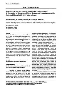
Concentration-dependent effect of Cl-IBMECA in WT/A3KO ⫹E/⫺E mice aorta. Incubation of WT aortic rings ⫹E to increasing concentration of Cl-IBMECA enhanced the contraction in a concentration-dependent manner with an EC50 ⫽ 2.9 ⫾ 0.2 ⫻ 10⫺9 M (Fig. 1). Cl-IBMECA (10⫺7 M) increased contraction significantly by 29% in ⫹E, while producing negligible effect (⬇6%) in endothelium-denuded (⫺E) WT mouse aorta, whereas these results were insignificant for both ⫹E and ⫺E A3KO mouse aorta. The Cl-IBMECA (10⫺7 M)-induced contraction in WT ⫹E aorta was completely abolished by MRS 1523 (10⫺5 M), a specific A3AR antagonist.
Fig. 1. Concentration-dependent contraction for Cl-IBMECA in endotheliumintact (⫹E) and -denuded (⫺E) wild-type (WT) and A3 knockout (KO) mice aorta. PE, phenylephrine. See MATERIALS AND METHODS for definition of MRS1523 and Cl-IBMECA. Values are mean ⫾ SE (n ⫽ 11). *P ⬍ 0.05 compared with WT ⫹E. AJP-Heart Circ Physiol • VOL
These results suggest that A3AR-mediated contraction is dependent on functional endothelium in mice aorta. To exclude the involvement of A1AR in A3AR-mediated aortic contraction, we carried out experiments using the selective A1AR agonist/antagonist, CCPA/DPCPX. CCPA-mediated contraction (25%) in WT ⫹E aorta was not affected by MRS 1523 (10⫺5 M), while this effect was inhibited by A1AR antagonist, DPCPX (10⫺5 M) (data not shown). This suggests that Cl-IBMECA-induced contraction of aortic rings was primarily due to activation of A3AR and not by A1AR in this tissue. A3AR gene expression by real-time PCR and Western blotting in ⫹E and ⫺E aorta of WT and A3KO mice. As can be seen in Fig. 2A, in WT ⫺E aorta, A3AR gene expression decreased significantly (74%) compared with WT ⫹E aorta, whereas this expression was undetected in A3KO ⫹E/⫺E aorta. The presence of A3AR protein in the ⫹E/⫺E aorta of WT and A3KO mice was detected with rabbit polyclonal A3 antiserum. As can be seen in Fig. 2B, Western blot analysis showed a decrease in A3AR protein expression by 72% in WT ⫺E aorta compared with WT ⫹E aorta. A3AR protein expression was undetected both in A3KO ⫺E and ⫹E aorta. These results demonstrate the presence of A3AR expression, mostly on the endothelium of WT mice aorta. Effects of indomethacin (a nonspecific COX blocker) on Cl-IBMECA-induced contraction in WT/A3KO ⫹E mice aorta. Figure 3 shows the response to Cl-IBMECA in the absence and presence of indomethacin (10⫺5 M) in WT ⫹E and A3KO ⫹E aortic rings. Indomethacin inhibited the CRC for Cl-IBMECA significantly in WT ⫹E aorta. Cl-IBMECA (10⫺7 M)-induced contraction was inhibited maximally by 70% in the presence of indomethacin (10⫺5 M) in WT ⫹E aorta. This inhibitory effect of indomethacin was insignificant in A3KO ⫹E aorta. Effects of SC-560 (COX-1 blocker) and NS-398 (COX-2 blocker) on Cl-IBMECA-induced contraction in WT/A3KO ⫹E mice aorta. As shown in Fig. 4A, SC-560 (10⫺8 M) inhibited Cl-IBMECA (10⫺7 M)-induced contractions by 60%, whereas NS-398 (10⫺6 M) had minimal inhibition (6%) in WT ⫹E aorta. On the other hand, the inhibitory effects of SC-560 (10⫺8 M) and NS-398 (10⫺6 M) were insignificant in A3KO ⫹E (Fig. 4B). In a separate experiment, addition of SC-560 (10⫺8 M) and NS-398 (10⫺6 M) together abolished completely the ClIBMECA-induced contraction in this tissue WT ⫹E (data not shown). These data suggest that COX-1 is mainly involved in A3AR-mediated endothelium-dependent contraction in WT mice aorta. Therefore, further studies were conducted to support the involvement of COX-1 pathway in A3AR contraction in this tissue. COX-1 gene expression by real-time PCR and Western blot in WT/A3KO ⫹E/⫺E mice aorta. Figure 5A shows COX-1 gene expression in WT and A3KO ⫹E/⫺E aorta. COX-1 gene expression decreased significantly by 96% (P ⬍ 0.05) in WT ⫺E compared with WT ⫹E aorta. In A3KO ⫺E aorta, COX-1 gene expression also decreased significantly by 85% (P ⬍ 0.05) compared with A3KO ⫹E aorta. These data were further confirmed by Western blotting using COX-1 antibody (Fig. 5B). Densitometric analysis showed that COX-1 expression in WT ⫺E aorta decreased by 94% (P ⬍ 0.05) compared with WT ⫹E aorta, whereas in A3KO ⫺E aorta, this expression decreased by 82% (P ⬍ 0.05) compared with A3KO ⫹E aorta.
293 • DECEMBER 2007 •
www.ajpheart.org
A3AR-MEDIATED ENDOTHELIUM-DEPENDENT VASOCONSTRICTION
H3451
Fig. 2. A3 adenosine receptor (AR) gene and protein expression, respectively, with real-time PCR (A) and Western blot (B) in ⫹E and ⫺E WT and A3KO mice aorta. ND, not detectable. Values are means ⫾ SE (n ⫽ 4). *P ⬍ 0.05 compared with WT ⫹E.
These data suggest that the COX-1 gene is mainly expressed on the aortic endothelium of WT/A3KO mice. Effect of Cl-IBMECA on COX-1 expression by Western blotting in WT/A3KO ⫹E mice aorta. The purpose of this experiment was to determine whether Cl-IBMECA-induced contraction is mediated through upregulation of COX-1. As can be seen in Fig. 6, left, the densitometric analysis of Western blot shows the increased COX-1 expression in the presence of Cl-IBMECA (10⫺7 M) by 43% (P ⬍ 0.05) over untreated WT ⫹E aorta (control, 100%). Pretreatment of aortic rings with MRS1523 (10⫺5 M) before Cl-IBMECA almost completely reversed the Cl-IBMECA-induced increase in COX-1 expression in WT ⫹E. Cl-IBMECA-induced COX-1 expression was insignificant in A3KO ⫹E aorta (Fig. 6, right).
Fig. 3. Effect of indomethacin on Cl-IBMECA-induced responses in WT/ A3KO ⫹E mice aorta. Values are means ⫾ SE (n ⫽ 7). *P ⬍ 0.05 compared with WT ⫹E. AJP-Heart Circ Physiol • VOL
Fig. 4. Effect of specific blockers of cyclooxygenase (COX)-1 (SC-560) and COX-2 (NS-398) on Cl-IBMECA-induced responses in WT ⫹E (A) and A3KO ⫹E (B) mice aorta. See MATERIALS AND METHODS for definition of SC-560 and NS-398. Values are means ⫾ SE (n ⫽ 6). *P ⬍ 0.05 compared with WT ⫹E.
Inhibition of Cl-IBMECA-induced contraction by TP receptor antagonist SQ 29548 in WT/A3KO ⫹E mice aorta. This experiment was conducted to gain further insight into the possible participation of vasoconstrictive prostanoids generated by COX-1 metabolism after activation of A3AR with the use of TP receptor antagonist (SQ 29548). SQ 29548 (10⫺6 M) significantly inhibited the CRC produced by Cl-IBMECA in WT ⫹E aorta. The contractile response produced by ClIBMECA at 10⫺7 M was reduced by 60.3% (P ⬍ 0.05) in the presence of SQ 29548 (10⫺6 M) in WT ⫹E, whereas this effect was insignificant in A3KO ⫹E aorta (Fig. 7). These results demonstrate that end products of COX-1 metabolism play an important role in A3AR-induced contraction in this tissue. Effect of TS inhibitor furegrelate on Cl-IBMECA-induced contraction. To further demonstrate involvement of TS in A3AR-mediated contraction in mice aorta, we used the TS inhibitor furegrelate on Cl-IBMECA-induced contraction. Furegrelate (10⫺5 M) inhibited Cl-IBMECA (10⫺7 M)-induced contraction by 61% (Fig. 8) in WT ⫹E mice aorta, whereas it produced no effects in A3KO mice aorta. These results demonstrate the involvement of Tx in A3AR-mediated contraction in mice aorta.
293 • DECEMBER 2007 •
www.ajpheart.org
H3452
A3AR-MEDIATED ENDOTHELIUM-DEPENDENT VASOCONSTRICTION
Fig. 5. COX-1 gene and protein expression, respectively, with real-time PCR (A) and Western blot (B) in ⫹E and ⫺E WT and A3KO mice aorta. Values are means ⫾ SE (n ⫽ 4). The densitometric values for COX-1 protein expression are shown as follows in parentheses: WT ⫹E (368 ⫾ 27)/⫺E (22 ⫾ 1.2) and A3KO ⫹E (335 ⫾ 22)/⫺E (60 ⫾ 4). *P ⬍ 0.05 compared with WT ⫹E/A3KO ⫹E.
Effect of furegrelate on Cl-IBMECA-induced TP receptor expression in WT ⫹E mice aorta. The effect of furegrelate on Cl-IBMECA (10⫺7 M)-induced TP receptor expression was also determined. Figure 9 shows representative immunoblots of TP receptor expression with densitometric analysis. As can be seen from Fig. 9, the treatment of WT ⫹E aortic rings with Cl-IBMECA for 60 min resulted in a 62% increase in TP receptor expression compared with untreated control. Cl-IBMECA-induced increase in TP receptor expression was markedly attenuated by preincubation of WT ⫹E aortic rings for 30 min with furegrelate (10⫺5 M) and MRS1523 (10⫺5 M). These results demonstrate that A3AR activation is linked to the upregulation of TP receptor, probably via activation of TS. Measurement of TxB2 levels in the presence of COX blockers and TS inhibitor. As shown in Table 1, the TxB2 (a stable TxA2 metabolite) levels increased by 71% with Cl-IBMECA treat-
Fig. 7. Effect of thromboxane prostanoid (TP) receptor antagonist (SQ 29548) on Cl-IBMECA-induced responses in WT/A3KO ⫹E mice aorta. See MATERIALS AND METHODS for definition of SQ 29548. Values are means ⫾ SE (n ⫽ 6). *P ⬍ 0.05 compared with WT ⫹E.
ment alone in WT ⫹E aorta compared with untreated control. The Cl-IBMECA-induced increase in TxB2 levels were attenuated significantly in the presence of indomethacin (10⫺5 M), SC-560 (10⫺8 M), and furegrelate (10⫺5 M) (Table 1). TxB2 levels were unaffected by A3AR agonist (Cl-IBMECA) or COXs, COX-1, and TS blockers (indomethacin, SC-560, and furegrelate) in A3KO ⫹E mice aorta (Table 1). These data suggest that major metabolite of COX-1 pathway could be TxB2 in A3AR-mediated contraction. Removal of endothelium ⫺E in WT mice aorta resulted in a 63% reduction in TxB2 levels compared with untreated control (data not shown), which further supports our molecular and organ bath data that A3AR-COX-1 signaling may be mediated through endothelium. DISCUSSION
In the present study, we have investigated A3AR-mediated endothelium-dependent contraction in relation to COX signaling pathway using WT/A3KO mice aorta. Our findings showed that the activation of A3AR by selective agonist Cl-IBMECA
Fig. 6. Cl-IBMECA-induced COX-1 protein expression and its inhibition with MRS1523 in WT ⫹E and A3KO ⫹E aorta. Results are shown from one experiment that is a representative of four independent experiments. Values are means ⫾ SE (n ⫽ 4) after densitometric analysis. *P ⬍ 0.05 compared with WT ⫹E untreated control. #P ⬍ 0.05 compared with WT ⫹E treated with Cl-IBMECA (10⫺7 M) alone.
AJP-Heart Circ Physiol • VOL
293 • DECEMBER 2007 •
www.ajpheart.org
H3453
A3AR-MEDIATED ENDOTHELIUM-DEPENDENT VASOCONSTRICTION
Table 1. Effects of indomethacin, SC-560, and furegrelate on TxB2 production in WT and A3KO endothelium-intact mice aorta Aortic Tissue TxB2, pg/mg protein Addition
Control Cl-IBMECA Cl-IBMECA Cl-IBMECA Cl-IBMECA
(10⫺7 (10⫺7 (10⫺7 (10⫺7
M) M) ⫹ indomethacin (10⫺5 M) M) ⫹ SC-560 (10⫺8 M) M) ⫹ furegrelate (10⫺5 M)
WT
A3KO
35.4⫾2.4 60.5⫾4.7* 38.4⫾3.1† 42.0⫾3.8† 40.6⫾3.5†
30.0⫾2.2 34.0⫾2.3 32.0⫾2.5 33.0⫾2.5 31.8⫾2.4
Values are means ⫾ SE; n ⫽ 4 – 6. TxB2, thromboxane B2; WT, wild-type mice; A3KO, A3 knockout mice. See MATERIALS AND METHODS for definitions of Cl-IBMECA and SC-560. *P ⬍ 0.05 compared with control. †P ⬍ 0.05 compared with Cl-IBMECA (10⫺7 M) alone. Fig. 8. Effect of thromboxane synthase inhibitor furegrelate on Cl-IBMECAinduced responses in WT/A3KO ⫹E mice aorta. Values are means ⫾ SE (n ⫽ 6). *P ⬍ 0.05 compared with WT ⫹E.
led to contraction and increase in TxB2 production in WT mice aorta that was dependent on the integrity of endothelium, since this response was almost completely abolished after removal of functional endothelium. This was also confirmed by real-time PCR and Western blot data that showed the presence of A3AR expression, mostly on aortic endothelium. Furthermore, our data showed that A3AR-mediated contraction in WT aorta is dependent mainly on endothelial COX-1, which was also confirmed by real-time PCR and Western blot data. COX-1 gene expression was significantly induced by A3AR activation in WT ⫹E aorta, whereas this increase in expression was negligible in A3KO. Activation of A3AR also resulted in increased TP receptor activation that was attenuated by
Fig. 9. Effect of thromboxane synthase inhibitor furegrelate and specific A3AR antagonist MRS1523 on Cl-IBMECA-induced TP-receptor expression with Western blotting in WT ⫹E mice aorta. Results are shown from one experiment that is a representative of four independent experiments. Values are means ⫾ SE (n ⫽ 4) after densitometric analysis. *P ⬍ 0.05 compared with WT ⫹E. #P ⬍ 0.05 compared with WT ⫹E treated with Cl-IBMECA (10⫺7 M) alone. AJP-Heart Circ Physiol • VOL
furegrelate (TS inhibitor). Cl-IBMECA-induced increase in COX-1 and TP receptor expression was markedly attenuated by MRS1523, a specific A3AR antagonist in WT ⫹E aorta. Nonspecific COX blocker, COX-1 specific blocker, and TS inhibitor also attenuated Cl-IBMECA-induced increase in TxB2 levels. Furthermore, contractile response to A3AR agonist and inhibitory effects produced by COX blockers were absent in A3KO ⫹E and ⫺E aorta. Our data demonstrated that Cl-IBMECA, a specific A3AR agonist, produced a concentration-dependent contraction in WT ⫹E mice aorta, which was completely abolished by its specific antagonist (MRS 1523). The contractile response was specific to A3AR activation, as it was confirmed by the use of the selective A1AR agonist (CCPA). The CCPA-induced aortic contraction was not affected by MRS 1523, while this effect was blocked by A1AR antagonist (DPCPX, data not shown). The antagonists for A3AR (MRS 1523) and A1AR (DPCPX) have been used previously in our laboratory (11, 36). Taken together, these findings suggest that Cl-IBMECA-induced contraction of aortic rings was primarily due to activation of A3AR and not due to the activation of A1AR in this tissue. Shepherd et al. (30) have previously reported the involvement of A3ARmediated vasoconstriction in hamster arterioles in vivo, which supports our present data. However, these authors did not address the role of endothelium in A3AR-mediated vasoconstriction (30). In support of our hypothesis that A3AR-mediated contraction is dependent on the integrity of endothelium, our data showed that the removal of endothelium in WT aorta resulted in the loss of contractile response to Cl-IBMECA. As further evidence that A3AR is indeed expressed on endothelium, we measured the gene expression in WT/A3KO ⫹E/⫺E aorta by real-time PCR and protein expression by Western blot analysis. The WT ⫺E aorta showed a significant decrease in A3AR expression compared with WT ⫹E aorta, whereas A3KO ⫹E/⫺E showed undetected levels of A3AR expression. A3AR expression in different vascular tissues has also been reported by other workers (43, 44). Recently, Zhao et al. (43) reported that the blood pressure in A3KO mice was similar to that in WT mice; however, when given adenosine infusion (intravenously), A3KO mice displayed a significant decrease in blood pressure compared with WT mice. The fall in blood pressure in A3KO mice could be due to the absence of the release of endothelium-derived contractile mediators, which may be
293 • DECEMBER 2007 •
www.ajpheart.org
H3454
A3AR-MEDIATED ENDOTHELIUM-DEPENDENT VASOCONSTRICTION
present in WT mice (43). Overall, our findings demonstrate that A3AR-mediated contraction in WT aorta is dependent on endothelium, which might play an important role in the regulation of blood pressure. The endothelium-derived contracting factors include superoxide and/or PGH2/TxA2 generated by COX-mediated metabolism of arachidonic acid (2, 13, 21, 31, 40). Pretreatment of aortic rings with a nonspecific COX blocker (indomethacin) and specific COX-1 blocker (SC-560) resulted in inhibition of Cl-IBMECA-induced contraction, whereas COX-2 blocker (NS-398) showed an insignificant inhibitory effect in WT ⫹E mice aorta. The findings in the present study, that SC-560 inhibited Cl-IBMECA-induced contractions almost completely compared with NS-398, indicate that vasoconstriction is mediated mainly through COX-1 in this tissue. These findings are further supported by the observation that pretreatment of WT ⫹E aortic rings with indomethacin, SC-560, and furegrelate significantly inhibited TxB2 levels compared with Cl-IBMECA alone (Table 1). An earlier report has also shown that COX-1 predominantly contributes to basal PG production, because COX-1 inhibitors significantly decrease total PG products in both kidney and aorta (27). Heymes et al. (20) have also demonstrated COX-1 as the major isoform of COXs in rat aorta. Our findings correlate with the above reports, suggesting that COX-1 is mostly involved in A3AR-mediated contraction. Further studies were carried out to confirm the location and involvement of COX-1 in A3AR-mediated contraction. COX-1 expression was located mainly on the endothelium, as its removal led to significant decreases in both gene and protein expression of COX-1. Treatment of WT ⫹E aorta with ClIBMECA resulted in upregulation of COX-1 expression, while producing a negligible effect in A3KO. Cl-IBMECA-induced increase in COX-1 expression was significantly blocked by MRS1523, a specific A3AR antagonist in WT ⫹E aorta. These data corroborate organ bath experiments and show that COX-1-mediated contraction after A3AR activation in WT ⫹E aorta may also require functional endothelium. Previous reports have also shown the expression of COXs on aortic endothelium of different species, which support our present studies (23, 24, 33). The observations that COX-1 and COX-2 blockers produce different responses to Cl-IBMECA-induced contraction in pharmacological studies suggest that these isoforms are regulated differentially after A3AR activation in the mice aorta. Clinical and animal studies also show that COX-1 and COX-2 are regulated differentially in the production of total prostanoids in both the renal and cardiovascular system (4, 26, 34, 42). The differential regulation could be due to the fact that COX-1, being a constitutive enzyme, plays an important role in the basal production of prostanoids and Tx (3, 9, 10, 19, 27), whereas COX-2, being an inducible enzyme, plays a significant role under inflammatory/disease conditions (7, 33). Additional studies on disease models are needed to further explore our understanding in this area of research. We further confirmed the involvement of end products of COX metabolism in A3AR-mediated contractions by using TP receptor antagonist (SQ 29548), COX blockers (indomethacin, SC-560), and TS inhibitor (furegrelate). SQ 29548 inhibited Cl-IBMECA-mediated contractions to a similar degree, as shown by nonspecific COX blocker (indomethacin) and by specific COX-1 blocker (SC-560), thus demonstrating that AJP-Heart Circ Physiol • VOL
COX-1 is indeed involved in A3AR-mediated aortic contraction. A3AR-mediated contraction and increase in TP receptor expression were markedly reduced by pretreatment with furegrelate, as well as MRS1523, a specific A3AR antagonist. Furthermore, one of major end metabolites of COX pathway, i.e., TxB2, was found to be increased after activation of A3AR with Cl-IBMECA, which was decreased by pretreatment with COX/COX-1 blockers and TS inhibitor (Table 1). These findings strongly suggest the involvement of A3AR-COX-1-TxA2 pathway in aortic contraction via endothelium. In conclusion, the present study demonstrated for the first time, utilizing WT and A3KO mouse aorta, that 1) activation of A3AR in WT leads to contraction, mainly via endothelium; and 2) A3AR-mediated contraction and TxB2 production are dependent mainly on COX-1 signaling pathway. Finally, our studies suggest that the A3AR-mediated contraction through endothelium may play a role in cardiovascular inflammation, including hypertension and atherosclerosis, by affecting COX signaling pathways. This could play an important role in the pathophysiology of vascular diseases and could provide newer therapeutic approaches. GRANTS This work was supported by National Heart, Lung, and Blood Institute Grants HL-027339 to S. J. Mustafa and HL-071802 to S. L. Tilley. REFERENCES 1. Ansari HR, Nadeem A, Talukder MAH, Sakhalkar S, Mustafa SJ. Evidence for the involvement of nitric oxide in A2B receptor-mediated vasorelaxation of mouse aorta. Am J Physiol Heart Circ Physiol 292: H719 –H725, 2007. 2. Badier M, Soler M, Mallea M, Delpierre S, Orehek J. Cholinergic responsiveness of respiratory and vascular tissues in two different rat strains. J Appl Physiol 64:323–328, 1988. 3. Bolla M, You D, Loufrani L, Levy-Toledano S, Habib A, Henrion D. Cyclooxygenase involvement in thromboxane-dependent contraction in rat mesenteric resistance arteries. Hypertension 43: 1264 –1269, 2004. 4. Bombardier C, Laine L, Reicin A, Shapiro D, Burgos-Vargas R, Davis B, Dar R, Ferraz MB, Hawkey CJ, Hochberg MC, Kvien TK, Schintzer TJ. Comparison of upper gastrointestinal toxicity of rofecoxib and naproxen in patients with rheumatoid arthritis VIGOR Study Group. N Engl J Med 343: 1520 –1528, 2003. 5. Breyer MD, Breyer RM. G protein-coupled prostanoid receptors and the kidney. Annu Rev Physiol 63: 579 – 605, 2001. 6. Catella-Lawson F, Reilly MP, Kapoor SC, Cucchiara AJ, DeMarco S, Tournier B, Vyas SN, FitzGerald GA. Cyclooxygenase inhibitors and the antiplatelet effects of aspirin. N Engl J Med 345: 1809 –1817, 2001. 7. Cipollone F, Fazia ML. COX-2 atherosclerosis. J Cardiovasc Pharmacol 47: S26 –S36, 2006. 8. Cong P, Xiao Z, Biancani P, Behar J. Prostaglandins mediate tonic contraction of the guinea pig and human gallbladder. Am J Physiol Gastrointest Liver Physiol 292: G409 –G418, 2007. 9. Elmhurst JL, Betti PA, Rangachari PK. Intestinal effects of isoprostanes: evidence for the involvement of prostanoid EP and TP receptors. J Pharmacol Exp Ther 282: 1198 –1205, 1997. 10. Ermert L, Ermert M, Duncker HR, Grimminger F, Seeger W. In situ localization and regulation of thromboxane A2 synthase in normal and LPS-primed lungs. Am J Physiol Lung Cell Mol Physiol 278: L744 –L753, 2000. 11. Fan M, Qin W, Mustafa SJ. Characterization of adenosine receptor(s) involved in adenosine-induced branchoconstriction in an allergic mouse model. Am J Physiol Lung Cell Mol Physiol 284: L1012–L1019, 2003. 12. Feoktistov I, Ryzhov S, Goldstein AE, Biaggoni I. Mast cell-mediated stimulation of angiogenesis: cooperative interaction between A2B and A3 adenosine receptors. Circ Res 92: 485– 492, 2003. 13. Furchgott RF, Vanhoutte PM. Endothelium-derived relaxing and contracting factors. FASEB J 3: 2007–2017, 1989.
293 • DECEMBER 2007 •
www.ajpheart.org
A3AR-MEDIATED ENDOTHELIUM-DEPENDENT VASOCONSTRICTION 14. Ge T, Vanhoutte PM, Boulanger CM. Increased response to prostaglandin H2 precedes changes in PGF-synthase 1 expression in the SHR aorta. Zhongguo Yao Li Xue Bao 20: 1087–1092, 1999. 15. Ge ZD, Peart JN, Kreckler LM, Wan TC, Jacobson MA, Gross GJ, Auchampach JA. Cl-IB-MECA [2-chloro-N6-(3-iodobenzyl) adenosine5⬘-N-methylcarboxamide] reduces ischemia/reperfusion injury in mice by activating the A3 adenosine receptor. J Pharmacol Exp Ther 319: 1200 – 1210, 2006. 16. Giannella E, Mochmann HC, Levi R. Ischemic preconditioning prevents the impairment of hypoxic coronary vasodilation caused by ischemia/ reperfusion: role of adenosine A1/A3 and bradykinin B2 receptor activation. Circ Res 81: 415– 422, 1997. 17. Gluais P, Paysant J, Badier-Commander C, Verbeuren T, Vanhoutte PM, Feletou M. In SHR aorta, calcium ionophore A-23187 releases prostacyclin and thromboxane A2 as endothelium-derived contracting factors. Am J Physiol Heart Circ Physiol 291: H2255–H2264, 2006. 18. Grbovic L, Radenkovic M. Analysis of adenosine vascular effect in isolated rat aorta: possible role of Na⫹/K⫹-ATPase. Pharmacol Toxicol 92: 265–271, 2003. 19. Greaves RR, O’Donnell LJ, Farthing MJ. Differential effect of prostaglandins on gallstone-free and gallstone-containing human gallbladder. Dig Dis Sci 45: 2376 –2381, 2000. 20. Heymes C, Habib A, Yang D, Mathieu E, Marotte F, Samuel J, Boulanger CM. Cyclo-oxygenase-1 and -2 contributions to endothelial dysfunction in ageing. Br J Pharmacol 131: 804 – 810, 2000. 21. Ihara E, Hirano K, Derkach DN, Nishimura J, Nawata H, Kanaide H. The mechanism of bradykinin-induced endothelium-dependent contraction and relaxation in the porcine interlobar renal artery. Br J Pharmacol 129: 943–952, 2000. 22. Livak KJ, Schmittgen TD. Analysis of relative gene expression data using real-time quantitative PCR and the 2[⫺delta delta C(T)] method. Methods 25: 402– 408, 2001. 23. Matz RL, de Sotomayor MA, Schott C, Stoclet JC, Andriantsitohaina R. Vascular bed heterogeneity in age-related endothelial dysfunction with respect to NO and eicosanoids. Br J Pharmacol 131: 303–311, 2000. 24. Molero L, Carrasco C, Marques M, Vaziri ND, Mateos-Caceres PJ, Casado S, Macaya C, Barrientos A, Lopez-Farre AJ. Involvement of endothelium and endothelin-1 in lead-induced smooth muscle cell dysfunction in rats. Kidney Int 69: 685– 690, 2006. 25. Potenza MA, Botrugno OA, DeSalvia MA, Lerro G, Nacci C, Marasciulo FL, Andriantsitohaina R, Mitolo-Cieppa D. Endothelial COX-1 and -2 differently affect reactivity of MVB in portal hypertensive rats. Am J Physiol Gastrointest Liver Physiol 283: G587–G594, 2002. 26. Qi Z, Hao CM, Langenbach RI, Breyer RM, Redha R, Morrow JD, Breyer MD. Opposite effects of cyclooxygenase-1 and -2 activity on the pressor response to angiotensin II. J Clin Invest 110: 61– 69, 2002. 27. Qi Z, Cai H, Morrow JD, Breyer MD. Differentiation of cyclooxygenase 1- and 2-derived prostanoids in mouse kidney and aorta. Hypertension 48: 323–328, 2006. 28. Salvatore CA, Tilley SL, Latour AM, Fletcher DS, Koller BH, Jacobson MA. Disruption of A3 adenosine receptor gene in mice and its effect on stimulated inflammatory cells. J Biol Chem 275: 4429 – 4434, 2000.
AJP-Heart Circ Physiol • VOL
H3455
29. Schiffrin EL. A critical review of the role of endothelial factors in the pathogenesis of hypertension. J Cardiovasc Pharmacol 38: S3–S6, 2001. 30. Shepherd RK, Linden J, Duling BR. Adenosine-induced vasoconstriction in vivo: role of mast cells and A3 adenosine receptor. Circ Res 78: 627– 634, 1996. 31. Shirahase H, Murase K, Kanda M, Kurahashi K, Nakamura S. Endothelium-dependent contraction induced by substance P in canine cerebral arteries: involvement of NK1 receptors and thromboxane A2. Life Sci 64: 211–219, 1999. 32. Smith WL, DeWitt DL, Garavito RM. Cyclooxygenases: structural, cellular, and molecular biology. Annu Rev Biochem 69: 145–182, 2000. 33. Stanfield KM, Khan KN, Gralinski MR. Localization of cyclooxygenase isozymes in cardiovascular tissues of dogs treated with naproxen. Vet Immunol Immunopathol 80: 309 –314, 2001. 34. Sullivan JC, Sasser JM, Pollock DM, Pollock JS. Sexual dimorphism in renal production of prostanoids in spontaneously hypertensive rats. Hypertension 45: 406 – 411, 2005. 35. Tabrizchi R, Bedi S. Pharmacology of adenosine receptors in the vasculature. Pharmacol Ther 91: 133–147, 2001. 36. Tawfik HE, Schnermann J, Oldenburg PJ, Mustafa SJ. Role of A1 adenosine receptors in regulation of vascular tone. Am J Physiol Heart Circ Physiol 288: H1411–H1416, 2005. 37. Tawfik HE, Teng B, Morrison RR, Schnermann J, Mustafa SJ. Role of A1 adenosine receptor in the regulation of coronary flow. Am J Physiol Heart Circ Physiol 291: H467–H472, 2006. 38. Thakali K, Davenport L, Fink GD, Watts SW. Cyclooxygenase, p38 MAPK, ERK MAPK, RHO kinase and SRC mediate hydrogen peroxideinduced contraction of rat thoracic aorta. J Pharmacol Exp Ther 320: 236 –243, 2007. 39. Vanhoutte PM, Feletou M, Taddei S. Endothelium-dependent contractions in hypertension. Br J Pharmacol 144: 477– 485, 2005. 40. Yang D, Feletou M, Boulanger CM, Wu HF, Levens N, Zhang JN, Vanhoutte PM. Oxygen-derived free radicals mediate endothelium dependent contractions to acetylecholine in aortas from spaontaneously hypertensive rats. Br J Pharmacol 136: 104 –110, 2002. 41. Yang D, Feletou M, Levens N, Zhang JN, Vanhoutte PM. A diffusible substance(s) mediates endothelium-dependent contractions in the aorta of SHR. Hypertension 41: 143–148, 2003. 42. Zewde T, Mattson DL. Inhibition of cyclooxygenase-2 in the rat renal medualla leads to sodium-sensitive hypertension. Hypertension 44: 424 – 428, 2004. 43. Zhao Z, Makaritsis K, Francis CE, Gavras H, Ravid K. A role for the A3 adenosine receptor in determining tissue levels of cAMP and blood pressure: studies in knock-out mice. Biochim Biophys Acta 1500: 280 –90, 2000. 44. Zhou QY, Li C, Olah ME, Johnson RA, Stiles GL, Civelli O. Molecular cloning and characterization of an adenosine receptor: the A3 adenosine receptor. Proc Natl Acad Sci USA 16: 7432– 4736, 1992. 45. Zhou Y, Mitra S, Varadharaj S, Parinandi N, Zweier JL, Flavahan NA. Increased expression of cyclooxygenase-2 mediates enhanced contraction to endothelin ETA receptor stimulation in endothelial nitric oxide synthase knockout mice. Circ Res 98: 1439 –1445, 2006.
293 • DECEMBER 2007 •
www.ajpheart.org