The effects of nutrition on the reproductive system of female mammals are discussed in an attempt to determine the importance of the role of the insulin-like ...
Involvement of insulin-like growth factors in the interactions between nutrition and reproduction in female mammals RMonget 13 and G.B.Martin2 , Station de Physiologie de la Reproduction des Mammiferes Domestiques, 37380 Nouzilly, France, and 2Faculty of Agriculture (Animal Science), The University of Western Australia, Nedlands, WA 6907, Australia 3
To whom correspondence should be addressed
The effects of nutrition on the reproductive system of female mammals are discussed in an attempt to determine the importance of the role of the insulin-like growth factor (IGF) system of proteins in the physiological mechanisms. The IGF system comprises IGF-I, IGF-II, their receptors and binding proteins. For this review, insulin and its receptors have been included in this system. The reproductive responses have been separated into two classes, based on fundamental differences in the reproductive biology and physiological mechanisms underlying them. The first involves the effects of nutrition on the induction of ovulation, at puberty or postpartum. In this case, the question is whether or not the animal will reproduce. The second class of response, changes in ovulation rate, involves considerations of reproductive efficiency in animals in which ovulation is inevitable. Key words: IGF/nutrition/metabolism/ovary/ hypothalamus/fertility
Introduction The ligands insulin, insulin-like growth factors (IGF) IGF-I and IGF-II, their binding proteins and receptors are found in a wide range of tissues throughout the body, where they control the anabolic and catabolic processes that constitute the general responses to changes in the level of nutrition. Nutritional treatments alter the expression of their genes in their tissues of origin and these effects lead to changes in their circulating concentrations. Many of these responses are correlated, some Human Reproduction Volume 12 Supplement 1 1997
positively and some negatively, with the effects of nutritional treatments on reproductive function. Most members of the IGF system are ubiquitous, being present and expressed in brain, pituitary and ovarian tissue. This raises the possibility that some of the reproductive responses are not only mediated by the classical endocrine actions of the products of the pancreas or liver, but also by autocrine and paracrine effects of the IGF family, perhaps at all levels of the brain-pituitary-ovarian axis. Follicular growth can be separated into two periods. The first period, up to 200 |im diameter in the rat, 2 mm in the ewe, and 5 mm in the cow and the human, seems to depend little, if at all, on the presence of gonadotrophins. Beyond this stage, growth and ovulation of follicles is strictly dependent on the presence of gonadotrophins. Hence, if we need to compare the relative importance of gonadotrophins and growth factors (such as the members of the IGF system), one may assume that the growth factors are more important in the early 'gonadotrophin-independent' period than in the strictly 'gonadotrophin-dependent' period. In the later stages, members of the IGF system might be considered 'permissive' of the action of gonadotrophins. Keeping these considerations in mind, the 'decision to ovulate' (i.e. reproduce) is effectively made at brain level and it is implemented via the hypothalamic gonadotrophin-releasing hormone (GnRH) signal that controls the secretion of pituitary gonadotrophins. The role of the IGF system in this process is not clear, but seems to include intra-cerebral autocrine or paracrine effects (due to changes in receptor or peptide expression)
© European Society for Human Reproduction & Embryology
33
P.Monget and G.B.Martin
as well as endocrine effects at brain level (due to changes in serum concentrations of insulin or IGFs). However, the importance of classical endocrine effects is difficult to estimate due to the lack of data concerning the passage of IGFs, insulin, or IGF binding proteins (IGFBPs) through the blood-brain barrier. At gonadal level, the IGF system seems to have only a permissive role because only the final stages of development of the ovarian follicles are involved and the progress of these very mature follicles over the final steps to ovulation is driven mainly by the gonadotrophic signal. In contrast, the 'decision to change ovulation rate' (i.e. increase the rate of reproduction) is only partly dependent on gonadotrophins, which seem to play a relatively passive role compared with that in the 'decision to ovulate'. In this case, we are considering a smaller, less mature class of antral follicles in the ovary, those that are several days away from being ready to ovulate. The IGF system affects the responsiveness of these follicles to gonadotrophins and thus helps determine the number of follicles that ultimately enter the gonadotrophin-driven stage. Again, there is evidence to suggest that these effects are exerted through classical hormonal pathways and, again, there is evidence that intra-ovarian paracrine and autocrine pathways are also involved. The elements of the IGF system are probably involved in the function of the reproductive axis at all levels, from brain to gonad. It is most likely that one or more members of this system mediate the responses to nutrition. However, much of our thinking is being guided by correlations between blood concentrations of the IGFs, insulin, or IGFBPs and reproductive responses to changes in nutrition, or by in-vitro studies of isolated brain, pituitary or ovarian tissues. This circumstantial evidence now needs support from experiments in which these elements are manipulated directly, or in which they are used in an attempt to alter brain and ovarian function. 4 Nutrition and reproduction The relationships between nutrition and reproduction have been subjected to intensive investigation since the turn of the century (for a review see 34
Clark, 1934) and it is now generally accepted that reproductive capacity cannot be optimized without adequate nutrition. Understanding the mechanisms linking these two aspects of physiology is of great interest for the treatment of menstrual dysfunction in women, as well as for the control of fertility in domestic animals. Many reviews have already been written on this subject and we will refer to them frequently because we do not intend to cover again the background to the various theories that have emerged. Rather, we will concentrate on the potential roles for the insulin-like growth factor (IGF) system in the nutrition-reproduction interaction. There are three reasons for this narrow focus: (i) the IGF system is presently one of the better understood systems of growth factors so we have a wealth of information draw on; (ii) the elements of the IGF system are known to be altered by nutritional status; and (iii) the IGF system is involved in many stages of the reproductive process. In this review, then, we will consider a range of situations in which female reproductive physiology is susceptible to metabolic perturbations and we will point out those in which the IGF system could be involved. In addition to IGF-I, IGF-II, their receptors and binding proteins, we have also included insulin and its receptor in the 'system'. We have also broken the reproductive responses up into two classes, based on fundamental differences in the reproductive biology and the physiological mechanisms that underlie them. The first involves the effects of nutrition on the induction of ovulation, particularly at puberty or postpartum. In this case, we are considering whether or not the animal will reproduce. The second class of response, change in ovulation rate, involves considerations of reproductive efficiency and rate of reproduction in animals in which ovulation is inevitable. Within this framework, we are able to assess our state of knowledge concerning the relative importance of the IGFs, the possible origins of IGF-based signals (humoral or local), and their probable sites of action. The IGF system For the sake of this review, we would like to include insulin as an element of the IGF system,
IGFs and interactions between nutrition and reproduction
if only because of the structural and functional similarity between this hormone and IGFs, and between the insulin receptor and the IGF type I receptor (LeRoith et al, 1995). Thus, our IGF system comprises three classes of component: the ligands, the receptors, and the binding proteins, each of which contains a range of elements: Three ligands IGF-I, IGF-II (Humbel, 1990) and insulin. Three receptors The IGF type I receptor mediates most (if not all) of the somatomedin-like actions of both IGF-I and IGF-II (Roth and Kiess, 1994; LeRoith et al, 1995); it has a slightly higher affinity for IGF-I than for IGF-II, and a much lower affinity for insulin, as evidenced by the fact that only pharmacological doses of insulin are able to activate it. The insulin receptor mediates most of the biological effects of insulin on metabolism and glucose uptake, for example, and has a low affinity for IGF-I or IGF-II. There is a strong structural and functional link between the insulin receptor and the IGF type I receptor. Both receptors are oc2p2 heterotetrameric complexes synthesized from two identical a(3 half-receptor precursors, and contain a tyrosine-kinase domain in the cytoplasmic portion. The binding of ligands to either receptor leads to the autophosphorylation of tyrosine residues and to the phosphorylation of insulin receptor subunit-1, which is then able to bind to SH-domain-containing proteins, such as PI3-kinase and Grb2 (Cheatham and Kahn, 1995; LeRoith et al, 1995). Moreover, recent data strongly suggest that non-identical a(3 half-receptor precursors can assemble to generate insulin/IGF-I hybrid receptor species both in vivo and in vitro (Frattali et al, 1992). Finally, the existence of 'atypical' type I receptors with altered affinity for IGFs or insulin has been suggested for some cell lines (Garofalo and Barenton, 1992; Milazzo et al, 1992). For all of these reasons, it is important to take into account both the insulin receptor and the IGF type I receptor when studying the mechanisms underlying alterations of metabolism, such as those following undernutrition. The IGF type II receptor, or IGF-II/mannose-6phosphate (IGF-II/M6P) receptor, binds IGF-II
and other molecules which possess a mannose-6phosphate residue such as lysosomal enzymes and TGF-pl (Roth and Kiess, 1994). This receptor does not bind insulin and binds IGF-I with very low affinity. It is therefore highly unlikely that the IGF-II/M6P receptor mediates any actions of IGF-I or insulin in vivo. Important functions of the type II receptor include mediation of the turn-over of lysosomal enzymes and degradation of IGF-II after internalization (Nolan et al, 1990). Moreover, experiments using IGF-II mutants have recently suggested that the IGF-II/M6P receptor may mediate some unexpected actions of IGF-II, such as those on cell motility, but not the 'classical' somatomedin-like actions such as those on mitogenesis. 5a: IGF-binding proteins These IGFBPs are present in all biological fluids and bind both IGF-I and IGF-II with high affinity (Jones and Clemmons, 1995). They can be arbitrarily classified into two groups: (i) IGFBP-1, -2, -4, -5 and -6 which are present in serum and in other fluids in a so-called 'small complex'; when visualized by Western ligand blotting, their apparent molecular weights range from 24-35 kDa, and their blood concentrations are either negatively (IGFBP-1 and -2) or not regulated by insulin, nutrition and growth hormone (GH); (ii) IGFBP-3, which is visualized as a AA-A2 kDa doublet by Western ligand blotting and is the most common IGFBP in serum, where it is mostly found as a 150 kDa 'large complex' comprising IGF-I or IGF-II plus an 85 kDa acid-labile subunit (ALS); the blood concentration of IGFBP-3 is positively regulated by GH and IGF-I. IGFBPs increase the half-life of IGFs and thus ensure a large pool of them in all the compartments of the organism. However, in theory, sequestration of IGFs by IGFBPs can also inhibit the actions of IGFs on the target cells because the IGFBPs and the type I receptor have similar affinities for IGF-I and IGF-II. This same difficulty plagues the interpretation of immunisation studies (Martensz, 1981; Martin, 1984). For IGFBPs, further complexity arises from the modulation of their affinity for IGF-I and IGF-II by post-translational changes, such as phosphorylation (IGFBP-1), binding to 35
P.Monget and G.B.Martin
extracellular matrix (IGFBP-5), or proteolysis (IGFBP-2, -3, -4 and -5). Proteolysis decreases the affinity of IGFBPs for their ligands and thus increases the bioavailability of IGFs. This phenomenon has been described in the serum of rodents and humans during pregnancy, after fasting, during severe illness, and following severe trauma (Jones and Clemmons, 1995). Limited proteolysis of IGFBPs has also been described in serum and lymph of normal adult humans (Lalou and Binoux, 1993) so it is likely that, in most target tissues, it plays a crucial role in modulating IGF bioavailability.
Consequences of negative energy balance on the IGF system One aim of this review is to consider the eonsequences of negative energy balance on female fertility. In general terms, negative energy balance can be caused by inadequate supply (a significant decrease in food intake) or by excessive consumption (e.g. during lactation, rapid growth or intense physical activity). The critical questions are: how does negative energy balance affect the IGF system, and do such responses affect reproductive function? Before attempting to answer these questions, we would like to point out a problem with the literature in this area. In discussion of nutrition-reproduction interactions, many authors fail to distinguish between 'fasting' (acute and complete removal of food) and chronic undernutrition (leading to sustained but normal losses of body weight). The physiological responses to these two types of 'nutritional treatment' probably differ significantly. During evolution, animals will have developed successful mechanisms to cope with long-term changes in food supply, allowing them to survive and reproduce. It is interesting to speculate on whether they would also have developed mechanisms to allow them to cope with fasting while reproducing, but arguably fasting is not a normal phenomenon and there is some danger in extrapolating from findings using such treatments to explain the more general relationships between diet and reproductive function. This is particularly so in laboratory animals that are normally fed ad libitum or at a regular time each day. In this situation, stress 36
responses might be confounded with nutritional responses (Cameron et al, 1993). Insulin secretion is acutely regulated by glucose availability so, under normal nutritional conditions, circulating concentrations of insulin fluctuate diurnally due to changes in food intake, especially in monogastrics. In contrast, there is little or no diurnal change in plasma IGF concentrations. Fasting and chronic undernutrition reduce blood concentrations of insulin, IGF-I and, to a lesser extent, IGF-II but, except in rodents, increase GH concentrations (Thissen et al, 1994). All of these responses are primarily due to changes in expression of the respective genes and may constitute an adaptive mechanism that favours lipolysis and increases the availability of fatty acids to peripheral tissues. Dietary restriction also affects circulating IGFBPs, increasing the concentrations of IGFBP-1 and IGFBP-2 and decreasing the concentrations of IGFBP-3. Again, these effects are essentially due to changes in gene expression in liver rather than changes in IGFBP-proteolytic activity, and they are believed to alter the availability of IGFs to the target tissues. In particular, experiments on fooddeprived guinea pigs have shown that the increase in the concentration of IGFBP-1 and -2 in serum is responsible for a decrease in IGF-I-stimulated collagen synthesis (Gosiewska et al., 1994). Finally, undernutrition is also accompanied by a peripheral resistance to the actions of insulin and IGF-I (Thissen et al, 1994). Overall, then, undernutrition is characterized by a decrease in tissue anabolism and an increase in tissue catabolism. Consequences of negative energy balance on reproductive function Even though follicular development and ovulation are not themselves energetically expensive, female mammals will not ovulate and risk committing themselves to the consequences of pregnancy and lactation until they are at an appropriate stage in their growth and have sufficient metabolic reserves to bear and care for their offspring. Thus, reproduction appears to be affected by changes in the level of nutrition, in body weight and in body composition. On the other hand, reproduction can
IGFs and interactions between nutrition and reproduction
be altered by nutrition in the absence of changes in body weight and composition, consistent with the hypothesis that metabolic status and energy balance is the link between nutrition and reproduction, rather than body mass or composition per se (Lindsay et al, 1993). The reproductive responses to a decrease in energy balance range from decreasing the number of ovulations, where diet is sub-optimal but largely adequate, to total shut-down where the nutritional signals are very unfavourable. Total shut-down can take the form of anovulation in adults and failure to reach puberty in young animals; in neither case is the reproductive process initiated. Before going further, it is important to point out that induction of ovulation and changes in ovulation rate are not different degrees of the same phenomenon. These two aspects of ovarian function are often discussed together but changes in ovulation rate are not simply a reflection of changes in the intensity of the mechanism that drives ovulation (Martin et al, 1981). In other words, anovulation is not zero ovulation rate. Very different physiological processes are responsible for changes in ovulation and changes in ovulation rate, arguably no surprise because the biological drives and consequences underlying these two aspects of reproductive activity are also very different. Initiation of ovulation is effectively a 'decision to reproduce' whereas, with changes in ovulation rate, the decision to reproduce has been made and it is now a question of the amount of 'risk' involved. Thus, to better understand the physiological mechanisms involved in the interactions between nutrition and reproduction, we will need to distinguish between the influences of nutrition on the change from zero to one (or more) ovulations and the influences on the change from one to several ovulations (whether that be in polytocus species or in monotocus species that have been submitted to superovulation treatment). We appreciate the anthropomorphic and teleological aspects of this concept of 'reproductive decisions', but will persist with our argument for the sake of continuity and clarity of expression. In terms of physiological mechanisms, the separation of the processes controlling ovulation from those controlling ovulation rate is most evident in the sequence of growth and development of ovarian
follicles. The decision to ovulate is implemented over the terminal stages of follicular growth, a period which includes the functional classes defined as 'gonadotrophin-sensitive' and 'ovulatory' by Scaramuzzi et al (1993). The maturation of follicles in these classes, and the process of ovulation itself, are strictly dependent on, and driven by, the pulsatile pattern of GnRH and the effects this signal has on the secretion of luteinizing hormone (LH) and follicle stimulating hormone (FSH) (Dufour et al, 1979; McNeilly et al, 1991). Thus, effects of undernutrition on the decision to ovulate (i.e. reproduce) are exerted primarily at hypothalamic level. By contrast, the decision to alter ovulation rate is implemented over the earlier stages of follicular growth, a period which includes the classes defined as 'primordial', 'committed' and 'gonadotrophin-dependent' (Scaramuzzi et al, 1993). These follicles are relatively, but not totally, independent of the presence of gonadotrophins and it is here that other factors, particularly the growth factors, can play an important role. Thus, when undernutrition leads to a decision to decrease ovulation rate, the effects are exerted primarily at ovarian level, where it reduces the number of follicles that enter the terminal stages and thus fall under the control of the GnRH signal.
Influence of nutrition on the decision to ovulate Peripubertal maturation of the hypothalamopituitary-gonadal axis Following neonatal activation of the hypothalamopituitary-ovarian axis, a prolonged hypogonadotrophic state ensues during which pulses of GnRH and LH are secreted but at a low frequency and amplitude. Just before the onset of puberty and menarche, all of the components of the hypothalamic-pituitary-ovarian axis are in place for oestrous cycle to be expressed. Indeed, the ovaries of heifers contain functional follicles and treatment with exogenous gonadotrophins can induce release of fertile ova before puberty. Similarly, the pituitary gland can respond adequately to exogenous GnRH long before puberty. However, secretion of endogenous gonadotrophins is inhibited by the high responsiveness of hypothalamus to inhibitory 37
P.Monget and G.B.Martin
effects of oestradiol on GnRH release (see Schillo, 1992; Kinder et al, 1995). It is currently not known what triggers the onset of puberty at an appropriate time. There are masses of data suggesting that the switch-on signal is related to stage of development, live weight, the ratio of adipose/lean tissue, or the presence of adequate reserves of nutrients in the body. These are empirical correlations and it has been difficult to turn them into cause-effect relationships. Whatever the signal, the primary event it switches on seems to be a decrease negative feedback by oestradiol on GnRH release in the hypothalamus. The subsequent increase in the frequency of GnRH (and hence LH) pulses promotes development of ovarian follicles and increases the secretion of oestradiol. This period during which the sensitivity to negative feedback by oestradiol appears to be changing is also a characteristic of the interval between menarche and first ovulation in menstrual primates. Kinder et al. (1995) proposed that modifications in binding of oestradiol to specific receptors present in hypothalamic neurons might be responsible for the decrease in negative feedback on GnRH release. Indeed, Day et al. (1987) have shown that the number of oestradiol receptors in the hypothalamus declines during the peripubertal period in heifers. Could the IGF system play a role in the process of puberty? During the peripubertal period, there are increases in the circulating concentrations of sex steroids, GH, IGF-I and insulin, but decreases in the concentrations of IGFBP-1 and sex hormonebinding globulin (Nobels and Dewailly, 1992; Renaville et al., 1993). These hormonal changes are probably responsible for the pubertal growth spurt and perhaps for the maturation of gonadal axis. In humans, clinical studies have shown that puberty is delayed by GH deficiency, but GH treatment of GH-deficient children does not advance the age of menarche, although it does shorten the interval between menarche and first ovulation (Wilson et al., 1989). Thus, during the peripubertal period, the GH axis may regulate the rate at which the body attains the capacity to reproduce rather than affect the switch-on signal of menarche (see below). On the other hand, this hypothesis is based on correlations and has not yet been directly tested. 38
In all species, the pubertal increase in GnRH/LH pulse frequency and the development of dominant oestrogenic follicles in the ovaries are delayed in young females that are underfed. For example, Foster's group has shown that growth retardation induced by severe dietary restriction results in a low frequency of LH pulses and a delay in onset of puberty in lambs. In this model, food restriction does not alter hypothalamic content of GnRH but strongly inhibits its release from the median eminence (Ebling et al, 1990), probably by prolonging the negative feedback effect of oestradiol (Kinder et al., 1987). The consequent decreases in pituitary synthesis and blood concentrations of FSH and LH can be reversed by 14 days of feeding ad libitum (Landefeld et al., 1989). Similarly, Kinder's group has shown that the frequency and amplitude of the LH pulses are greatly decreased in heifers that have been underfed for 120 days before puberty (Kinder et al., 1987). Food restriction of 75 kg prepubertal gilts also decreased pituitary responsiveness to exogenous GnRH, the mean volume of follicular fluid in the 20 largest ovarian follicles, and mean uterine weight (Booth etal, 1994). Importantly, these experiments clearly show that alterations of reproductive function caused by nutritional manipulation are more closely related to the metabolic status of the animals than to their live weight or other widely used indices of body composition. Reproductive function and the indices of body composition are both nutritional endpoints which are merely correlated and thus do not necessarily reflect cause-effect relationships (Lindsay et al, 1993). Reproductive dysfunction in women undergoing intensive physical activity In women from Western countries, reproductive failure has been related to intensive sports training, dieting with the intention of losing weight (the extreme version of which is anorexia), and vegetarian diets (Rosetta, 1993). In the case of chronic strenuous exercise (marathon running, ballet dancing), a wide range of menstrual disturbances are observed, ranging from complete amenorrhoea, through oligomenorrhoea (less than nine menses per year with cycles length varying from 7-11 weeks) to irregular menstruation. In women from
IGFs and interactions between nutrition and reproduction
the third world, seasonal food shortages and poor quality diets (high concentrations of carbohydrates, low concentrations of animal proteins, fat and zinc) are responsible for chronic malnutrition and reproductive disturbance. In both cases, amenorrhoeic women generally have a low index of body mass and a low percentage of body fat. Moreover, both amenorrheic and cyclic female athletes have a lower LH pulse frequency than sedentary controls (Veldhuis et al, 1985; Fisher et al, 1986). Serum FSH concentrations are also slightly decreased (Fisher et al, 1986) but may be within the normal range (Veldhuis et al, 1985). In addition, in cyclic sedentary women, experimental short-term fasting for three days in the mid-follicular phase (between day 7 and day 9) significantly reduces LH pulse frequency and circulating concentrations of LH, IGF-I and tri-iodothyronine, compared with well-fed controls (Olson et al, 1995). Nutrition and postpartum anoestrus in cows In cattle, reproductive efficiency is often limited by prolonged periods of postpartum anovulation (Jolly et al, 1995). In addition to the effects of suckling and social interactions with the calf, the delay to resumption of oestrus is due to an inadequate metabolic status. Due to the very high nutritional requirements for initiation of lactation, the peripartum period is characterized by a decrease in energy balance and in the circulating concentrations of IGF-I, and an increase in GH and free fatty acid concentrations (Rutter et al, 1989; Holland et al, 1988). Even under intensive husbandry, the metabolic status of such cows is suboptimal for the resumption of cyclicity. This is reflected in the fact that body fatness at calving is inversely correlated with interval between parturition and return to oestrus (see Schillo, 1992). Within this context, it is not surprising that a moderate level of undernutrition in the last trimester of pregnancy exacerbates delays in the resumption of ovulation. It does this without affecting the regular patterns of growth and regression of dominant follicles, or the ability of these follicles to ovulate following an injection of LH or GnRH. On the other hand, severe restriction of food intake during the pre- and postpartum period is often
characterized by a decrease in the maximum size of preovulatory follicles as well as in the persistence of dominant anovulatory follicles, and a further increase in the interval between calving and oestrus. Ultimately, extreme undernutrition over this period results in the absence of ovarian follicles >5 mm diameter (Jolly et al, 1995). Once again, the primary endocrine deficit associated with the delay to resumption of ovarian cyclicity is the failure of the hypothalamus to resume a normal pattern of pulsatility for GnRH and LH, and thus support the development of preovulatory follicles. Moreover, in a recent study with suckled cows, Grimard et al (1995) have shown that, with underfeeding at parturition, a decrease in LH pulse frequency was observed at day 30 of energy restriction, when energy balance was strongly negative (low concentrations of insulin and glucose, and high free fatty acid values), but not at day 70 by which time the animals had restored their energy balance. Hence in this model, GnRH/LH pulsatility is strictly related to metabolic status. Nutrition and post-weaning anoestrus in sows In contrast to the dairy cow, the release of GnRH by the hypothalamus of the pig is strongly inhibited during suckled lactation. Under intensive husbandry, piglets are weaned after 3-5 weeks and the sows are thought to resume normal cyclicity within 1 week after weaning. Once again, due the high nutritional requirements for lactation, sows are often in a suboptimal metabolic status at the moment when the frequency of pulses of GnRH is supposed to increase. Moreover, several investigators have demonstrated that excessive losses of body weight during lactation accentuate the decrease in energy balance and extend the interval from weaning to oestrus, so that there is a significant positive correlation between these variables. This is particularly important in primiparous sows which have low reserves of body fat (Einarsson and Rojkittikhun, 1993). Indeed, these authors reported data from Baidoo and Aherne (1988) which shows that serum gonadotrophin concentrations after weaning were significantly higher in sows fed ad libitum during lactation than in sows fed 50% of the ad-libitum intake. Among the consequences of 39
P.Monget and G.B.Martin
the low level of feeding were a greater loss of backfat, higher GH and cortisol concentrations, and lower insulin concentrations. This is supported by the data of Tokach et al. (1992) who found that the insulin concentrations on day 7 of lactation were correlated with the number of LH pulses on Days 14, 21 and 28 of lactation, and that sows with higher mean LH concentrations during lactation returned to oestrus earlier than sows with lower LH concentrations. Nutritional anoestrus and putative central actions of insulin-like growth factors In all the cases presented above, undernutrition is responsible for anovulation and the negative energy balance is associated with a decrease in the frequency of pulses of GnRH and LH. Low FSH concentrations are also observed in the case of amenorrhoeic human athletes and when puberty is delayed in underfed females. This low rate of secretion of LH and FSH blocks the final maturation and ovulation of dominant oestrogenic follicles. Metabolic hormones have not been systematically measured in many of these situations but, where they have been, the decrease in gonadotrophin secretion is generally associated with an increase in the circulating concentrations of GH, cortisol and free fatty acids, and a decrease in the concentrations of insulin, IGF-I and glucose. For example, concentrations of both LH and IGF-I decrease during feed restriction in non-pregnant and non-lactating cows, and in women (Richards et al, 1991; Olson et al, 1995), suggesting that secretion of these hormones may be linked physiologically. Although this association clearly needs to be tested further, it suggests a central effect of elements of the IGF system or other metabolic signals on the reproductive endocrine axis. Several arguments favour a direct role of insulin and/or IGF-I on GnRH release in the hypothalamus. Presence of insulin-like peptides and their receptors in the hypothalamus and pituitary gland Insulin and the IGF type I and II receptors are all present in brain and pituitary tissue. In the rat, insulin receptors have been detected by autoradiography in the choroid plexus, the suprachiasmatic nucleus, the paraventricular nucleus, the arcuate 40
nucleus and in lateral hypothalamus, as well as in the pituitary gland (Lesniak et al, 1988). Membrane binding studies have also shown that insulin receptors are present in medial and lateral hypothalamus (Marks and Eastman, 1989), in the arcuate nucleus and in the median eminence (Van Houten et al, 1980). By autoradiography, immunohistochemistry and in-situ hybridization, IGF type I receptors have been detected in the pituitary gland, the choroid plexus, the olfactory bulb, the suprachiasmatic nucleus, the paraventricular nucleus and the median eminence (Lesniak et al, 1988; Marks et al, 1991; Bondy et al, 1992; de Keyser et al, 1994). Finally, IGF type II receptors have been detected in the pituitary gland, the suprachiasmatic nucleus, the paraventricular nucleus and the median eminence, and are synthesised in the choroid plexus (Nilsson et al, 1992). In-situ hybridization studies have also shown that the median eminence expresses high concentrations of IGFBP-2. Moreover, in addition to their receptors, insulin, IGF-I and IGF-II are themselves present in brain tissue. It is not clear whether this insulin is of endocrine or paracrine origin, but some investigators have detected specific insulin mRNA as well as C-peptide immunoreactivity in brain tissue (see Wosniak et al, 1993). The possibility that blood insulin could enter the brain across the bloodbrain barrier through an insulin receptor-mediated intracellular transport (at the level of the choroid plexus for example) has also been suggested. Finally, Livingstone et al. (1995) suggested that endocrine insulin can exert acute effects on gluco-regulation and food intake by gaining direct access to the hypothalamus via the median eminence, a brain region that is located outside the blood brain-barrier. Indeed, inhibition of food intake by peripherally administered insulin is accompanied by alteration of content and gene expression of hypothalamic IGF-II in a region-specific manner (see below; Lauterio et al, 1990), and also by the suppression of neuropeptide Y release from nerve terminals in the paraventricular nucleus in the rat (Sahu et al, 1995). It seems likely that IGF-I and IGF-II found in brain tissue have both systemic and local origins, because in-situ hybridization experiments have detected mRNA for them in different areas of the
IGFs and interactions between nutrition and reproduction
central nervous system (Bondy et al, 1992). In the hypothalamus, IGF-II mRNA and peptide are relatively abundant, but there seems to be little expression of IGF-I (Lauterio et al, 1990; Bondy et al, 1992) so the IGF-I peptide detected by immunohistochemistry in the hypothalamus of the male rat (Aguado et al, 1992) was probably transported there. Modulation of insulin and IGF-I receptors in hypothalamus by food restriction Starvation of male rats to induce a 58% loss of weight provokes a decrease in the concentration of insulin receptors in the medial but not the lateral hypothalamus (Melnyk and Martin, 1984). The satiety centre is located in the medial hypothalamus so this observation is 'consistent with the view that insulin acts in this region to control food intake. In another experiment, it was shown that starvation of female rats (41% loss of live weight) decreases the amount of insulin receptor in the olfactory bulb but not in the hypothalamus (Marks and Eastman, 1989). Finally, autoradiographic experiments have shown that starvation of rats increases the concentration of IGF-I receptors in the median eminence (Bohannon et al, 1988). It is interesting to note that, in the study by Marks and Eastman (1989), the number of insulin receptors increased in the liver in parallel with the decrease in the olfactory bulb. More generally, there seem to be important structural and functional differences between insulin and IGF-I receptors present in the brain and in other peripheral compartments, such as liver and adipose tissue (Heidenreich and Brandenburg, 1986; Lowe and LeRoith, 1986; Simon et al, 1986; for review, see LeRoith et al, 1995). The physiological significance of these differences is not yet clear, but they might reflect the absolute requirement for glucose by brain tissue in contrast with other tissues. This has long been considered a survival strategy. Stimulation of GnRH and gonadotrophin release by insulin and IGF-I Insulin: many experiments have shown that LH pulse frequency is closely related to energy balance, which is itself related to mean insulin concentrations and the amplitude of post-prandial increase in circulating insulin concentrations. Booth (1990) has shown that injection of glucose into feed-
restricted gilts can increase LH secretion in parallel with an increase in circulating insulin concentrations. Also for the pig, Einarsson and Rojkittikhun (1993) referred to an abstract by Cox (1989) describing an increase in LH pulse frequency following intracerebroventricular administration of insulin, though this observation has not been substantiated in a full report. In the mature ram, increases in the level of nutrition can increase GnRH pulse frequency within a few days (for review see Martin and Walkden-Brown, 1995), and these responses are correlated with increases in the concentrations of glucose and insulin in both peripheral blood and cerebrospinal fluid (Boukhliq et al, 1996). However, when glucose is infused into underfed rams, GnRH pulse frequency is not affected, despite large increases in the blood concentrations of both glucose and insulin (Boukhliq et al, 1996). On the other hand, infusion of insulin directly into the third cerebral ventricle does increase GnRH pulse frequency and the effect appears to be independent of glucose (Miller et al, 1995). Taken together, these observations suggest that the relationships between blood and brain insulin concentrations are correlation, rather than cause and effect, and that the insulin which affects the function of the brain reproductive centres of the mature ram is produced locally. This role for brain insulin is supported by in-vitro studies showing that low concentrations of insulin dramatically increase GnRH release by perifused hypothalamic fragments (Arias et al, 1992), although this effect occurred only when glucose was available. However, we cannot dismiss the possibility that insulin in the peripheral circulation is also involved in nutrition-reproduction interactions. Indeed, at pituitary level, Adashi et al (1981) have shown that physiological doses of insulin enhance basal and GnRH-stimulated release of FSH and LH by rat pituitary cells in vitro. Moreover, as stated above, peripheral insulin is also able to alter gene expression or release of various substances in the hypothalamus (e.g. IGF-II, neuropeptide Y). Some of these may themselves affect GnRH secretion, so systemic insulin could also indirectly modulate GnRH release at brain level. IGF-I: immunohistochemical studies by Duenas et al (1994) have suggested that IGF-I concentra41
P.Monget and G.B.Martin
Negative feedback
Hypothalamic-Preoptic Area Continuum
"Small" antral ! "Large" antral follicles ! follicles
Ovary
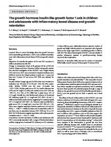
Corpus luteum Figure 1. Possible sites of action of the insulin-like growth factor (IGF) system in the hypothalamo-pituitary-ovarian axis; the normal situation. Growth of 'small' antral follicles is influenced by interactions between the gonadotrophins and members of the IGF system (the IGFs, their binding proteins, or insulin). These effects control the numbers of such follicles that survive atresia and reach the preovulatory stage of development. These preovulatory follicles are then driven through the final stages of maturation and the process of ovulation by the gonadotrophins, and thus by the gonadotrophin-releasing hormone (GnRH) signal arising from the central nervous system. The endocrine action of systemic insulin and IGFs at brain level is difficult to assess due to the lack of data concerning their passage through the blood-brain barrier, though they may affect pituitary function. On the other hand, intracerebrally synthesized members of the IGF system might be involved, perhaps interacting with the ovarian steroids that exert negative feedback on the activity of the GnRH system. In this way, they may influence the tonic secretion of gonadotrophins and thus exert further influence over the development of both small and large antral follicles.
tions in the arcuate nucleus increase markedly at puberty in male and female rats. Moreover, IGF-I concentrations are higher in this region in the afternoon of pro-oestrus and in the morning of oestrus, compared with other periods of the cycle. The intensity of IGF-I immunoreactivity in the arcuate nucleus was enhanced by oestradiol. Expression of IGF-I mRNA increases in the median eminence during the first pro-oestrus, in parallel with the increase in expression in the liver and increases in serum concentrations of IGF-I, and IGF-I stimulates the release of GnRH by fragments 42
of median eminence in vitro (Hiney et al, 1991). In addition, injections of IGF-I clearly decrease the sensitivity of hypothalamo-pituitary axis to oestradiol negative feedback, and accelerate the process of puberty in the Rhesus monkey (Wilson, 1995). The pituitary gland may also be affected, because expression of IGF-I mRNA in pituitary tissue is also stimulated by oestradiol (Michels et al, 1993). In the pituitary gland of the cow, the concentrations of IGFBP change in association with serum concentrations of progesterone during the oestrous cycle (Funston et al, 1995a). Thus,
IGFs and interactions between nutrition and reproduction
Negative feedback
Hypothalamic-Preoptic Area Continuum
Negative feedback
^.Adipose tissue"^
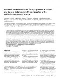
Figure 2. Possible sites of action of the insulin-like growth factor (IGF) system in the hypothalamo-pituitary-ovarian axis; undernutrition and the 'decision to ovulate'. Alteration of intracerebral expression and action of elements of the IGF system may lead to increases in responsiveness to negative feedback and thus reductions in the tonic secretion of gonadotrophinreleasing hormone (GnRH). The consequent lack of gonadotrophins, in association with the low circulating concentrations of IGFs and insulin, act at ovarian level to reduce the number of antral follicles that survive atresia and reach the preovulatory stage of development. Under extreme conditions (severe undernutrition), it would prevent ovulation because no follicles at all would enter the ovulatory pool or, if some do, they would not be stimulated to secrete sufficient oestrogen to induce the preovulatory luteinizing hormone (LH) surge.
there may be changes in the bioavailability of IGFs in pituitary cells during the cycle, despite lack of variation in circulating concentrations of IGF-I and IGFBPs. Together, all these observations strongly support a role for insulin and IGF-I in the control of GnRH neurosecretion in the hypothalamus and median eminence, and gonadotrophin secretion by the pituitary gland. These substances should therefore be considered as major candidates for linking the metabolic status with reproductive function,
particularly with regard to the 'decision to ovulate' (Figures 1 and 2). Possible mechanisms of action of insulin and IGF-I on GnRH neurosecretion Experiments on both peripubertal and undernourished adult animals suggest that part of the mechanism through which insulin and IGF-I affect GnRH secretion would involve modulation of the sensitivity of hypothalamic reproductive centres to oestradiol (see above). This is supported by studies in rats and Syrian hamsters showing that experimental 43
P.Monget and CB.Martin
diabetes decreases the concentration of oestradiol receptors in the hypothalamus and pituitary gland (Gentry et al, 1977; Siegel and Wade, 1979; Weisenberg et al, 1983; Tesone et al, 1985; Li et al, 1994). This might explain the alteration of oestrous behaviour in diabetic rodents. As reported above, central insulin, whether it be of peripheral or central origin, may be acting on metabolism, food intake and neurohormone secretion via IGF-II produced locally in the hypothalamus. Indeed, in the rat, peripheral as well as intracerebroventricular injections of insulin, but not glucose, lead to increases in the IGF-II content of the ventromedial hypothalamus and paraventricular nucleus, and to decreases in the arcuate nucleus and the neurointermediate lobe of the pituitary gland (Lauterio et al, 1990; Ahmed and Lauterio, 1992). At cellular level, several mechanisms can be proposed for the effects of insulin in the hypothalamus. It could act directly via a cascade of phosphorylations from its own receptor to the oestradiol receptor or IGF-II genes. It might also act indirectly simply by increasing the availability of metabolic fuel on hypothalamic neurons. Of particular interest is the fact that, although glucose alone is not able to increase GnRH secretion in rams (Miller et al, 1995), peripheral injection of 2-deoxy-glucose, a blocker of glycolysis, into ewes decreases LH secretion despite an increase in serum insulin concentrations (Funston et al, 1995b). Similarly, injection of both 2-deoxy-glucose and methyl palmoxirate, a blocker of fatty acid oxidation, into female Syrian hamsters has the same effects as food deprivation or streptozotocin-induced diabetes on the hypothalamus: it suppresses steroid-induced oestrous behaviour and decreases the number of immunoreactive oestradiol receptors in the ventromedial hypothalamus and the lateral hypothalamus, whereas it increases the number of oestradiol receptors in the medial preoptic area (Li et al, 1994). Moreover, it is important to note that, in the central nervous system, the insulin-responsive glucose transporter, GLUT-4, is expressed only in the hypothalamus and the cerebellum (as well as the pituitary gland), whereas other transporters (GLUT-1, -2 and -3) are widely expressed in the brain (Brant et al, 1993; Livingstone et al, 1995). Once again, these specific localizations of the 44
various glucose transporters may contribute to the adaptive distribution of energy within the brain during nutrient restriction. A large body of data has shown that the ventromedial hypothalamus is a key location for many homeostatic systems, including regulation of body temperature, water and electrolyte balance, and food intake, as well as oestrous behaviour and GnRH secretion. The fact that this region is more exposed to peripheral plasma (and therefore to variations in concentrations of insulin, IGF-I, and metabolic fuels) than many other regions in the brain, and that it also has a relatively high content of insulin-regulated GLUT-4, leads to the hypothesis that the ventromedial hypothalamus is at least partly involved in the effects of negative energy balance, whether it is induced by food deprivation or increased energy expenditure, on the liberation of GnRH. Moreover, of particular interest is the fact that leptin produced by adipocytes, recently cloned and identified in the ob/ob mouse model as a 'fat-derived satiety factor', acts at the level of the hypothalamus to regulate food intake and energy expenditure (Zhang et al, 1994). Expression of this factor is stimulated by insulin administration and by food intake (Saladin et al, 1995). Considering the often cited correlation between the amount of adipose tissue and GnRH pulsatility, it would be interesting to test whether this leptin is also involved in reproductive physiology. Influence of nutrition on ovulation rate When all the metabolic and hormonal conditions are compatible with the decision to reproduce, and thus ovulate, the role played by nutrition has not ended because it modulates other, more subtle aspects of ovarian physiology. In polytocus species (sheep, pig), the level of nutrition affects ovulation rate (the number of ovulations per female ovulating). In monotocus species, such as the human, it will modulate the length of the sexual cycle (as discussed above for female athletes), the number of growing follicles, and thus the ovulation rate following ovulation stimulation treatment. Influence of flushing on ovulation rate In female sheep, short periods of improved nutrition before and during mating will increase the propor-
IGFs and interactions between nutrition and reproduction
tion of ewes bearing twins. This practice of 'flushing' need last as little as 4 days (Stewart and Oldham, 1986) and is particularly efficient for ewes that are in fairly poor body condition and are maintained under extensive systems of husbandry. Ewes in good body condition already have a high ovulation rate, usually close to their genetic potential for this character, so they respond poorly to flushing (Downing and Scaramuzzi, 1991). An excellent experimental model here involves feeding a supplement of lupin grain (a legume high in digestible energy and protein) to ewes during the luteal phase. This treatment reliably increases ovulation rate and blood insulin concentrations, but has no significant effect on GnRH/LH pulse frequency or the circulating amounts of IGF-I or FSH (Downing et al, 1995). Other experimental models are more chronic in nature, and attempt to relate whole body condition to ovulation rate. In particular, Rhind and McNeilly (1986, 1989) fed two groups of ewes differentially to achieve either a high or a low body condition score, and then to maintain that difference. Ewes with a high condition score had more growing follicles and a higher ovulation rate than ewes with a low score. Similar results were obtained by Xu et al. (1989). None of these studies included detailed analysis of the metabolic status of the animals, but energy balance was probably equilibrated under both nutritional treatments because body condition scores and live weights were maintained at relatively constant levels. In the pig, Cosgrove et al. (1992) studied the effects of short-term ad-libitum refeeding during the luteal phase of gilts that had been maintained on 30% of ad-libitum feed intake for 7 days. This treatment led to slight increases in the diameter of the follicles and the basal rate of oestradiol synthesis by the granulosa cells from those follicles. As in the sheep studies described above, no changes in FSH and LH secretion were observed following refeeding. Similarly, Cox's group has shown that, in the gilt treated with pregnant mare's serum gonadotrophin (PMSG), experimentally-induced diabetes provokes an increase in rate of atresia of small follicles (Cox et al, 1994). Previously, this laboratory had shown that cyclic gilts fed low levels of dietary energy at the end of the luteal
phase and during the follicular phase had lower ovulation rates than pigs fed with a high energy diet (Cox et al, 1987). Moreover, injections of insulin also increased ovulation rate. In this experiment, concentrations of FSH and LH were only increased during the first 24 h of sampling in gilts receiving insulin and high energy diet. Furthermore, the same group has shown that withdrawal of insulin from diabetic gilts during the luteal phase decreases ovulation rate and increases the number of atretic follicles in cyclic pigs and in pigs treated with PMSG (Cox et al, 1994). Curiously, LH pulse frequency was increased in this experiment. These observations agree with those of Matamaros et al. (1990) who found that injection of insulin during the follicular phase of the oestrous cycle increases the number of medium-sized follicles by reducing atresia. They also found that peripheral injections of insulin into prepubertal gilts increased IGF-I values in the antral fluid of medium-sized follicles (Matamaros et al, 1991). In these particular experiments, peripheral FSH and LH concentrations were not systematically studied but, in experiments in sheep and pigs, with either flushing or insulin injections, it is rare to find clear correlations between ovulation rate and circulating gonadotrophin concentrations. It is tempting to consider this lack of a positive result as conclusive evidence that ovulation rate is controlled purely by gonadotrophin-independent mechanisms. However, it is difficult to be sure that either the negative observation (no change in LH or FSH secretion) or the conclusion (gonadotrophin independence) is definitive. Returning to the basic principles of negative feedback, we would expect an increase in ovarian follicular growth to lead to the secretion of extra oestrogen and inhibin which would, in turn, decrease gonadotrophin secretion. Thus, when ovulation rate is changed acutely (by nutritional treatments, for example), the normal concentrations of gonadotrophin we generally observe must, in fact, reflect a shift in the equilibrium of the feedback system because there are extra ovarian follicles producing extra negative feedback. In other words, the lack of a decrease in the secretion of the gonadotrophins despite extra feedback may itself be evidence of participation by the gonadotrophic axis, perhaps through resist45
P.Monget and CB.Martin
ance to the inhibitory effects of oestradiol (Martin and Thomas, 1990). The only way to resolve this apparent paradox is to find a variable which describes the equilibrium of the feedback system. One possibility is ratios of the concentrations of the gonadotrophic and ovarian hormones. This brings us up against the technical barrier of measurement of plasma inhibin and oestradiol; very few reproductive endocrinologists have complete confidence in the assays currently in use for sheep and cattle, especially for the basal concentrations we need to consider here. To make matters worse, Adams et al. (1993) have shown that serial blood sampling to measure GnRH/LH pulse frequency seems to affect gonadotrophin secretion as well as ovulation rate. Importantly, ovulation rate can be either reduced or stimulated, depending on the time of the cycle that the samples are taken. In other words, many studies might have been compromised by a physiological version of Heisenberg's Uncertainty Principle: the phenomenon being measured is altered by the act of measuring it. In this light, reports that describe a relationship (or lack of one) between LH pulse frequency and ovulation rate need to be carefully re-examined. Interestingly, a sampling frequency appropriate for measurement of FSH patterns does not have this effect and this might explain the success of some studies showing a positive relationship between FSH concentrations and ovulation rate (e.g. McNatty et al, 1988). Is there a role for the ovarian IGF system in the effects of nutrition on ovulation rate? The role of the IGF system in ovarian folliculogenesis has been extensively studied over the past 10 years (reviews: Adashi et al, 1992; Giudice, 1992; Monget and Monniaux, 1995). Here, we will only outline the observations which might help explain the possible role of the members of this system in the interaction between nutrition and reproduction at ovarian level. In the sheep, rat, pig and human, IGF-I stimulates both the proliferation and the differentiation of granulosa cells, as well as steroidogenesis in thecal cells (Adashi et al, 1992; Giudice, 1992). In the sheep, IGF-I stimulates proliferation of granulosa cells mainly in small follicles (1-3 mm diameter). 46
In contrast, the stimulatory effect of IGF-I on secretion of progesterone is observed in granulosa cells from large (>5 mm diameter) rather than small follicles (Monget and Monniaux, 1995). Thus, it seems that whether IGF-I stimulates proliferation or differentiation of granulosa cells depends on the stage of development of the follicle. The origin of insulin in the ovary is likely to be endocrine, whereas ovarian IGFs seem to have both paracrine and endocrine origins: Paracrine In mammals, there is a great deal of variety in the ovarian sites in which IGFs are expressed. For example, in the human, IGF-I is expressed in thecal cells from small antral follicles (5-7 mm diameter), whereas IGF-II is highly expressed in granulosa from preovulatory follicles (El Roeiy et al, 1993). By contrast, in the rat and the ewe, IGF-II is exclusively expressed in thecal cells. In the rat, IGF-I is expressed in granulosa cells from growing antral follicles (see Monget and Monniaux, 1995; Perks et al, 1995). In the sheep, IGF-I mRNA is mainly found in luteal cells whereas, in the pig, it is mostly found in granulosa cells from growing antral follicles. Endocrine In all of these species, several arguments favour an at least partly endocrine origin for IGFs found in the ovary. First, in the woman, cow, ewe and mare, the concentrations of IGF-I in large healthy follicles are positively correlated with serum concentrations (see Monget and Monniaux, 1995). In the woman and the ewe, significant amounts of IGF-I are found in preovulatory follicles whereas 'local' synthesis is very low (El Roeiy et al, 1993; Perks et al, 1995). Moreover, concentrations of intrafollicular IGFBP-3 (the main IGFBP in serum and follicular fluid) are similar to or slightly lower than those in the corresponding serum as assessed by Western ligand blotting. Since little or no expression of IGFBP-3 was found either in granulosa or in thecal cells (see Monget and Monniaux, 1995), and as the systemic 150 kDa IGFBP-IGF complex has been identified in ovine and human follicular fluid (Hodgkinson et al, 1989; Kubota etal, 1993), one may postulate that the intrafollicu-
IGFs and interactions between nutrition and reproduction
lar IGFs/IGFBP-3 complex is partly derived from the circulatory pool. According to these data, is there a role for intraovarian insulin or IGFs (synthesized locally or elsewhere) in the effects of nutrition on ovulation rate? Two questions have to be answered: firstly, do changes in nutritional status affect intrafollicular concentrations or expression of IGFs in the ovary? Few and conflicting results are available. In the pig, the decrease in ovulation rate following experimentally induced diabetes (Cox et al, 1994; see above) was accompanied by a concomitant decrease in both systemic and intrafollicular IGF-I concentrations. By contrast, Spicer et al. (1992) found that a 48 h fast in heifers decreased IGF-I values in serum but not in follicular fluid. In this experiment, fasting decreased the intrafollicular concentration of oestradiol, but gonadotrophins were not assayed. Also using heifers, Vandeharr et al. (1995) found that the imposition of negative energy balance for four consecutive oestrous cycles decreases peripheral IGF-I concentrations and expression of IGF-I and GH receptor in the liver, but does not affect expression of IGF-I and GH receptor in luteal tissue, despite a decrease in the growth of the corpus luteum and the secretion of progesterone. Collectively, these data suggest that intraovarian concentrations of IGF-I may be maintained longer than systemic concentrations, at least during short periods of dietary restriction in ruminants. More importantly, if elements of the IGF system mediate the adverse effect of negative energy balance on ovarian function, the mode of action seems more likely to be endocrine than paracrine. In this case, it is difficult to distinguish the consequences of such endocrine changes at the ovarian level from those at the hypothalamopituitary level. In particular, one may hypothesize that a flushing-induced increase in serum insulin (and, possibly, IGF-I) concentrations would do both, namely enhance the growth of mainly small follicles (see below) through a direct action at ovarian level, and sustain gonadotrophin secretion on the hypothalamo-pituitary compartment by increasing its threshold of sensitivity to negative feedback by the extra oestradiol and inhibin produced by the extra follicular growth. The second question is: what could be the site
of action of the IGF system in the ovary? As described above, the influence of the IGF system on the decision to ovulate is mainly central, directly affecting FSH and LH secretion and thereby the final maturation and ovulatory ability of large, antral gonadotrophin-dependent follicles. In contrast, in the case of change of ovulation rate following an alteration of energy balance, several lines of evidence suggest that the main target tissue for insulin and IGFs is small follicles (diameters of 1-3 mm in the sheep, or 4-6 mm in the pig), rather than large preovulatory follicles (i) in both ewes and sows, short-term flushing affects ovulation rate when treatment is confined to the end of the luteal phase, the time when small follicles are recruited for the next ovulation; (ii) the absence of a strong correlation between gonadotrophin concentration and ovulation rate seems to exclude dramatic effects on large preovulatory follicles whose development is strictly dependent on FSH and LH; rather, the evidence suggests that nutrition affects smaller follicles in which development is less sensitive to gonadotrophin concentrations (Dufour et al, 1979; McNeilly et al, 1991); (iii) in heifers, Gong et al. (1991) have shown that daily injections of recombinant bovine GH over two oestrous cycles can double the number of 2—5 mm diameter follicles, without modifying the number of 5-10 mm and preovulatory follicles; GH treatment increased serum concentrations of IGF-I, and probably insulin, but did not affect the amounts of FSH and LH. Thus, it seems likely that the action of GH was mediated by IGF-I (and/or insulin) since bovine ovarian follicles, in contrast to corpus luteum, contain very low levels of GH receptor (Lucy et al, 1993). Overall, then, the effects of nutrition on ovulation rate are determined primarily at ovarian level, and small to medium-sized, antral follicles are the main target tissue for the nutritional signals. The evidence that members of the IGF system are a component of those signals is compelling, but not definitive (Figures 1 and 3). Conclusion Insulin, IGF-I and IGF-II, and their binding proteins and receptors are found in a wide variety of tissues throughout the body, where they control 47
P.Monget and G.B.Martin
Negative feedback
Hypothalamic-Preoptic Area Continuum
"Small" antral j "Large" antral follicles j follicles
Local Production?
IGF-I, IGF-II
Ovary
Corpus luteum Figure 3. Possible sites of action of the insulin-like growth factor (IGF) system in the hypothalamo-pituitary-ovarian axis; good nutrition and the 'decision to increase ovulation rate'. The smaller antral follicles are stimulated by interactions at ovarian level between the gonadotrophins and the IGFs, their binding proteins, or insulin. This increases the number of such follicles that survive atresia and reach the preovulatory stage of development. At the hypothalamo-pituitary level, locally-produced elements of the IGF system might also stimulate gonadotrophin-releasing hormone (GnRH) and gonadotrophin secretion thus maintaining their concentrations in the circulation despite the increased amount of inhibitory feedback produced by the extra follicles. Ultimately, more preovulatory follicles develop and fall under the control of the gonadotrophins, and thus the GnRH signal arising from the central nervous system. The gonadotrophic axis then drives all of these follicles through to ovulation.
the anabolic and catabolic processes that constitute the general responses to changes in level of nutrition. Nutritional treatments alter the expression of their genes in their tissues of origin and these effects lead to changes in their circulating concentrations. Many of these responses are correlated, some positively and some negatively, with the effects of these nutritional treatments on reproductive function. There is also good evidence that most members of the IGF system (ligands as well as receptors) are also found in brain and ovarian tissue, and the presence of their mRNAs shows that several of them are synthezised there. This 48
raises the possibility that some of the reproductive responses are not only mediated by the classical hormonal actions of the products of the pancreas or liver, but also by autocrine and paracrine effects of the IGF system, at all levels of the brainpituitary-ovarian axis. The role of the IGF family in the 'decision to ovulate' is not clear, but seems to mainly include endocrine (change in plasma hormone concentrations) as well as intracerebral autocrine or paracrine effects (changes in receptor or peptide expression) at brain level. It is difficult to determine the importance of classical endocrine effects because
IGFs and interactions between nutrition and reproduction
it is difficult to assess the importance of bloodbrain barrier permeability. This contrasts with the 'decision to change ovulation rate' which seems to depend to a large degree on the endocrine action of the IGF system directly on small ovarian follicles. Whether intra-ovarian paracrine pathways have a major role in this decision is difficult to judge without further research. Much of the evidence for involvement of the IGF family in the effects of nutrition on reproductive function is derived from correlations between blood concentrations of the IGFs and reproductive responses to changes in nutrition, or by in-vitro studies of isolated brain, pituitary or ovarian tissues. This circumstantial evidence now needs support from experiments in which the IGFs are manipulated directly, or in which they are used in an attempt to alter brain and ovarian function.
References Adams, N.R., Atkinson, S., Martin, G.B. et al (1993) Frequent blood sampling changes the plasma concentration of LH and FSH and the ovulation rate in Merino ewes. J. Reprod. Fertil, 99, 689-694. Adashi, E.Y., Hsueh, A.J.W. and Yen, S.S.C. (1981) Insulin enhancement of luteinizing hormone and follicle-stimulating hormone release by cultured pituitary cells. Endocrinology, 108, 1441-1449. Adashi, E.Y., Resnick, C.E., Hurwitz, A. et al. (1992) The intra-ovarian IGF system. Growth Reg., 2, 10-15. Aguado, F., Fernandez, T, Martinez-Murillo, R. et al. (1992) Immunocytochemical localization of insulinlike growth factor I in the hypothalamo-hypophyseal system of the adult rat. Neuroendocrinology, 56, 856-863. Ahmed, I. and Lauterio, T.J. (1992) Intracerebroventricular injection of insulin or glucose alters insulin-like growth factor II (IGF-II) concentrations in specific hypothalamic nuclei. Brain Res., 595, 242-248. Arias, P., Rodriguez, M , Szwarcfarb B. et al. (1992) Effect of insulin on LHRH release by perifused hypothalamic fragments. Neuroendocrinology, 56, 415-418. Bohannon, N.J., Corp, E.S., Wilcox, B.J. et al. (1988) Characterization of insulin-like growth factor I receptors in the median eminence of the brain and their modulation by food restriction. Endocrinology, 122, 1940-1947. Bondy, C , Werner, H., Roberts, C.T. Jr and LeRoith, D. (1992) Cellular pattern of type-I insulin-like growth factor receptor gene expression during maturation of
the rat brain: comparison with insulin-like growth factors I and II. Neuroscience, 46, 909-923. Booth, P.J., Craigon, J. and Foxcroft, G.R. (1994) Nutritional manipulation of growth and metabolic and reproductive status in prepubertal gilts. J. Anim. Set, 72, 2415-2424. Boukhliq, R., Miller, D.W. and Martin, G.B. (1996) Relationship between nutritional stimulation of gonadotrophin secretion and the peripheral and cerebrospinal fluid (CSF) concentrations of glucose and insulin in rams. Anim. Reprod. Sci., 41, 201-214. Brant, A.M., Jess, T.J., Milligan, G. et al. (1993) Immunological analysis of glucose transporters expressed in different regions of the rat brain and central nervous system. Biochem. Biophys. Res. Comm., 192, 1297-1302. Cheatham, B. and Kahn, C.R. (1995) Insulin action and the insulin signaling network. Endocr. Rev., 16, 117-142. Clark, R.T. (1934) Studies of reproduction in sheep. I. The ovulation rate of the ewe as affected by the plane of nutrition. Anat. Rec, 60, 125-134. Cosgrove, J.R., Tilton, J.E., Hunter, M.G. and Foxcroft, G.R. (1992) Gonadotropin-independent mechanisms participate in ovarian responses to realimentation in feed-restricted prepubertal gilts. Biol. Reprod., 47, 736-745. Cox, N.M., Stuart, M.J., Althen, T.G. et al. (1987) Enhancement of ovulation rate in gilts by increasing dietary energy and administering insulin during follicular growth. J. Anim. Sci., 64, 507-516. Cox, N.M., Meurer, K.A., Carlton, C.A. et al. (1994) Effect of diabetes mellitus during the luteal phase of the oestrus cycle on preovulatory follicular function, ovulation and gonadotrophins in gilts. J. Reprod. Fertil, 101, 77-86. Day, M.L., Imakawa, K., Wolfe, P.L. etal. (1987) Endocrine mechanisms of puberty in heifers: role of hypothalamo-pituitary estradiol receptors in the negative feed-back of estradiol on luteinizing hormone secretion. Biol. Reprod., 37, 1054-1065. de Keyser, J., Wilczak, N., de Backer, J.-P. et al. (1994) Insulin-like growth factor-I receptors in human brain and pituitary gland: an autoradiographic study. Synapse, 17, 196-202. Downing, J.A. and Scaramuzzi, R.J. (1991) Nutrient effects on ovulation rate, ovarian function and the secretion of gonadotrophic and metabolic hormones in sheep. /. Reprod. Fertil, 43 (Suppl.), 209-227. Downing, J.A., Joss, J., Connell, P. and Scaramuzzi, R.J. (1995) Ovulation rate and the concentrations of gonadotrophic and metabolic hormones in ewes fed lupin grain. J. Reprod. Fertil, 103, 137-145. Duenas, M., Luquin, S., Chowen, J.A. et al. (1994) Gonadal hormone regulation of insulin-like growth factor-I-like immunoreactivity in hypothalamic 49
P.Monget and G.B.Martin astroglia of developing and adult rats. Neuroendocrinology, 59, 528-538. Dufour, J., Cahill, L.P. and Mauleon, P. (1979) Short- and long-term effects of hypophysectomy and unilateral ovariectomy on ovarian follicular populations in sheep. J. Reprod. Fertil, 57, 301-309. Ebling, F.J.P., Wood, R.I., Karsch F.J. et al (1990) Metabolic interfaces between growth and reproduction. III. Central mechanisms controlling pulsatile luteinizing hormone secretion in the nutritionally growth-limited female lamb. Endocrinology, 126, 2719-2727. Einarsson, S. and Rojkittikhun, T. (1993) Effects of nutrition on pregnant and lactating sows. J. Reprod. Fertil, 48 (Suppl.), 229-239. El-Roeiy, A., Chen, X., Roberts, V.J. et al. (1993) Expression of insulin-like growth factor-I (IGF-I) and IGF-II and the IGF-I, IGF-II, and insulin receptor genes and localization of the gene products in the human ovary J. Clin. Endocrinol. Metab., 11, 1411— 1418. Fisher, E.C., Nelson, M.E., Frontera, W.R. et al. (1986) Bone mineral content and levels of gonadotropins and estrogens in amenorrheic running women. J. Clin. Endocrinol. Metab., 62, 1232-1236. Frattali, A.L., Treadway, J.L. and Pessin, J.E. (1992) Insulin/IGF-1 hybrid receptors: implications for the dominant-negative phenotype in syndromes of insulin resistance. J. Cell. Biochem., 48, 43-50. Funston, R.N., Moss, G.E. and Roberts, A.J. (1995a) Insulin-like growth factor-I (IGF-I) and IGF-binding proteins in bovine sera and pituitaries at different stages of the estrous cycle. Endocrinology, 136,62-68. Funston, R.N., Roberts, A.J., Hixon, D.L. et al. (1995b) Effect of acute glucose antagonism on hypophyseal hormones and concentrations of insulin-like growth factor (IGF)-I and IGF-binding proteins in serum, anterior pituitary, and hypothalamus in ewes. Biol. Reprod, 52, 1179-1186. Garofalo, R.S. and Barenton, B. (1992) Functional and immunological distinction between insulin-like growth factor I receptor subtypes in KB cells. J. Biol. Chem., 267, 11470-11475. Gentry, R.T., Wade, G.N. and Blaustein, J.D. (1977) Binding of [3H]estradiol by brain cell nuclei and female rat sexual behavior: inhibition by experimental diabetes. Brain Res., 135, 130-146. Giudice, L.C. (1992) Insulin-like growth factors and ovarian follicular development. Endocr. Rev., 13, 641-669. Gosiewska, A., Wilson, S., Kwon, D., and Peterkofsky, B. (1994) Evidence for an in vivo role of insulin-like growth factor-binding protein-1 and -2 as inhibitors of collagen gene expression in vitamin C-deficient and fasted guinea pigs. Endocrinology, 134, 1329-1339. Grimard, B., Humblot, P., Ponter, A.A. et al. (1995) Influence of postpartum energy restriction on energy
50
status, plasma LH and oestradiol secretion and follicular development in suckled beef cows. J. Reprod. Fertil, 104, 173-179. Harel, Z. and Tannenbaum, G.S. (1992) Centrally administered insulin-like growth factor II fails to alter pulsatile growth hormone secretion or food intake. Neuroendocrinology, 56, 161-168. Heidenreich, K.A. and Brandenburg, D. (1986) Oligosaccharide heterogeneity of insulin receptors. Comparison of JV-linked glycosylation of insulin receptors in adipocytes and brain. Endocrinology, 118, 1835-1842. Hiney, J.K., Ojeda, S.R. and Les Dees, W. (1991) Insulinlike growth factor I: a possible metabolic signal involved in the regulation of female puberty. Neuroendocrinology, 54, 420—123. Hodgkinson, S.C., Moore, L., Napier, J.R. et al. (1989) Characterization of insulin-like growth factor binding proteins in ovine tissue fluids. /. Endocrinol, 120, 429^38. Holland, M.D., Hossner, K.L., Tatum, J.D. et al. (1988) Serum insulin-like growth factor I profiles in beef heifers with single and twin pregnancies. J. Anim. Sci., 66, 3190-3196. Humbel, R.E. (1990) Insulin-like growth factors I and II. Eur. J. Biochem., 190, 445-462 Jolly, P.D., McDouglas, S., Fitzpatrick, L.A. et al. (1995) Physiological effects of undernutrition on postpartum anoestrus in cows. J. Reprod. Fertil, 49 (Suppl.), 477^92. Jones, J.I. and Clemmons, D.R. (1995) Insulin-like growth factors and their binding proteins: biological actions. Endocr. Rev. 16, 3-34. Kinder, J.E., Day, M.L. and Kittok, RJ. (1987) Endocrine regulation of puberty in cows and ewes. J. Reprod. Fertil, 34 (Suppl.), 167-186. Kinder, J.E., Bergfeld, E.G.M., Wehrman, M.E. et al. (1995) Endocrine basis for puberty in heifers and ewes. J. Reprod. Fertil, 49 (Suppl.), 393-407. Kubota, T., Sakamoto, S., Kamada, S. et al (1993) Insulin-like growth factor II in follicular fluid of the patients with in vitro fertilization and embryo transfer. Fertil Steril, 59, 844-849. Lalou, C. and Binoux, M. (1993) Evidence that limited proteolysis of Insulin-like Growth Factor Binding Protein-3 (IGFBP-3) occurs in the normal state outside of the bloodstream. Regul Peptides, 48, 179-188. Landefeld, T.D., Ebling, F.J.P., Suttie, J.M. et al. (1989) Metabolic interfaces between growth and reproduction. II. Characterization of changes in messenger ribonucleic acid concentrations of gonadotropin subunits, growth hormone, and prolactin in nutritionally growth-limited lambs and the differential effects of increased nutrition. Endocrinology, 125, 351—356. Lauterio, T.J., Aravich, P.F. and Rotwein, P. (1990) Divergent effects of insulin on insulin-like growth
IGFs and interactions between nutrition and reproduction
factor II gene expression in the rat hypothalamus. Endocrinology, 126, 392-398. LeRoith, D., Werner, H., Beitner-Johnson, D. and Roberts Jr, C.T. (1995) Molecular and cellular aspects of the insulin-like growth factor I receptor. Endocr. Rev., 16, 143-163. Lesniak, M.A., Hill, J.M., Kiess, W. et al (1988) Receptors for insulin-like growth factors I and II: autoradiographic localization in rat brain and comparison to receptors for insulin. Endocrinology, 123, 2089-2099. Li, H.-Y., Wade, G.N. and Blaustein, J.D. (1994) Manipulations of metabolic fuel availability alter estrous behaviour and neural estrogen receptor immunoreactivity in Syrian hamsters. Endocrinology, 135, 240-247. Lindsay, D.R., Martin, G.B. and Williams, I.H. (1993) Nutrition and reproduction. In King, G.J. (eds), Reproduction in Domesticated Animals. World Animal Science Series, Elsevier Science Publishers, pp. 459^91. Livingstone, C , Lyall, H. and Gould, G.W. (1995) Hypothalamic GLUT 4 expression: a glucose- and insulin-sensing mechanism? Mol. Cell. Endocrinol, 107, 67-70. Lowe, W. and LeRoith, D. (1986) Insulin receptors from guinea pig liver and brain: structural and functional studies. Endocrinology, 118, 1669-1677. Lucy, M.C., Collier, R.J., Kitchell, M.L. et al. (1993) Immunohistochemical and nucleic acid analysis of somatotropin receptor populations in the bovine ovary. Biol. Reprod, 48, 1219-1227. McNatty, K.P., Hudson, N., Gibb and Collins, F.L. (1988) Plasma concentrations of FSH, LH and progesterone in sheep immunized against an androstenedione-protein conjugate. J. Reprod. Fertil, 82, 63-69. McNeilly, A.S., Picton, H.M., Campbell, B.K. and Baird, D.T. (1991) Gonadotrophic control of follicle growth in the ewe. J. Reprod. Fertil, 43 (Suppl), 177-186. Marks, J.L. and Eastman, C.J. (1989) Effect of starvation on insulin receptors in rat brain. Neuroscience, 30, 551-556. Marks, J.L., Porte, D. Jr and Baskin, D.G. (1991) Localization of type I Insulin-like growth factor receptor messenger RNA in the adult rat brain by in situ hybridization. Mol. Endocrinol, 5, 1158-1168. Martensz, N.D. (1981). The biological effects of immunization against steroids in female mammals. Oxford Rev. Reprod. Biol, 2, 232-262. Martin, G.B. (1984). Factors affecting the secretion of luteinizing hormone in the ewe. Biol. Rev., 59, 1-87. Martin, G.B., Scaramuzzi, R.J. and Lindsay, D.R. (1981) Induction of ovulation in seasonally anovular ewes by the introduction of rams: effects of progesterone and active immunization against androstenedione. Aust. J. Biol ScL, 34, 569-575. Martin, G.B. and Thomas, G.B. (1990) Roles of
communication between the hypothalamus, pituitary gland and ovary in the breeding activity of ewes. In Oldham, CM., Martin, G.B. and Purvis, I.W. (eds), Reproductive Physiology of Merino Sheep - Concepts and Consequences. School of Agriculture (Animal Science), The University of Western Australia, Perth, pp. 2 3 ^ 0 . Martin, G.B. and Walkden-Brown, S.W. (1995) Nutritional influences on reproduction in mature male sheep and goats. J. Reprod. Fertil, 49 (Suppl.), 437^49. Matamaros, I.A., Cox, N.M. and Moore, A.B. (1990) Exogenous insulin and additional energy affect follicular distribution, follicular steroid concentrations, and granulosa cell human chorionic gonadotropin binding in swine. Biol. Reprod., 43,1-7. Matamaros, LA., Cox, N.M. and Moore, A.B. (1991) Effects of exogenous insulin and body condition on metabolic hormones and gonadotropin-induced follicular development in prepubertal gilts. J. Anim. ScL, 69, 2081-2091. Melnik, R.B. and Martin, J.M. (1984) Starvation-induced changes in insulin binding to hypothalamic receptors in the rat. Acta Endocrinol, 107, 78-85. Michels, K.M., Lee, W.-H., Seltzer, A. et al. (1993) Up-regulation of pituitary [125I]Insulin-like Growth Factor-I (IGF-I) binding and IGF Binding Protein-2 and IGF-I gene expression by estrogen. Endocrinology, 132, 23-29. Milazzo, G., Yip, C.C., Maddux, B.A. et al. (1992) High-affinity insulin binding to an atypical insulinlike growth factor-I receptor in human breast cancer cells. J. Clin. Invest., 89, 899-908. Miller, D.W., Blache, D. and Martin, G.B. (1995) Insulin stimulates LH secretion in rams when infused into the third cerebral ventricle: comparison with the effect of improved nutrition. J. Endocrinol, 147, 321-329. Monget, P. and Monniaux, D. (1995) Growth factors and the control of folliculogenesis. /. Reprod. Fertil, 49 (Suppl.), 321-333. Nilsson, C , Blay, P., Nielsen, F.C. and Gammeltoft, S. (1992) Gene expression and receptor binding of insulin-like growth factor-II in pig choroid plexus epithelial cells. J. Neurochem., 58, 923-930. Nolan, CM., Kyle, J.W., Watanabe, H. and Sly, W.S. (1990) Binding of Insulin-like Growth Factor II (IGF-II) by human cation-dependent mannose 6phosphate receptor/IGF-II receptor expressed in receptor-deficient mouse L cells. Cell Reg., 1197-213. Olson, B.R., Cartledge, T., Sebring, N. et al. (1995) Short-term fasting affects luteinizing hormone secretory dynamics but not reproductive function in normal-weight sedentary women. /. Clin. Endocrinol. Metab., 80, 1187-1193. Perks, CM., Denning-Kendall, P.A., Gilmour, R.S. and Wathes, D.C. (1995) Localization of messenger ribonucleic acids for insulin-like growth factor I
51
P.Monget and G.B.Martin (IGF-I), IGF-II, and the type 1 IGF receptor in the ovine ovary throughout the estrous cycle. Endocrinology, 136, 5266-5273. Renaville, R., Devolder, A., Massart, S. et al. (1993) Changes in hypophysial-gonadal axis during the onset of puberty in young bulls. J. Reprod. Fertil, 99, 443^49. Rhind, S.M. and McNeilly, A.S> (1986) Follicle populations, ovulation rates and plasma profiles of LH, FSH and prolactin in Scottish Blackface ewes in high and low levels of body condition. Anim. Reprod. ScL, 10, 105-115. Rhind, S.M., Goddard, P.J., McMillen, S.R. and McNeilly, A.S. (1993) FSH-stimulated follicle development in ewes in high and low body condition and chronically treated with gonadotrophin-releasing hormone agonist. J. Reprod. Fertil., 97, 451-456. Rosetta, L. (1993) Female reproductive dysfunction and intense physical training. In Oxford Rev. Reprod. Biol, 15, 113-141. Roth, R.A. and Kiess, W. (1994) Insulin-like growth factor receptors: recent developments and new methodologies. Program of the Third International Symposium on Insulin-like Growth Factors, Sydney. Growth Reg., 4 (Suppl. 1), 31-38. Rutter, L.M., Snopek, R. and Manns, J.G. (1989) Serum concentrations of IGF-I in postpartum beef cows. J. Anim. ScL, 67, 2060-2066. Sahu, A., Dube, M.G., Phelps, C.P. et al. (1995) Insulin and insulin-like growth factor II suppress neuropeptide Y release from the nerve terminals in the paraventricular nucleus: a putative hypothalamic site for energy homeostasis. Endocrinology, 136, 57185724. Saladin, R., De Vos, P., Guerre-Millo, M. et al. (1995) Transient increase in obese gene expression after food intake or insulin administration. Nature, 377,527-529. Scaramuzzi, R.J., Baird, D.T., Campbell, B.K. et al. (1993) A model for follicle selection and the determination of ovulation rate in the ewe. Reprod.
Fertil. Dev., 5, 459^78. Schillo, K.K. (1992) Effects of dietary energy on control of luteinizing hormone secretion in cattle and sheep. J. Anim. ScL, 70, 1271-1282. Siegel, L.I. and Wade, G.N. (1979) Insulin withdrawal impairs sexual receptivity and retention of brain cell nuclear estrogen receptors in diabetic rats. Neuroendocrinology, 29, 200-206. Simon, J., Rosebrough, R.W., McMurtry, J.P. et al. (1986) Fasting and refeeding alter the insulin receptor kinase in chicken liver but fail to affect brain insulin receptors. J. Biol. Chem., 261, 17081-17088. Spicer, L.J., Crowe, M.A., Prendiville, D.J. et al. (1992) Systemic but not intraovarian concentrations of
52
insulin-like growth factor-I are affected by short-term fasting. Biol. Reprod., 46, 920-925. Stewart, R. and Oldham, CM. (1986) Feeding lupins to ewes for four days during the luteal phase can increase ovulation rate. Anim. Prod. Aust., 16, 367-369. Tesone, M., Ladenheim, R.G. and Charreau, E.H. (1985) Alterations in the prolactin secretion in streptozotocininduced diabetic rats. Correlation with pituitary and hypothalamus estradiol receptors. Mol. Cell. Endocrinol, 43, 135-140. Thissen, J.-P, Ketelslegers, J.-M. and Underwood, L.E. (1994) Nutritional regulation of the insulin-like growth factors. Endocr. Rev., 15, 80-101. Tokach, M.D., Pettigrew, J.E., Dial, G.D. et al. (1992) Characterization of luteinizing hormone secretion in the primiparous, lactating sow: relationship to blood metabolites and return-to-estrus interval. J. Anim. ScL, 70, 2195-2201. Vandehaar, M.J., Sharma, B.K. and Fogwell, R.L. (1995) Effect of dietary energy restriction on the expression of insulin-like growth factor-I in liver and corpus luteum of heifers. J. Dairy ScL, 78, 832-841. van Houten, M., Posner, B., Kopriwa, B. and Brawer, J.R. (1980) Insulin binding sites localized to nerve terminals in rat median eminence and arcuate nucleus. Science, 207, 1081-1083. Veldhuis, J.D., Evans, W.S., Demers, L.M. et al. (1985) Altered neuroendocrine regulation of gonadotropin secretion in women distance runners. /. Clin. Endocrinol. Metab., 61, 557-563. Weisenberg, L., Fridman, O., Libertun, C. and de Nicola, A.F. (1983) Changes in nuclear translocation of estradiol-receptor complex in anterior pituitary and uterus of rats with streptozotocin diabetes. J. Steroid. Biochem., 19, 1737-1741. Wilson, M.E. , Gordon, T.P., Rudman, C.G. and Tanner, J.M. (1989) Effects of growth hormone on the tempo of sexual maturation in female rhesus monkeys. J. Clin. Endocrinol. Metab., 68, 29-38. Wilson, M.E. (1995) IGF-I administration advances the decrease in hypersensitivity to oestradiol negative feedback inhibition of serum LH in adolescent female rhesus monkeys. J. Endocrinol., 145, 121-130. Wozniak, M., Rydzewski, B., Baker, S.P and Raizada, M.K. (1993) The cellular and physiological actions of insulin in the central nervous system. Neurochem. Int., 22, 1-10. Xu, Z., McDonald, M.F. and McCutcheon, S.N. (1989) The effects of nutritionally-induced liveweight differences on follicular development, ovulation rate, oestrous activity and plasma follicle-stimulating hormone levels in the ewe. Anim. Reprod. ScL, 19, 67-78. Zhang, Y., Proenca, R., Maffei, M. et al. (1994) Positional cloning of the mouse obese gene and its human homologue. Nature, 372, 425^32.