0 and Ibias = 0.392 pA/cm2: integration is started from initial conditions appropriate to ...... 375 t 41.4 (SD) ms in 12 isolated SAN preparations ...... 304-318,1985.
Three ways of abolishing automaticity in sinoatrial ionic modeling and nonlinear dynamics
node:
MICHAEL R. GUEVARA AND HABO J. JONGSMA Department of Physiology, McGill University, Montreal, Quebec H3G 1 Y6, Canada; and Physiological Laboratory, University of Amsterdam, 1105 AZ Amsterdam, The Netherlands Guevara, Michael R., and Habo J. Jongsma. Three ways of abolishing automaticity in sinoatrial node: ionic modeling and nonlinear dynamics. Am. J. Physiol. 262 (Heart Circ. Physiol. 31): H1268-H1286,1992.-A review of the experimental literature reveals that there are essentially three qualitatively different ways in which spontaneous activity in the sinoatrial node can be abolished. We show that these three ways also occur in an ionic model of space-clamped nodal membrane. In one of these three ways, injection of a current pulse abolishes (“annihilates”) spontaneous action potential generation. In the other two ways, as some parameter is changed, one sees a sequence of qualitative changes in the behavior of the membrane as it is brought to quiescence. In one of these two ways there are incrementing prepotentials intermixed with action potentials, with a maintained small-amplitude subthreshold oscillation being the limiting case of such behavior. Thus both experimental and modeling work indicate that the number of ways in which spontaneous activity can be abolished, or initiated, in the sinoatrial node is limited. The classification into three ways is based on ideas drawn from the qualitative theory of differential equations, which are introduced. The classification scheme can be extended to encompass behaviors seen in other cardiac oscillators. annihilation; single-pulse triggering; afterpotentials; delayed afterdepolarizations; subthreshold oscillations; oscillatory prepotentials; bifurcations; chaos CAUSE of the potentially life-threatening cardiac arrhythmia called sinoatrial arrest is the cessation of spontaneous action potential generation in the sinoatrial node (SAN), the principal natural pacemaker of the mammalian heart. In a search of the literature we have turned up many traces of the transmembrane potential showing cessation of spontaneous activity in the SAN as the result of some experimental intervention. Perusal of these recordings has led us to classify these tracings into three groups, showing that there are essentially three qualitatively different ways in which the normal spontaneous activity of the node can be abolished. In the first way, there is a gradual progressive decline in the amplitude of the action potential until quiescence occurs. In the second way, injection of a brief stimulus pulse annihilates spontaneous activity, which can then be restarted or triggered by injecting a second stimulus pulse. In the third way, before the membrane becomes quiescent as the result of some intervention that gradually takes hold, one sees skipped-beat runs in which spontaneously occurring action potentials are preceded by one or more small-amplitude subthreshold oscillatory prepotentials. Because all the traces mentioned come from experiONE
HE68
0363-6135/92
$2.00
Copyright
ments carried out on isolated right atria1 preparations or on small pieces of tissue isolated from the SAN, the extent to which the behaviors seen might be accounted for solely by membrane properties of SAN cells is uncertain. For example, subthreshold deflections resembling pre- or afterpotentials recorded in one cell might actually be electrotonic potentials reflecting occurrence of block of propagation into that area of the SAN (10, 44). To avoid this complication in the interpretation of the results, we carried out simulations using an ionic model of an isopotential patch of membrane, where spatial factors of this sort cannot occur. In addition, numerical investigation of an ionic model allows one to probe the ionic basis underlying the particular behavior observed. The model of isopotential SAN membrane studied here is that of Irisawa and Noma (36). Because it is a HodgkinHuxley-type model, it is formulated as a system of nonlinear ordinary differential equations. A major point of this paper is that a branch of nonlinear mathematics, bifurcation theory, can be used to obtain significant insights into qualitative aspects of the various behaviors displayed by this class of model. Indeed, it is our claim that one cannot fully appreciate the results of the numerical simulations presented below and the corresponding experimental findings without also at least a passing acquaintance with concepts stemming from the qualitative theory of differential equations. We therefore interweave presentation of the results of numerical simulation of the model with interpretations of those simulations in terms of the qualitative dynamics of the system. For readers wishing to obtain further details about bifurcation theory, the qualitative theory of differential equations, and related aspects of nonlinear mathematics, several textbooks that are readable by someone with a biological sciences background are now available (1, 25, 71, 74, 84). METHODS We investigated the effect of changing, one at a time, many different parameters in the Irisawa-Noma (36) ionic model of isopotential SAN membrane. In several instances, when a parameter was altered sufficiently from its normal value, termination of spontaneous activity resulted. The Irisawa-Noma model is based on voltage-clamp data recorded from small pieces of tissue taken from the rabbit SAN and provides a quantitative description of five currents: the fast inward sodium current (&), the slow inward calcium current (Is), the delayed rectifier potassium current (IK), the hyperpolarization-acti-
0 1992 the American
Physiological
Society
BIFURCATIONS
AND SINUS
vated pacemaker current (Ih; commonly termed If), and a timeindependent leakage current (11).We used two different methods of investigation, direct numerical integration of the equations and bifurcation analysis. Numerical integration. We numerically integrated the Irisawa-Noma equations in a manner identical to that employed in a recent study that showed that this model accounts very well for experimentally observed phase-resetting phenomenology (30). We use a variable time-step algorithm that is much more efficient than fixed time-step algorithms, yielding equivalent accuracy with much less computation (79). In addition, the convergence of the algorithm for equations of the HodgkinHuxley type used in this model can be mathematically proven (79). By adjusting the integration time step At at any time t to be 1 of the 10 values 2N(0.016) ms with 0 5 N 5 9, the change in the transmembrane potential AV in iterating from time t to time t + At can be kept ~0.4 mV in the simulations shown below (with one exception, see Fig. 15). When AV is >0.4 mV, the time step is successively halved and the calculation redone until a value of A V of
-54
c
-591 -541 D
-54
E
-595 0
t (set)
20
Fig. 14. Subthreshold potentials during quiescence. 1bias= 0.814 (A), 0.82 (B), 0.83 (C), 0.85 (D), and 0.90 (E) pA/cm2. A maintained subthreshold oscillation is present in A. Membrane is quiescent in BE, with a hyperpolarizing pulse of amplitude 0.05 PA/cm2 and duration 20 ms injected at t = 10 s. Initial conditions in each case are close to equilibrium point, which is unstable in A but stable in B-E.
The traces presented above are not the first to show abolition of spontaneous activity in an SAN model as some intervention is made: for example, cessation of activity has been previously demonstrated as a result of adding a bias current (86), decreasing 1s (12, 75), adding acetylcholine (75, 86), increasing the internal sodium concentration (52), or blocking either of the calcium currents IL or IT (51). However, in these previous studies, the parameter under consideration was changed in steps too coarse to allow precise determination of how spontaneous activity would cease. In fact, the point of these other simulations was simply that spontaneous activity would disappear if the change made was sufficiently great. The present study is the first to probe finely the various routes leading to quiescence in an ionic model of SAN isopotential membrane. First way. The first way of abolishing activity (Figs. 2
HE80
BIFURCATIONS
AND
SINUS
NODE
AUTOMATICITY
and 3) has been described as a result of several different in the sites from which the SAN specimens were taken. interventions in the SAN (10,40,50,63, 70). A common Recent work has shown that many of the electrophysiofeature of these interventions is a gradual progressive logical differences in small pieces of tissue taken from different sites are largely intrinsic, being local properties primary reduction in either I, or 1k, producing a continual of the cell membrane, and are not due to electrotonic smooth fall in the size of the upstroke or repolarization phase of the action potential, respectively. A fall in the interactions (39,41,61). This conclusion is reinforced by size of either of these two phases also leads to a fall in more recent work using single SAN cells (59). the other: a primary decrease in 1s leads to a fall in In some pieces of tissue isolated from the SAN, voltovershoot potential, decreased activation of 1k, and a age-clamp experiments indicate that 1h is present only in more depolarized maximum diastolic potential; a primary relatively small amounts in the pacemaker range of podecrease in 1k produces a more depolarized maximum tential, and so 1h would be expected to play a negligible diastolic potential, increased resting inactivation of I, role in generating spontaneous diastolic depolarization in those pieces (11,12). More recent work on single cells during diastole, and a fall in the overshoot potential. Thus, in both cases, the action potential amplitude (the isolated from electrophysiologically unidentified areas of difference between overshoot and maximum diastolic the SAN indicates that there is significant variability in potentials) declines. As in Figs. 2 and 3, where Is is the amount of 1h (pA/pF) present in different cells (59). blocked, blocking 1k in the Irisawa-Noma model also In fact, blocking 1h with Cs2+ in some small-piece preparations produces only a slight decrease in rate (12, 41, produces a gradual fall in action potential amplitude. Second way. Annihilation and single-pulse triggering 57). In contast, in other pieces, a significant amount of become possible when there is coexistence of a stable 1h is activated by a voltage-clamp step into the pacemaker range of potentials (11, 12, 48), and relatively large equilibrium point and a stable limit cycle (Fig. 7). Indeed, it was this theoretical concept that led Appleton and van changes in beat rate can be produced when Cs2+ is applied der Pol (4) in 1922 to search for and find single-pulse (12, 41, 57). In the Irisawa-Noma model in which a bias current is applied, our work inditriggering in a simple electronic oscillator. Indeed, oscil- hyperpolarizing lators with the topology shown in Fig. 7A are referred to cates that 1h must be absent (or sufficiently reduced) for in the engineering literature as “hard” oscillators, since annihilation to be seen. The exact degree of reduction is very delicate, depending on the details of the model. For they must be excited in some way with a shock of finite amplitude to be started up (in contrast to the “soft” example, in the predecessor of the Irisawa-Noma model, which also has a small contribution of &, it is not oscillator of Fig. 3G). Teorell (see Ref. 73 and references to earlier work contained therein) invoked the concept necessary to reduce 1h to see annihilation (43). In this of a hard oscillator to explain the “single-pulse initiation” respect, it is interesting to note that in both experimental and “brief-pulse annihilation” that he had observed in and modeling work on two other cardiac tissues in which analogue computer simulations of a physical model of a annihilation has been demonstrated [depolarized Purmechanoreceptor. It was also theoretical work, based on kinje fibre (15, 38, 68), depolarized ventricular muscle consideration of the topology of phase resetting and the (1%24,68)1,one would also expect a negligible contriimplied existence of a “singular stimulus,” that led Winbution of 1b to the generation of diastolic depolarization. free (83) some years later to independently propose the In addition, in all three of these tissues, lNa is substanconcept of annihilation. This pioneering work of Winfree tially reduced as a consequence of inactivation secondary directly led experimentalists to conduct the first systemto depolarization of the membrane, a feature shared with atic searches for annihilation in several biological prep- the Irisawa-Noma model. In a very recent report using a arations, including cardiac tissue (35, 37, 38). Although fourth preparation, the embryonic chick atria1 heart cell aggregate, which does not possess &, annihilation could several examples of a premature stimulus annihilating not be produced unless I Napwhich is relatively large in spontaneous activity had appeared in the cardiac literature before these systematic searches were made (see this preparation, was at least partially blocked with discussion in chapt. VIII of Ref. 20), no theoretical tetrodotoxin (72). Computer simulation using an ionic model of this preparation is also in agreement with this interpretation of those isolated results was offered. Annihilation. Although there are many experimental experimental finding (72). Once again, different areas of studies showing at least some features of the first and the SAN show evidence of intrinsically possessing radithird routes to quiescence in the SAN, we know of only cally different amounts of INa (39, 41,61). Finally, in the one report in which this second route (annihilation) has one reported case of annihilation in the SAN, the interbeen described in the SAN (37). As previously noted, the beat interval was quite long for a kitten C-390 ms in Fig. results of another experimental study indicate that an- 1 and -670 ms in Fig. 2 of Ref. 37 vs. a mean interval of nihilation should not be possible (56). The reasons for 375 t 41.4 (SD) ms in 12 isolated SAN preparations the diametrically opposing results in these two studies from cats weighing 2.5-4.5 kg in Ref. 601. This is conare unknown; they might, for example, simply involve sistent with our finding in the model (Fig. 5C) that annihilation is only seen when the interbeat interval is species differences [cat (37) vs. rabbit (56)] or differences in the experimental preparation [sucrose-gap (37) vs. prolonged significantly by, for example, injecting a hysmall piece (56)]. However, it must be kept in mind that perpolarizing bias current. The fact that annihilation is not seen in the model with 1h present in sufficiently large the SAN is an inhomogeneous structure with, for example, action potentials of many different morphologies amounts might indicate that 1h is playing a protective role in the peripheral areas of the SAN, where it is being recorded in different areas of the node (7). Thus the contrasting results of Jalife and Antzelevitch (37) present in considerable amounts, in that it prevents and Noma and Irisawa (56) might be due to differences annihilation from occurring in the primary natural pace-
BIFURCATIONS
AND
SINUS
maker of the mammalian heart in response to an otherwise suitably timed premature atria1 contraction. The study with atria1 aggregates mentioned above might also indicate that lNa is playing a similar protective role in peripheral areas of the node where it is also present in large amounts. One other point that might have some bearing on the differences in the two studies is the fact that the equilibrium point that is created anew (at voltage VI in Fig. 4C) as 1biasis changed can be stable, thus allowing annihilation to be seen. For this point to be created, the steadystate I-V characteristic curve must be N-shaped, having a region of negative slope. However, most published I-V curves from the SAN have an N-shaped region that is either very shallow or even entirely absent (48, 53, 57). Indeed, in the Irisawa-Noma model, either modified (by removing 1h) or unmodified, the I- Vcurve is very shallow, with the region of negative slope conductance disappearing if, for example, the lNa window current is removed. Therefore three equilibria exist over only a very small range of the parameter being changed (0.3897 PA/cm2 < 1biasC 0.6085 PA/cm2 in the case of Fig. 6A), and the possibility of annihilation exists only over a much smaller part of this already small range (0.3909 PA/cm2 < 0.3916 PA/cm2 in Fig. 6B). In Purkinje fiber < Ibias and ventricular muscle, the N-shaped region of negativeslope conductance is much more prominent. Single-pulse triggering and annihilation resembling that shown in Fig. 5, i.e., with the resting potential lying in the pacemaker and not the plateau range of potentials, can be seen in a model of Purkinje fiber with 1h removed (Guevara, unpublished data) and in models of ventricular muscle (16, SO), which do not possess 1h, in response to injection of a depolarizing bias current. In both cases, the I- Vcurve has three zero-current crossings when these phenomena occur (Guevara, unpublished data and Refs. 16,80, respectively). Because voltage-clamp studies were not carried out in the one reported case of annihilation (37), one cannot say at this time whether the existence of an N-shaped I-V curve is crucial in allowing annihilation to be seen in the SAN, as Figs. 4 and 5 might indicate. Nevertheless, in the one reported case of annihilation in the kitten SAN (Fig. 1 of Ref. 37), the membrane potential did come to rest at about -53 mV. This zero-current potential is -15-20 mV hyperpolarized with respect to that in the rabbit SAN (56) but is consistent with the scenario in Fig. 4, B and C. In all of the above, we have used the term annihilate to mean the permanent cessation of spontaneous action potential generation following injection of a brief-duration stimulus. By permanent, we mean that the membrane remains quiescent indefinitely in the absence of any subsequent externally applied stimulation. Thus we are not using the term to refer to a rather long, but temporary, arrest or pause of action potential generation, a sense in which the term annihilation has been used (65). Although such very long pauses, of the order of several cycle lengths, have been described in response to delivery of a current pulse in an ionic model of the SAN (65), we are not aware of any corresponding description in experimental work on the SAN. However, temporary pauses have been described experimentally in depolarized Purkinje fiber (38) and in aggregates of embryonic
NODE
AUTOMATICITY
H1281
chick ventricular cells (17, 32), as well as in models of both of these tissues (17, 32). During such a pause, one often sees an incrementing small-amplitude oscillation (e.g., see Fig. lB2 of Ref. 38 and Fig. 5 of Ref. 32). Such behavior is expected if the state point of the system is perturbed into a neighborhood of the stable manifold of an unstable equilibrium point possessing a pair of complex conjugate eigenvalues with positive real parts. In modeling work, starting numerical integration from initial conditions close to the equilibrium point itself produces incrementing small-amplitude oscillatory activity (Fig. 4A; see also Fig. 9 of Ref. 31 for the case of Purkinje fiber). The corresponding experiment has been carried out in the SAN (see Fig. 8 of Ref. 56). When the system admits a stable equilibrium point, only certain critical combinations of amplitude, duration, and timing [“portal” of entry (6)] for a given polarity (i.e., depolarizing or hyperpolarizing) stimulus will result in annihilation. This property was probably first remarked on in a biological context by Teorell (73), who noted that the pulse had to be placed “strategically,” with the “vulnerable” region for a depolarizing stimulus coinciding with the refractory period. At a given polarity and pulse duration, the combination of stimulus amplitude and timing defines the black hole (15,16,84) in the two-dimensional (amplitude, timing) stimulation parameter space, with the size of the black hole depending on stimulus duration (73). At this point, we wish to repeat a cautionary note initially sounded by Teorell (73): inability to produce annihilation by executing phase-resetting runs over a wide range of stimulus amplitude at a given pulse duration is not conclusive proof that the equilibrium point is unstable, since the possibility exists that annihilation could occur at some other pulse duration. In such instances one can traverse the type l-type 0 phase-resetting border by crossing a part of the stable manifold of the equilibrium point that is not a part of the full-dimensional null space, obtaining a direct transition from type 1 to type 0 phase resetting without producing annihilation. Thus, in instances where this direct transition is recorded experimentally (32, 78), there is no guarantee that a full-dimensional null space is not present, which might be accessible should some other pulse duration be used. However, in the former study (32), modeling work shows the equilibrium point to be unstable (17). Thus inability to produce annihilation in some fraction of preparations within a given study (e.g., see refs. in chapt. VIII of Ref. 20 and Refs. 37, 38, 72) might be at least partly due to inappropriate choice of pulse duration for a given preparation. Indeed there is no a priori reason to suppose that, should the equilibrium point be stable, there must be some physiological combination of stimulation parameters that would allow annihilation to be manifested. About the only unequivocal way to determine that annihilation is impossible is to find all equilibrium points by constructing an I-V curve and then to demonstrate that all these points are unstable by clamping the membrane to each of these voltages in turn, allowing sufficient time for transients to pass, releasing the voltage clamp, and seeing that spontaneous activity resumes (e.g., see Fig. 8 of Ref. 56). Single-pulse triggering, triggered activity, and afterpotentiak The converse of annihilation, single-pulse trig-
H1282
BIFURCATIONS
AND
SINUS
gering (Fig. 5B), has been reported in SAN tissue made quiescent by reduction of the external sodium concentration (81), by overdrive suppression following partial blockade of 1B(19), by application of papaverine (69), or by annihilation itself (37). However, in the first study, spontaneous activity was only transiently reestablished, which was also sometimes the case in the second and third studies. Transient single-pulse triggered activity can also be seen in Purkinje fiber (19, 20). We stress here that single-pulse triggering of the sort shown in Fig. 5B is not identical with one form of activity commonly described as “triggered activity” in which it is necessary to apply a train of two or more stimuli during quiescence (18, 20), since a single suprathreshold stimulus is then not sufficient to trigger activity. Perhaps the simplest way to induce triggered activity is to apply two closely coupled suprathreshold stimuli and then progressively decrease the coupling interval. One then sees a gradual growth i.n the height of the delayed afterdepo la.rization following the second action potential as the coupling interval is reduced. Eventually, this afterpotential attains threshold, producing one or more nondriven action potentials, perhaps even a sustained rhythm. No such effect is seen in the Irisawa-Noma model. This is not surprising, given that this growth in the afterpotential size is attributed to the transient inward or oscillatory current (1ti or Ios, respectively), which is not incorporated in this model. We are not aware of any unambiguous reports of triggered activity in the SAN; in reports in which a train of stimuli starts up activity (e.g., see Ref. 69), it is not clear that a single stimulus would not have had the same effect. We propose that the term singlepulse triggering be used to describe the form of triggering seen in Fig. 5B to discriminate this form of initiation of spontaneous activity from triggered activity. Delayed afterdepolarizations following stimulation of an action potential during quiescence have been described in the SAN when the external Na’ concentration is decreased (56, 81) or when papaverine is added (69) and in other areas of the heart in response to a wide variety of interventions (18, 20). These afterpotentials are usually attributed to the presence of 1ti, which has recently been described in voltage-clamp experiments on the SAN following exposure to a low-K+ solution (13). However, it is possible to see delayed afterdepolarizations in the Irisawa-Noma model, as well as in a model of ventricular muscle (Guevara, unpublished data), neither of whi .ch i ncludes this current. Afterdepo larizations occur in the model when the membrane is quiescent, but on the verge of becom ing spontaneously active (Fig. 9F), wh .en the equilibrium point possesses a pair of complex conjugate eigenvalues wi th negative real part. The existence of such eigenvalu .es produces a stable focus or spiral point, as described above, and therefore results in the damped oscillatory approach to the resting potential shown in Fig. 14, B-E (see Refs. 14, 47 for a detailed analysis of the analogous situation in squid giant axon). This finding suggests that in situations where 1ti is assumed to be implicated in generating triggered activity, it might very well be that afterpotentials seen before the onset of such activity stem, at least in part, from IIOn-lt; mechanisms similar to those shown in Fig. 13, left. In a similar vein, in the many circumstances in which one
NODE
AUTOMATICITY
sees rhythms consisting of incrementing prepotentials intermixed with action potentials (20), the presence of 1ti might not be needed to account for these rhythms (e.g., Fig. 9). Third way. In contrast to annihilation, there have been many reports in which skipped-beat runs similar in appearance to those shown in Fig. 9, B-D, have been described in sinus tissue. Interventions that produce skipped-beat runs include increasing the external K+ concentration (9, 53, 56), decreasing the external Na+ concentration (55, 81)) decreasing catecholamine levels (76), adding papaverine (69) or acetylcholine (9, 39) to the bathing solution, blocking the Na’-K+ pump by reducing the extracellular K+ concentration (54), and increasing the osmolarity of the bathing fluid (58). A common feature of all of these interventions is that there is a dramatic slowing in the rate of rise of the pacemaker potential. Skipped-beat runs can also be seen in simulations when changes corresponding to several of the above interventions are made in various SAN models (Gue vara, unpublished data). Thus skipped-beat runs have been found in a variety of very different situations: in fact, our search of the literature leads us to conclude that the third way of abolishing spontaneous activity is the most pervasive of the three, which is perhaps u nfortu nate, since it is also the most complicated of the three. Unlike skipped-beat runs, there are only rare experimental reports of a maintained small-amplitude oscillation in sinus tissue following a sequence of skipped-beat runs (9, 54). Figure 9E shows that a maintained subthreshold oscillation, and not quiescence, can indeed be regarded in some sense as a limiting case of skipped-beat runs of arbitrarily long period. The fact that the range of the control parameter (1biasin this case) over which th .is oscillation is seen is very narrow might explain why it has been so rarely described in experimental work. In fact, in our own experimental and modeling work in which the external K+ concentration is gradually raised, one must adjust the K+ concentration exquisitely to obtain a maintained subthreshold oscillation (Guevara et al., unpublished data). A similar proviso holds in other experimental and modeling work in which the oscillation is seen in response to a variety of different interventions (Guevara, unpublished data). The isolated observation of a small-amplitude oscillation is not enough to guarantee that the third route is being followed, since such an oscillation can also be seen in the first way (e.g., see Fig. 3E). In addition, one must not confuse the waveforms of Fig. 9, B-E, with those due to block of propagation, which produces well-separated electrotonic “bumps,” often showing clearly defined rising and falling exponentially shaped phases superimposed on the resting potential or the pacemaker potential (10, 44, 64). Chaotic
dynamics
and period-doubling
bifurcations.
Waveforms strikingly similar to those shown in Fig. 9 have been described in other biological, chemical, and electronic oscillators as well as in simplified low-dimensional models of such systems (see Refs. 5, 16, 20, 23, 26-28, 33, 46, 71,82 and references therein). In many of these reports the existence of deterministically irregular or “chaotic ” dynamics is claimed. A s previously mentioned, the trace shown in Fig. 9D indicates that the
BIFURCATIONS
AND SINUS
system is close to possessing a homoclinic orbit biasympotic to a saddle-focus equilibrium point. It is now well known that the breakup of this kind of homoclinic orbit can produce chaotic dynamics, the Shil’nikov phenomenon (23, 26, 82). The condition on the real parts of the eigenvalues of the saddle focus at homoclinicity that are sufficient (but not necessary [see Fig. 4.9(ii) of Ref. 26 and associated description]) to guarantee the existence of Shil’nikov chaos, real part of complex conjugate pair less than absolute value of all other (real) eigenvalues, are indeed fulfilled in the system of Fig. 9. This chaotic dynamics arises via a cascade of period-doubling bifurcations (e.g., see Figs. 4.2 and 4.8 of Ref. 26). However, we have not observed period-doubling rhythms or irregular dynamics in our numerical integration of the Irisawa-Noma equations in single precision (Fig. 9) even when 1biaswas changed in the sixth significant decimal place in regions where such behavior is to be expected (e.g., between the values of 1biasused in Fig. 9, B and C and D and E). This is not surprising, given that the range of the bifurcation parameter (Ibias) over which these behaviors are expected to occur is very narrow (e.g., see Fig. 4.8 of Ref. 26) and that the chaotic behavior in this form of chaos has a very small-scale or fine-grained nature, being apparent only if traces are examined at quite high magnification (see Fig. 6 of Ref. 5). However, we do have evidence in the double-precision calculations of Fig. 11 for two successive period-doubling bifurcations of the small-amplitude orbit of Fig. 9E. The first perioddoubled orbit is stable over a range of 1bias of only -0.00017 pA/cm2, and one expects any higher order period-doubled orbits to be stable over increasingly smaller intervals of 1bias.From the point of view of an experimentalist, the argument becomes largely academic, since it would be exceedingly unlikely that one could observe such rhythms in the corresponding experimental work due to the presence of membrane noise, which would especially corrupt the low-amplitude subthreshold activity (see Fig. 6a of Ref. 5). Indeed, we have only infrequently observed a transient rhythm similar to the period-doubled rhythm of Fig. 11D in our experiments involving change of external K+ concentration. In one very careful experimental study on a chemical oscillator in which patterns similar to those shown in Fig. 9 were described, no trace of aperiodic dynamics could be found (46). A subsequent modeling study showed evidence of small-scale chaos over very narrow ranges of the bifurcation parameter; however, the chaos was judged to be on “a scale too small to be observable in laboratory experiments” (see Fig. 6 of Ref. 5). Further discussion about the possible existence of chaotic dynamics can be found in Ref. 33, where a simplified three-dimensional version of the Irisawa-Noma model is investigated, with rhythms similar to some of those shown in Fig. 9 being seen as 11is increased. Although we have not observed period-doubling bifurcations in the single-precision numerical integrations of Fig. 9, as would be expected since Shil’nikov chaos exists (23,26,82), we have found a period-doubling bifurcation in the Irisawa-Noma model when the maximal conductance of IS is increased (Fig. 15) instead of decreased as earlier in Figs. 2 and 3 (which produced a Hopf bifurcation). Note the beat-to-beat alternation in action poten-
H1283
NODE AUTOMATICITY
63
---___
te’
X
1
-.
*\
,’
\\
X
,’
‘\
P
,’
&.-,*”
‘.-____-
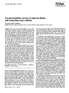
@ /
Fig. 15. A and B: period-doubling bifurcation in ionic model. Effect of increasing maximal conductance of 1s to 4.9 (A) and 5.0 (B) times its standard value. A period-doubling bifurcation occurs for maximal conductance somewhere between 4.9 and 5.0 times its normal value. Note transient alternation in A. Minimum value of At was decreased to 0.008 ms for this simulation to allow AV to remain CO.4 mV. C and Lk period-doubling bifurcation in S-dimensional system. As a parameter is changed, preexisting stable limit cycle (solid closed curve in C) becomes unstable (dashed curve in D) and spawns in its immediate vicinity a stable limit cycle (solid closed curve in D) that has twice the period of original stable limit-cycle oscillation in C.
tial parameters, most noticeably the maximum diastolic potential, in Fig. 15B. Alternation in action potential morphology has previously been described in the SAN at least once, in an experiment in which isoproterenol, which augments 18, was applied (81). As is the case with a homoclinic orbit, a period-doubled orbit can exist only in a system with dimension greater than or equal to three, since in a two-dimensional system the perioddoubled orbit would have to cross itself, thus violating uniqueness of solution. Figure 15C shows a projection of a limit cycle in such a high-dimensional system onto the plane of the paper. This limit cycle undergoes a perioddoubling bifurcation producing a period-doubled orbit (solid curve in Fig. 150). Note that the original orbit is still preserved but has become unstable (dashed curve in Fig. 15D), repelling nearby trajectories onto the stable period-doubled orbit. Other biological oscillators. Although we have concentrated above on the SAN, our proposed classification scheme can be extended to include other cardiac oscillators. For example, the second way of stopping (annihilation) and the associated single-pulse triggering have been described in experiments on depolarized Purkinje fiber (38, 68; see other refs. in Ref. 20), depolarized ventricular muscle (24, 68), aggregates of embryonic atria1 cells (72), the mitral valve (20), and in ionic models of the first three tissues (15, 16, 72, 80). In fact, a bifurcation diagram qualitatively equivalent to that shown in Fig. 6 can be obtained in a reduced twodimensional model of ventricular muscle subjected to a depolarizing bias current (see Fig. 8 of Ref. 80). However, in the full ventricular model (16,80) there is a subcritical rather than a supercritical Hopf bifurcation with depolarizing Ibias, leading to annihilation with the resting potential lying in the plateau, and not the pacemaker, range of potentials (see also Refs. 15, 43).
H1284
BIFURCATIONS
AND SINUS
Patterns of activity resembling some of those shown in Fig. 9 can be seen in atria1 muscle (64, 67), Purkinje fiber (see refs. in Ref. 27), and heart-cell aggregates (49), as well as in ionic models of the latter two tissues (Ref. 27 and J. R. Clay and A. Shrier, personal communication, respectively). However, unlike the case of the SAN, this modeling work shows that the existence of the maintained subthreshold oscillation and the skipped-beat runs, which occur in a more hyperpolarized range of potentials. hinges on the presence of 1h. A mirror image of the third way, in which there are afterpotentials in the plateau range of potentials (“early afterdepolarizations”), can be seen in Purkinje fiber and ventricular muscle and their models (15, 16, 18, 20,80). In that case, the maintained small-amplitude oscillation (“IX oscillation”) lies in the plateau range of potentials, and the membrane becomes quiescent with the membrane potential lying in that range. In one of these modeling studies (16), the existence of chaotic dynamics was claimed. Two reports show that this inversion of the third way (i.e., early afterdepolarizations) can be seen in experiments on the SAN (50, 58). Our classification scheme can be applied to biological oscillators originating from tissues other than the heart. For example, patterns of activity similar to those shown in Fig. 3, which are generated by a single Hopf bifurcation, have been described in the giant axon of the squid (2), as well as in the Hodgkin-Huxley equations that model the squid axon (2). Homoclinic orbits occur in the Hodgkin-Huxley equations when the Nernst potential for K+ is changed in conjunction with injection of 1bias (42). Annihilation has been described experimentally in the squid axon (35), following its theoretical prediction in the Hodgkin-Huxley model (6, 35, 66). Skipped-beat runs [34 (“skip runs”), 451 and a maintained smallamplitude oscillation (45) have been described in neural tissue, as well as in an electronic analogue of the Hodgkin-Huxley equations (28). Skipped-beat runs (22) and a “slow-wave” subthreshold pacemaker oscillation are also commonly seen in smooth muscle. Spatial effects. We have not dealt above with rhythms, such as sinoatrial exit block, intranodal reentry, or atria1 fibrillation, in which the usual more-or-less concentric spread of activation out of the node and into the right atrium (7) is abolished and replaced by a different spatiotemporal organization of the activation sequence. Because propagation is involved, modeling of such rhythms involves simulation of a partial differential equation, and we have confined ourselves in the above to isopotential situations described by systems of ordinary differential equations. In addition, due to the highly complex nature of the coupling between cells in the SAN, it would be difficult to construct such a model in a realistic way, since, among other things, the usual cable-model approach used in such work would not be appropriate (e.g., see Fig. 4 of Ref. 8). Clinical significance. Although injection of a single stimulus can indeed initiate nonsustained reentrant activity in a pathway including the SAN (3), Fig. 5B suggests that activity can also be initiated by a nonreentrant mechanism (see also discussion in chapt. VIII of Ref. 20). Figure 9, B-D, demonstrates a novel way of producing very long interbeat intervals. Should similar
NODE AUTOMATICITY
activity occur in the intact heart, analysis of the electrocardiogram would lead to the diagnosis of sinoatrial pause or exit block, even though the SAN would still be active, generating incrementing subthreshold prepotentials during the pause. Indeed in an arrhythmia that is presently diagnosed as one form of second-degree sinoatria1 exit block (Wenckebach or Mobitz I), the prolonged R-R interval is less than twice the duration of the preceding R-R interval (77); similar rhythms can be seen at values of I bias lying between those used in Fig. 9, A and B. In addition, should similar activity occur in a parasystolic focus [e.g., Purkinje fiber (27)], long interectopic intervals that would be approximately multiples of some basic interval would be manifest on the electrocardiogram. This would be attributed to intermittent exit block from the parasystolic focus, even though block of conduction would not be involved. Nonlinear dynamics. In summary, consideration of a large body of experimental and modeling work in the SAN and other cardiac tissues points to the conclusion that the number of ways in which spontaneous activity in a cardiac oscillator can be stopped or started is quite small. In fact, the presently available experimental evidence indicates that this number might be as small as three in the SAN. It is not yet entirely clear why a particular intervention results in one way and not another. In particular, conditions necessary and sufficient for annihilation to occur are yet to be determined. Because annihilation can be seen in a reduced three-dimensional ( V, f, p) version of the Irisawa-Noma model (33), we anticipate that further investigation of that model will help in sorting out these conditions. Finally, as in the case of periodically stimulated cardiac cells (e.g., see Ref. 29), the above work provides further evidence that concepts drawn from a branch of nonlinear mathematics, bifurcation theory, can be used to form a classification scheme for a variety of behaviors seen in experimental cardiac electrophysiology. NOTE
ADDED
IN PROOF
Annihilation has been reported recently in an isolated SAN cell subjected to a depolarizing bias current (J. M. B. Anumonwo, et al. Circ. Res. 68: 1138-1153, 1991). Annihilation was not seen in the second cell studied, which was not subjected to a bias current.
We thank Dr. E. Doedel for providing us with the AUTO program, Dr. A. Vinet for help with installing AUTO, Dr. J. S. Outerbridge for use of the graphics package and general help with computers, Dr. A. van Ginneken for helpful comments on the manuscript, and Dr. H. Fozzard for directing us to the work of Teorell. We also thank Christine Pamplin and Sandra James for typing the manuscript, A. van Horssen for drafting some of the figures, and Robert Lamarche and Robert Thomson for photographing the figures. M. R. Guevara, as a postdoctoral fellow, was supported by the Canadian Heart Foundation and the Natural Sciences and Engineering Research Council of Canada (1984-86). The study was supported by grants to H. J. Jongsma from the Dutch Organization for Pure Research and to M. R. Guevara from the Medical Research Council of Canada. Present address of H. J. Jongsma: Fysiologisch Laboratorium, Universiteit van Amsterdam, Faculteit der Geneeskunde, Academisch Medisch Centrum, Meibergdreef 15, 1105 Amsterdam, The Netherlands. Address for reprint requests: M. R. Guevara, Dept. of Physiology,
BIFURCATIONS McGill University, 3655 Drummond Canada.
St., Montreal,
AND SINUS NODE
Quebec H3G lY6, 26.
Received 19 September 1990; accepted in final form 27 November 1991.
27 .
REFERENCES R. M., and C. D. Shaw. Dynamics-77~ Geometry of Santa Cruz, CA: Aerial, 1983, pt. l-3. 2. Aihara, K., and G. Matsumoto. Temporally coherent organization and instabilities in squid giant axon. J. Theor. Biol. 95: 6971.
Abraham, Behavior.
720,1982.
Allessie, M. A., sinus node reentry 4. Appelton, E. V., tion-hysteresis in 3.
and F. I. M. Bonke. Direct demonstration of in the rabbit heart. Circ. Res. 44: 557-568,1979. and B. van Der Pol, Jr. On a type of oscillaa simple triode generator. Philos. Mag. 43: l77-
28.
29. 30.
193,1922. 5.
Argoul, F., A. Arneodo, P. Richetti, J. C. Roux, and H. L. Swinney. Chemical chaos: from hints to confirmation. Act. Chem. Res. 20: 436-442,
1987.
Best, E. N. Null space in the Hodgkin-Huxley equations. A critical test. Biophys. J. 27: 87-104, 1979. 7. Bleeker, W. K., A. J. C. Mackaay, M. Masson-P&et, L. N. Bouman, and A. E. Becker. Functional and morphological organization of the rabbit sinus node. Circ. Res. 46: ll-22,198O. 8. Bouman, L. N., J. J. Duivenvoorden, F. F. Bukauskas, and H. J, Jongsma. Anisotropy of electrotonus in the sinoatrial node of the rabbit heart. J. Mol. Cell. Cardiol. 21: 407-418,1989. 9. Bowler, E. The initiation of impulses in cardiac muscle. Am. J. 6.
Physiol.
138: 273-282,
1943.
C. M., and H.-H. Lu. The SinoatriuZ Pacemaker of the Heart. Springfield, IL: Thomas, 1972. 11. Brown, H., J. Kimura, and S. Noble. The relative contributions of various time-dependent membrane currents to pacemaker activity in the sino atria1 node. In: Cardiac Rate and Rhythm, edited by L. N. Bouman and H. J. Jongsma. The Hague: Nijhoff, 1982, p. 10.
Brooks,
54-68.
Brown, H. F., J. Kimura, D. Noble, S. J. Noble, and A. Taupignon. The ionic currents underlying pacemaker activity in rabbit sino-atria1 node: experimental results and computer simulations. Proc. R. Sot. Lond. B Biol. Sci. 222: 329-347, 1984. 13. Brown, H. F., D. Noble, S. J. Noble, and A. I. Taupignon. Relationship between the transient inward current and slow inward currents in the sino-atria1 node of the rabbit. J. Physiol. Land. 370: 299-315,1986. 14. Chandler, W. K., R. Fitzhugh, and K. S. Cole. Theoretical stability properties of a space-clamped axon. Biophys. J. 2: 10512.
Problems
in Ordinary
Differential
Equations
With
Applications.
Pasadena: California Institute of Technology, 1986. (Technical Report, Applied Mathematics) 22. Droogmans, G., and G. Callewaert. Ca++-channel current and its modification by the dihydropyridine agonist BAY k 8644 in isolated smooth muscle cells. Pfluegers Arch. 406: 259-265, 1986. 23. Gaspard, P., R. Kapral, and G. Nicolis. Bifurcation phenomena near homoclinic systems: a two-parameter analysis. J. Stat. Phys.
35: 697-727,1984.
Gilmour, R. F., Jr., J. J. Heger, E. N. Prystowsky, and D. P. Zipes. Cellular electrophysiologic abnormalities of diseased human ventricular myocardium. Am. J. Cardiol. 51: 137-144,1983. 25. Glass, L., and M. C. Mackey. From Clocks to Chaos: The 24.
Rhythms of Life. Princeton, NJ: Princeton Univ. Press, 1988. Glendinning, P., and C. Sparrow. Local and global behaviour near homoclinic orbits. J. Stat. Phys. 35: 645-695, 1984. Guevara, M. R. Afterpotentials and pacemaker oscillations in an ionic model of cardiac Purkinje fibres. In: Temporal Disorder in Human Oscillatory Systems, edited by L. Rensing, U. an der Heiden, and M. C. Mackey. Berlin: Springer, 1987, p. 127-133. Guevara, M. R. Displaced reinjection attractors in an electronic analogue of the membrane of the squid giant axon. In: A Chaotic Hierarchy, edited by M. Klein and G. Baier. Singapore: World Scientific, 1991, p. 153-164. Guevara, M. R., L. Glass, and A. Shrier. Phase locking, perioddoubling bifurcations, and irregular dynamics in periodically stimulated cardiac cells. Science Wash. DC 214: 1350-1353, 1981. Guevara, M. R., and H. J. Jongsma. Phase resetting in a model of sinoatrial nodal membrane: ionic and topological aspects. Am. J. Physiol.
258 (Heart
Circ.
Physiol.
27): H734-H747,
1990.
Guevara, M. R., and A. Shrier. Phase resetting in a model of cardiac Purkinje fiber. Biophys. J. 52: 165-175,1987. 32. Guevara, M. R., A. Shrier, and L. Glass. Phase resetting of spontaneously beating embryonic ventricular heart cell aggregates. 31.
Am.
J. Physiol.
251 (Heart
Circ.
Physiol.
20): H1298-H1305,1986.
Guevara, M. R., A. C. G. van Ginneken, and H. J. Jongsma. Patterns of activity in a reduced ionic model of a cell from the rabbit sinoatrial node. In: Chaos in Biological Systems, edited by H. Degn, A. V. Holden, and L. F. Olsen. London: Plenum, 1987, p. 5-12. 34. Guttman, R., and R. Barnhill. Oscillation and repetitive firing in squid axons. J. Gen. Physiol. 55: 104-118,197O. R., S. Lewis, and J. Rinzel. Control of repetitive 35. Guttman, firing in squid axon membrane as a model for a neuroneoscillator. 33.
J. Physiol.
Land.
305: 377-395,198O.
Irisawa, H., and A. Noma. Pacemaker mechanisms of rabbit sinoatrial node cells. In: Cardiac Rate and Rhythm, edited by L. N. Bouman and H. J. Jongsma. The Hague: Nijhoff, 1982, p. 35-51. 37. Jalife, J., and C. Antzelevitch. Phase resetting and annihilation of pacemaker activity in cardiac tissue. Science Wash. DC 206: 36.
695-697,1979. 38.
Jalife, J., and C. Antzelevitch. Pacemaker annihilation: diagnostic and therapeutic implications. Am. Heart J. 100: 128-130,
39.
Kodama, I., and M. R. Boyett. Regional differences in the electrical activity of the rabbit sinus node. Pfluegers Arch. 404:
40.
Kohlhardt, M., H.-R. Figulla, and 0. Tripathi. The slow membrane channel as the predominant mediator of the excitation process of the sinoatrial pacemaker cell. Basic Res. Curdiol. 71: 17-
41.
Kreitner, D. Electrophysiological study of the two main pacemaker mechanisms in the rabbit sinus node. Cardiouusc. Res. 19:
42.
Labouriau, I. S. Homoclinic and periodic solutions of nerve impulse equations. In: Chaos in Biological Systems, edited by H. Degn, A. V. Holden, and L. F. Olsen. London: Plenum, 1987, p.
1980.
214-226,1985.
127,1962.
15. Chay, T. R., and Y. S. Lee. Impulse responses of automaticity in the Purkinje fiber. Biophys. J. 45: 841-849, 1984. 16. Chay, T. R., and Y. S. Lee. Phase resetting and bifurcation in the ventricular myocardium. Biophys. J. 47: 641-651,1985. 17. Clay, J. R., M. R. Guevara, and A. Shrier. Phase resetting of the rhythmic activity of embryonic heart cell aggregates. Experiment and theory. Biophys. J. 45: 699-714,1984. 18. Cranefield, P. F. Action potentials, afterpotentials and arrhythmias. Circ. Res. 41: 415-423, 1977, P. F. Does spontaneous activity arise from phase 4 19. Cranefield, depolarization or from triggering? In: The Sinus Node, edited by F. I. M. Bonke. The Hague: Nijhoff, 1978, p. 348-356. 20. Cranefield, P. F., and R. S. Aronson. Cardiac Arrhythmias: The Role of Triggered Activity and Other Mechanisms. Mount Kisco, NY: Futura, 1988. 21. Doedel, E. J., and J. P. Kernevez. Software for Continuation
H1285
AUTOMATICITY
26, 1976.
304-318,1985.
105-111.
Landau, M., P. Lorente, D. Michaels, and J. Jalife. Bistabilities and annihilation phenomena in electrophysiological cardiac models. Circ. Res. 66: 1658-1672, 1990. 44. Lipsius, S. L. Electrotonic interactions in delayed propagation and block within the guinea pig SA node. Am. J. Physiol. 245 43.
(Heart
Circ.
Physiol.
14): H7-H16,
1983.
Llinas, R., and Y. Yarom. Oscillatory properties of guinea-pig inferior olivary neurones and their pharmacological modulation: an in vitro study. J. Physiol. Lond. 376: 163-182, 1986. A complex transition sequence 46. Maselko, J., and H. L. Swinney. in the Belousov-Zhabotinskii reaction. Phys. Ser. T9: 35-39,1985. A., F. Conti, F. Dodge, and R. Schor. Subthreshold 47. Mauro, behaviour and phenomenological impedance of the squid giant axon. J. Gen. Physiol. 55: 497-523,197O. J., and M. Morad. Ionic currents responsible for the 48. Maylie, generation of pace-maker current in the rabbit sino-atria1 node. J. 45.
Physiol.
Lond.
355: 215-235,
1984.
McDonald, T. F., and H. G. Sachs. Electrical activity in embryonic heart cell aggregates. PfZuegers Arch. 354: 165-176, 1975. 50. Miyamae, S., and K. Goto. The effects of extracellular calcium removal on sino-atria1 node cells treated with potassium-depleted solutions. Jpn. J. Physiol. 36: 403-409, 1986. 49.
H1286
BIFURCATIONS
AND
SINUS
B. Possible functional significance of a novel type of 1986. cardiac Ca channel. Biomed. Biochim. Acta 45: K37-K45, 52. Noble, D., and S. J. Noble. A model of sino-atria1 node electrical activity based on a modification of the DiFrancesco-Noble (1984) equations. Proc. R. Sot. Lond. B Biol. Sci. 222: 295-304, 1984. 53. Noma, A. Mechanisms underlying cessation of rabbit sinoatrial node pacemaker activity in high potassium solutions. Jpn. J. 51. Nilius,
54. 55.
Physiol. Noma,
26: 619-630, 1976. A., and H. Irisawa. sinoatrial node cell. PfZuegers Noma, A., and H. Irisawa.
Electrogenic Arch.
sodium pump in rabbit
351: 177-182,
1974.
The effect of sodium ion on the initial phase of the sinoatrial pacemaker action potential in rabbits.
Jpn. J. Physiol. A., and 56. Noma,
24: 617-632, H. Irisawa.
membrane potential Physiol. 57. Noma,
1974.
Effects of Na+ and K+ on the resting of the rabbit sinoatrial node cell. Jpn. J.
25: 287-302, 1975. A., M. Morad, and
H. Irisawa. Does the “pacemaker current” generate the diastolic depolarization in the rabbit SA node cells? Pfluegers Arch. 397: 190-194, 1983. 58. Ohba, M. Effects of tonicity on the pacemaker activity of guineapig sino-atria1 node. Jpn. J. Physiot. 36: 1027-1038, 1986. 59.
Oei, H. I., A. C. G. van Ginneken, H. J. Jongsma, and L. N. Bouman. Mechanisms of impulse generation in isolated cells from the rabbit sinoatrial node. J. Mol. Cell. CardioZ. 21: 1137-1149, 1989. 60. Opthof, T., B. de Jonge, M. Masson-Pevet, H. J. Jongsma, and L. N. Bouman. Functional and morphological organization of the cat sinoatrial node. J. Mol. CeZZ. Curdiol. 18: 1015-1031, 1986. 61. Opthof, T., A. C. G. van Ginneken, L. N. Bouman, and H. J. Jongsma. The intrinsic cycle length in small pieces from the rabbit sinoatrial node. J. Mol. Cell Cardiol. 19: 923-934, 1987. 62. Osterrieder, W., D. Pelzer, Q.-F. Yang, and W. Trautwein.
The electrophysiological basis of the bradycardic action of AQA 39 on the sinoatrial node. Naunyn-Schmiedeberg’s Arch. Pharmacol. 317: 233-237,198l. 63. Osterrieder, W.,
Q.-F. Yang, and W. Trautwein. Effects of barium on the membrane currents in the rabbit S-A node. PfZuegers
Arch. 64. Paes
394: 78-84, 1982. de Carvalho, A.
Slow and subliminal responses: their mechanism and relationship to other oscillatory phenomena in cardiac muscle. In: Cardiac Electrophysiology and Arrhythmias, edited by D. P. Zipes and J. Jalife. Orlando, FL: Grune & Stratton, 1985, p. 89-96. 65. Reiner, V. S., and C. Antzelevitch. Phase resetting and annihilation in a mathematical model of sinus node. Am. J. Physiol. 66.
249 (Heart Circ. Physiol. Rinzel, J., and R. N.
18): H1143-H1153, Miller. Numerical
1985.
calculation of stable and unstable periodic solutions to the Hodgkin-Huxley equations.
Math. 67. Rosen,
Biosci. 49: 27-59, 1980. M. R., and A. J. Hordof. atrium. In: The Slow Inward Current
NODE 69.
70.
71.
and
Cardiac
Arrhythmias,
Influence of papaverine on Sanguinetti, M. C., and T. C. West. spontaneous activity of isolated right atria from small mammals. J. Pharmacol. Senami, M.,
Exp. and
Ther. 228: 500-509, 1984. H. Irisawa. Effect of procaine
amide on the membrane currents of the sino-atria1 node cells of rabbits. Jpn. J. Physiol. Seydel, Stability Shrier,
31: 225-236, 1981. R. From Equilibrium to Chaos: Amsterdam: Elsevier, Analysis. A., J. R. Clay, and R. Brochu.
Practical 1988.
Bifurcation
and
Effects of tetrodotoxin on heart cell aggregates. Phase resetting and annihilation of activity. Biophys. J. 58: 623-629, 1990. T. A biophysical analysis of mechano-electrical transduc73. Teorell, tion. In: Principles of Receptor Physiology, edited by W. R. Loewenstein. Berlin: Springer, 1971, vol. 1, p. 291-339. 72.
74. Thompson, and Chaos. 75. Trautwein,
J. M.
T.,
and
H. B. Stewart.
Nonlinear
Dynamics
Chichester, UK: Wiley, 1986. W. Effect of acetylcholine on the S-A node of the heart. In: Cellular Pacemakers, edited by D. 0. Carpenter. New York: Wiley, 1982, vol. 1, p. 127-160. 76. Tuganowski, W., M. Krause, and K. Korczak. The effect of dibutyryl3’5’~yclic AMP on the cardiac pacemaker, arrested with reserpine and cY-methyl-tyrosine. Naunyn-Schmiedeberg’s Arch. Phurmacol. 77. Vaiciulyte,
280: 63-70, 1973. R., and J. Kaukenas.
Clinical and computer diagnosis of the second degree sinoauricular block by the method of rhythmography. In: Electrophysiology and Surgery of Cardiac Arrhythmias, edited by J. Bredikis. Vilnius: Mokslas, 1987, p. 102108.
78. Van Meerwijk, W. P. M., G. de Bruin, A. C. G. van Ginneken, J. van Hartevelt, H. J. Jongsma, E. W. Kruyt, S. S. Scott, and D. L. Ypey. Phase resetting properties of cardiac pacemaker cells. Biophys. J. 83: 613-629, 1984. 79. Victorri, B., A. Vinet, F. A. Roberge, and J.-P. Drouhard.
Numerical integration in the reconstruction of cardiac action potentials using Hodgkin-Huxley-type models. Comp. Biomed. Res. 18: 20-23,1985. A., and 80. Vinet,
F. A. Roberge. A model study of stability and oscillations in the myocardial cell membrane. J. Theor. Biol. 147:
377-412,199O. 81. West, T. C.
Effects of chronotropic influences on subthreshold oscillations in the sino-atria1 node. In: The Specialized Tissues of the Heart, edited by A. Paes de Carvalho, W. C. de Mello, and B. F. Hoffman. Amsterdam: Elsevier, 1961, p. 81-94.
82. Wiggins,
S. Global
Bifurcation
and
Chaos.
Analytical
Methods.
New York: Springer, 1988. 83. Winfree, A. T. Phase control of neural pacemakers. Science Wash. 84.
The slow response in human
edited by D. P. Zipes, J. C. Bailey, and V. Elharrar. The Hague: Nijhoff, 1980, p. 295-308. 68. Rosenthal, J. E., and G. R. Ferrier. Contribution of variable entrance and exit block in protected foci to arrhythmogenesis in 67: l-8, 1983. isolated ventricular tissues. Circulation
AUTOMATICITY
85.
DC 197: 761-763,1977. Winfree, A. T. When Time Dynamics of Electrochemical
Breahs Down: The Three-Dimensional Waves and Cardiac Arrhythmias. Princeton, NJ: Princeton Univ. Press, 1987. Woods, W. T., F. Urthaler, and T. N. James, Electrical activity
in canine sinus node cells during arrest produced by acetylcholine. 86.
J. Mol. Cell. Cardiol. 13: 349-357, 1981. Yanagihara, K., A. Noma, and H. Irisawa.
Reconstruction of sino-atria1 node pacemaker potential based on the voltage clamp experiments. Jpn. J. Physiol. 30: 841-857, 1980.