MATBIO-01086; No of Pages 10 Matrix Biology xxx (2014) xxx–xxx
Contents lists available at ScienceDirect
Matrix Biology journal homepage: www.elsevier.com/locate/matbio
Ionic osmolytes and intracellular calcium regulate tissue production in chondrocytes cultured in a 3D charged hydrogel Nikki L. Farnsworth a, Benjamin E. Mead a, Lorena R. Antunez a, Amy E. Palmer b,c, Stephanie J. Bryant a,c,d,⁎ a
Department of Chemical and Biological Engineering, University of Colorado Boulder, Boulder, CO, United States Department of Chemistry and Biochemistry, University of Colorado Boulder, Boulder, CO, United States BioFrontiers Institute, University of Colorado Boulder, Boulder, CO, United States d Material Science and Engineering Program, University of Colorado Boulder, Boulder, CO, United States b c
a r t i c l e
i n f o
Article history: Received 1 March 2014 Received in revised form 3 August 2014 Accepted 6 August 2014 Available online xxxx Keywords: PEG Osmolarity Chondroitin-sulfate Dynamic load Anabolic activity
a b s t r a c t The goal of this study was to investigate the role of fixed negative charges in regulating cartilage-like tissue production by chondrocytes under static and dynamic three-dimensional culture, and to determine whether intracellular calcium ([Ca2+]i) is involved in mediating this response. Initial experiments using the 3D neutral hydrogel were conducted in static isotonic culture with ionic and non-ionic osmolytes added to the culture medium. Tissue production by bovine chondrocytes with non-ionic osmolytes was 1.9-fold greater than with ionic osmolytes, suggesting that the ionic nature of the osmolyte is an important regulator of tissue production. To investigate fixed negative charges, a 3D culture system containing encapsulated chondrocytes was employed based on a synthetic and neutral hydrogel platform within which negatively charged chondroitin sulfate was incorporated in a controlled manner. Incorporation of negative charges did not affect the mechanical properties of the hydrogel; however, intracellular ion concentration was elevated from the culture medium (330 mOsm) and estimated to be similar to that in ~400 mOsm culture medium. With dynamic loading, GAG synthesis decreased by 26% in neutral hydrogels cultured in 400 mOsm medium, and increased by 26% in charged gels cultured in 330 mOsm. Treatment of chondrocyte-seeded hydrogels with the Ca2+ chelator BAPTA-AM decreased GAG synthesis by 32–46% and was similar among all conditions, suggesting multiple roles for Ca2+ mediated tissue production including with ionic osmolytes. In conclusion, findings from this study suggest that a dynamic ionic environment regulates tissue synthesis and points to [Ca2+]i signaling as a potential mediator. © 2014 Published by Elsevier B.V. This is an open access article under the CC BY-NC-ND license (http://creativecommons.org/licenses/by-nc-nd/3.0/).
1. Introduction Cartilage remodeling and homeostasis in vivo is mediated, in part, by mechanical signals (Grodzinsky et al., 2000). Mechanical forces produce several changes in the local environment surrounding cartilage cells (i.e. chondrocytes) such as fluid flow, movement of mobile ions and cellular deformation. Chondrocytes (cartilage cells) sense these extracellular cues and translate them into intracellular signals that mediate downstream processes through mechanotransduction. Chondrocytes regulate their metabolism in a load-dependent manner (Tsuang et al., 2008), suggesting that mechanical loading may play an important role in transmitting mechanical cues to cells. While progress has been made in elucidating mechanotransduction pathways in chondrocytes, the mechanisms are still not well understood (Ramage et al., 2009). The extracellular matrix of cartilage has two main components: a dense cross-linked collagen matrix, mainly type II collagen; and aggrecan, ⁎ Corresponding author at: 3415 Colorado Avenue, Campus Box 596, Boulder, CO 80309, USA. Tel.: +1 303 735 6714; fax: +1 303 492 4341. E-mail address:
[email protected] (S.J. Bryant).
which is composed of highly negatively charged glycosaminoglycans (GAGs) (Poole, 1986). The negatively charged GAGs in aggrecan give cartilage a high fixed charge density, allow the tissue to imbibe large amounts of water, and give cartilage its ability to withstand large compressive loads (Urban et al., 1993; Mow et al., 1999). These charges also affect the local concentrations of ions, attracting Na+ and K+ from the interstitial fluid (Maroudas and Evans, 1972; Mow et al., 1999; Oswald et al., 2008). This leads to a high osmolarity in cartilage ranging from ~ 350 to 450 mOsm, which is substantially higher than most tissues (Yellowley et al., 2002; Le et al., 2006). During dynamic loading of cartilage, fluid is expelled from the tissue causing dynamic changes in the ionic and osmotic microenvironment (Grodzinsky et al., 2000; Bush and Hall, 2001; Wuertz et al., 2007). These dynamic changes can activate intracellular signaling pathways (Chao et al., 2005) and therefore may be important cues involved in mechanotransduction in chondrocytes. Intracellular calcium ([Ca2+]i) is a dynamic signaling ion that regulates many processes within the cell including gene expression, differentiation, and tissue synthesis (Berridge et al., 2000, 2003). While Ca2+ signaling has not been linked to metabolic changes in the chondrocyte, several studies have shown that osmotic loading stimulates
http://dx.doi.org/10.1016/j.matbio.2014.08.002 0945-053X/© 2014 Published by Elsevier B.V. This is an open access article under the CC BY-NC-ND license (http://creativecommons.org/licenses/by-nc-nd/3.0/).
Please cite this article as: Farnsworth, N.L., et al., Ionic osmolytes and intracellular calcium regulate tissue production in chondrocytes cultured in a 3D charged hydrogel, Matrix Biol. (2014), http://dx.doi.org/10.1016/j.matbio.2014.08.002
2
N.L. Farnsworth et al. / Matrix Biology xxx (2014) xxx–xxx
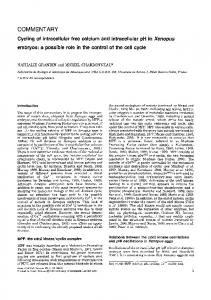
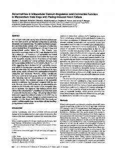
calcium signaling in a frequency-dependent manner (Chao et al., 2006). Studies have also linked extracellular osmotic environment to tissue production in chondrocytes (Hall et al., 1991; Urban, 1994; Villanueva et al., 2009) whereby medium osmolarity that closely matches the osmolarity in cartilage enhanced tissue synthesis over sub- or supraphysiological osmolarity (Urban et al., 1993; Xu et al., 2010). Taken together these prior findings led us to hypothesize that changes in osmolarity, induced by dynamic loading, affect chondrocyte tissue synthesis via Ca2+-mediated events. The overall aim of this study was to investigate the role of fixed negative charges on tissue production in chondrocytes and to establish a link between osmolarity, [Ca2+]i and tissue production. A 3D culture model system was employed based on neutral poly(ethylene glycol) (PEG) hydrogels and incorporated chondroitin sulfate (CHS) (Bryant et al., 2004), the most abundant negatively charged GAG in cartilage. A PEG-based hydrogel was chosen for the ability to control the mechanical properties of the gel and independently copolymerize chondroitin sulfate molecules into the gel to create fixed negative charges that mimic to a certain degree native cartilage. The PEG–CHS co-polymer hydrogel has been previously shown to support encapsulated chondrocytes, although down-regulations in aggrecan and collagen II gene expression over time (Bryant et al., 2005) as well as proteoglycan synthesis (Villanueva et al., 2010) were reported. However, under dynamic loading, the PEG–CHS hydrogel enhanced tissue production by encapsulated chondrocytes implicating the importance of a dynamic environment (Villanueva et al., 2010). This model system was chosen because it enabled decoupling between ionic and osmotic osmolytes on tissue production using neutral PEG-Only hydrogels. On the other hand, PEG–CHS hydrogels, when cultured under dynamic compressive loading, enabled investigation into the effects of dynamic changes in mobile ions in a manner similar to that in cartilage. Studies were also performed to establish whether dynamic changes in osmolarity influenced [Ca2+]i and subsequently whether [Ca2+]i was involved in regulating tissue production in chondrocytes in response to changes in ionic osmolytes under dynamic compressive loading. Findings from this study suggest that a dynamic ionic environment regulates tissue synthesis and point to [Ca2+]i signaling as a potential mediator. 2. Results The aim of this study was to investigate the role of ionic osmolytes in regulating tissue production in chondrocytes under physiological loading. The effect of ionic (salts) and non-ionic (sucrose) osmolytes in regulating tissue production by encapsulated chondrocytes cultured under static culture was first investigated. Standard chondrocyte medium, described in the methods, at 330 mOsm was adjusted with either salts or sucrose to 400, 430, or 460 mOsm. Of the osmolarities investigated, GAG content was highest at 430 mOsm, where for ionic osmolytes it was 50% higher and for non-ionic osmolytes 190% higher than the 330 mOsm condition (Fig. 1A). On average, GAG content with nonionic osmolytes was 1.9-fold higher than hydrogels cultured with ionic osmolytes. Total collagen content was highest at 430 mOsm with ionic osmolytes (by 100% over 330 mOsm) and highest at 460 mOsm with non-ionic osmolytes (by 85% over 330 mOsm) (Fig. 1B). On average, total collagen content was similar with ionic and non-ionic osmolytes. The role of ionic osmolytes in a charged matrix was investigated using co-polymerized PEG–CHS hydrogels (Fig. 2A). The weight percent of the total macromer in solution was adjusted to 15% w/v for a PEG– CHS ratio of 80:20 (by weight) resulting in a compressive modulus of 122 ± 6 kPa, which was statistically similar to that of PEG-Only gels (126 ± 2 kPa). A similar compressive modulus will ensure that the stress and strain imparted on the chondrocytes during loading will be similar in both hydrogels. By varying ion concentration in the culture medium for the PEG-Only hydrogels and comparing intracellular pH (pHi), the osmolarity in the PEG–CHS gels was estimated. The intracellular dye
2′,7′-bis-(-carboxyethyl)-5-(and-6)-carboxyfluorescein acetoxymethyl (BCECF AM) was used to quantify intracellular pH. Initial experiments performed in suspension culture showed that intracellular pH (pHi) decreased with increasing ionic osmolyte concentration (Fig. 2B). Similar results were observed in 3D PEG-Only hydrogels cultured in medium with different ionic osmolyte concentration (Fig. 2C), confirming a correlation with pHi and extracellular osmolarity. To assess changes in osmolarity with the addition of fixed negative charges, intracellular pHi measurements were performed on chondrocytes encapsulated in PEG– CHS hydrogel (Fig. 2C), and the osmolarity in the gel was estimated based on this established correlation. The pHi in chondrocytes encapsulated in PEG–CHS gels cultured in 330 mOsm medium was statistically similar to that of the PEG-Only gels in 400 mOsm medium. To assess the role of dynamic changes in ionic and osmotic environments on tissue production, PEG-Only hydrogels cultured in 400 mOsm media and PEG–CHS hydrogels cultured in 330 mOsm media were subjected to dynamic compressive loading for 6 h (Fig. 3). The total combined amount of newly synthesized proteoglycans that were measured in the gel and in the media per ng DNA for the loaded gels was normalized to the respective free swelling gels (Fig. 3A). No significant differences in DNA content were observed between PEG and PEG–CHS gels under free swelling conditions (p = 0.66) or with loading (p = 0.14) (data not shown). Dynamic loading inhibited proteoglycan synthesis by 26.3% in the PEG-Only gels, while under the same conditions proteoglycan synthesis was stimulated by 26.0% in the PEG–CHS gels. Tissue deposition was further assessed 12 h after the application of loading to allow sufficient time for tissue to be deposited and detected. Aggrecan and collagen II depositions were assessed by immunohistochemistry (Fig. 3B–D). As shown in representative images, semi-quantitative analysis revealed that loading led to a ~40% decrease in aggrecan (Fig. 3C) and collagen (Fig. 3D) depositions in PEG-Only gels; while in PEG–CHS gels, loading did not affect aggrecan deposition (Fig. 3C), but did lead a ~23% decrease in collagen deposition. To investigate the role of [Ca2+]i in the regulation of chondrocytes mediated by changes in ionic osmolytes, [Ca2+]i levels were measured in 2D cultures upon changing the concentration of ionic osmolytes (Fig. 4). Fig. 4A shows representative traces of the change in the Fura2 AM 350/380 ratio upon manipulation of osmolarity, where the Fura-2 ratio reports on changes in [Ca2+]i. When the culture medium was changed from 330 to 400 mOsm with ionic osmolytes, a spike in [Ca2+]i was observed. Similarly, when the culture medium was changed back to 330 mOsm, another spike in [Ca2+]i was observed. After perturbation, the range of fluorescence intensity of the dye was confirmed by treatment with EGTA, which chelates the intra-/extracellular Ca2+ and resulted in the lowest ratio. This was followed by treatment with high levels of calcium chloride, which resulted in the maximum fluorescence signal shown by a large increase in the ratio. Any cell not exhibiting these characteristics with EGTA and calcium chloride was excluded from the analysis. Treatment with 10 μM of the Ca2+ chelator 1,2-bis(2aminophenoxy)ethane-N,N,N′,N′-tetraacetic acid tetrakis(acetoxymethyl ester) (BAPTA-AM) abrogated the [Ca2+]i transients. Representative images of the Fura-2 350/380 ratio for randomly selected chondrocytes exhibiting [Ca2+]i transients in response to dynamic changes in osmolarity are shown in Fig. 4B. Transients were identified by a sharp increase in the 350/380 excitation ratio (~ 10 to 30 s) above 10% of the baseline, followed by a slower decrease in ratio (~30 to 60s). The percentage of cells exhibiting endogenous [Ca2+]i transients in the absence of a stimulus was 40% (Fig. 4C). An initial change in osmolarity from 330 to 400 mOsm with ionic osmolytes resulted in ~ 65% of cells exhibiting [Ca2+]i transients, while a second change in osmolarity from 400 to 330 mOsm resulted in ~ 85% of the chondrocytes responding with [Ca2+]i transients. Given that [Ca2+]i can change upon perturbation of medium osmolarity, we next investigated the role of [Ca2+]i in regulating tissue production upon dynamic loading of a charged matrix. Chondrocytes encapsulated in PEG-Only and PEG–CHS gels were treated with
Please cite this article as: Farnsworth, N.L., et al., Ionic osmolytes and intracellular calcium regulate tissue production in chondrocytes cultured in a 3D charged hydrogel, Matrix Biol. (2014), http://dx.doi.org/10.1016/j.matbio.2014.08.002
N.L. Farnsworth et al. / Matrix Biology xxx (2014) xxx–xxx
A
3
B
Fig. 1. Sulfated glycosaminoglycans (sGAG) (A) and total collagen (B) content in chondrocyte-laden PEG-Only hydrogels and cultured for 7 days in standard 330 mOsm culture medium or medium supplemented with ionic osmolytes (salts) or non-ionic osmolytes (sucrose). Data are normalized to DNA content and day 0 time points for the respective conditions. Data represent the mean of n = 4 samples and error bars represent a 95% confidence interval.
BAPTA-AM to chelate the [Ca2+]i prior to and during dynamic loading (Fig. 5). Regardless of hydrogel formulation or culture condition, proteoglycan synthesis was inhibited by 39% with BAPTA treatment on average (Fig. 5A). Tissue deposition in the BAPTA treated chondrocytes was further assessed 12 h after the application of loading to allow sufficient time for tissue to be deposited and detected. Aggrecan and collagen II deposition was assessed by immunohistochemistry (Fig. 5B, C). Minimal aggrecan deposition was detected in the hydrogels regardless of gel formulation and culture condition with BAPTA treatment, compared with untreated samples. Aggrecan deposition decreased with dynamic loading in PEG-Only gels, while aggrecan deposition was unaffected in PEG–CHS gels with loading (Fig. 5B). Collagen II deposition was detected in the free swelling conditions for the PEG-Only and PEG– CHS gels; however less collagen II deposition was detected in the loaded hydrogels regardless of hydrogel formulation. 3. Discussion Findings from this study confirm that ionic osmolytes influence tissue production by chondrocytes encapsulated in 3D hydrogels through both osmolarity and ionic strength and that dynamic oscillations in ion concentration mediate load-induced tissue production. Our findings also point towards [Ca2 +]i as being a key signaling messenger in the regulation of tissue production by chondrocytes arising from a 3D culture environment, loading, and dynamic changes in ion concentration. These studies support our overarching hypothesis that changes in ion concentration, induced by dynamic loading, affect chondrocyte tissue synthesis likely through Ca2+-mediated events, and therefore, warrant further study into uncovering the mechanisms involved. The first goal of this study was to investigate whether ionic osmolytes influence chondrocytes primarily through osmotic environment or by a combination of the osmotic environment and ionic strength. Our results indicate that both the osmotic environment and ionic strength are capable of influencing tissue deposition in 3D chondrocytes cultures. However, differences were observed between ionic and osmotic environments, suggesting that the ionic strength of the extracellular environment differentially contributes to the regulation of tissue production. This finding is consistent with other studies investigating chondrocytes in 2D (Urban et al., 1993) and more recently 3D (Xu et al., 2010) cultures. While the exact mechanism remains to be elucidated, studies have pointed towards rapid changes in cell volume as one of the main effects resulting from changes in osmolarity (Lang et al., 1998; Xu et al., 2010; Lewis et al., 2011) and activation of membrane ion transporters such as Na+/K+ ATPase in response to
ionic, but not non-ionic, osmolytes (Mobasheri, 1998). Both have been shown to lead to downstream events that alter protein synthesis (Horowitz and Lau, 1988; Lang et al., 1998; Lewis et al., 2011). The trends for collagen production mirrored that for GAG production for the ionic osmolytes, although not for the non-ionic osmolytes. Studies have shown that ionic osmolytes can affect the transcription factor SOX9, which regulates collagen II expression, and also protein production, suggesting a potential regulatory mechanism (Tew et al., 2008). In cartilage, ionic osmolytes arise from a high density of fixed negative charges which attracts ions from the surrounding fluid. Therefore to mimic the ionic environment in cartilage, fixed charges were incorporated into the hydrogel. A change in intracellular pH occurs as water, ions and protons are transported across the cell membrane in response to changes in extracellular osmolarity (Wilkins and Hall, 1995; Xu et al., 2010). Therefore, by varying ion concentration of the culture medium in the neutral PEG hydrogels and comparing intracellular pHi, the osmolarity in the PEG–CHS gels was estimated and determined to be approximately 400 mOsm. It is important to note the fluorescence intensity of the BCECF dye in chondrocytes encapsulated in PEG hydrogels was lower than that of chondrocytes in suspension, leading to a higher estimate of the intracellular pH. We suspect this difference is attributed to light attenuation by the hydrogel. For hydrogel systems with similar osmolarity, as determined by intracellular pH, under dynamic compressive loading, proteoglycan synthesis and aggrecan deposition were dramatically increased for the PEG–CHS gels compared to the neutral hydrogel. Studies with cartilage explants subjected to dynamic compressive loading demonstrated that ions flow in and out of the tissue with loading, as measured by electrical streaming potentials generated from the flow of these ions (Kim et al., 1995). We therefore attribute our findings to dynamic oscillations in the local ion concentration, which appears to have a significant and positive effect on proteoglycan/aggrecan synthesis. This finding agrees well with previous results from our group (Villanueva et al., 2010) and with Chao et al. (2006) who through the use of a microfluidic device showed that aggrecan gene expression was higher under oscillatory changes in ion concentration compared to a single change in ion concentration. Interestingly, collagen II deposition was downregulated with loading, regardless of the culture system. Previous studies have reported that collagen II gene expression steadily decreased over 14 days in culture in PEG–CHS gels under free swelling conditions (Bryant et al., 2005). Other studies have shown that exogenous proteoglycans can inhibit collagen synthesis (Handley et al., 1980). Our studies suggest that mechanical loads (e.g., stress or strain applied to a cell) may be a stronger regulator of collagen II, which over powers any effects of osmolarity
Please cite this article as: Farnsworth, N.L., et al., Ionic osmolytes and intracellular calcium regulate tissue production in chondrocytes cultured in a 3D charged hydrogel, Matrix Biol. (2014), http://dx.doi.org/10.1016/j.matbio.2014.08.002
4
N.L. Farnsworth et al. / Matrix Biology xxx (2014) xxx–xxx
A
C p p=0.01 2
i
I t Intracellular ll l pH H ((pH H)
i
8
Intracellular t ace u a p pH (pH (p )
B
7..5 p