From the Mallinckrodt Institute of Radiology (C.P.D., W.J.P.) and the Department of Neurosurgery and Neurology (W.J.P.), Washington. University School of ...
AJNR Am J Neuroradiol 19:488 –490, March 1998
Isolated Cortical Venous Thrombosis and Ulcerative Colitis Colin P. Derdeyn and William J. Powers signal intensity on T1-weighted sequences was identified on several images superficial to the cortex overlying the hemorrhage. A focal structure of low signal intensity adjacent to the presumed thrombosed cortical vein and at the junction of the vein and the hematoma was also identified, which may have represented flow void from partial recanalization of the thrombosed vessel. An angiogram obtained the day after the MR examination showed a filling defect in a sylvian vein in a location corresponding to the MR abnormality (Fig 1E and F). Dural sinuses filled normally. These findings supported the diagnosis of an isolated cortical venous thrombosis. The patient was started on phenytoin and warfarin sodium. She has continued to experience episodes of tongue quivering but without generalization.
Summary: Aseptic cortical venous thrombosis is rare without concomitant dural sinus thrombosis. Ulcerative colitis is associated with both dural sinus thrombosis and isolated cortical venous thrombosis. We describe a 26-year-old woman with ulcerative colitis who had a spontaneous cerebral hemorrhage. An overlying thrombosed cortical vein was identified on spin-echo MR images and confirmed with angiography. Signal characteristics of thrombosed cortical veins are similar to those described in dural sinus thrombosis.
Isolated cortical venous thrombosis is a rare but recognized cause of parenchymal hemorrhage. Positive identification of the thrombosed cortical vein may be difficult, particularly if the clinical suspicion is low; for this reason, knowledge of associated predisposing conditions is important. Ulcerative colitis is commonly associated with dural sinus thrombosis and less frequently with isolated cortical venous thrombosis (1–3). Two cases of isolated cortical venous thrombosis without dural sinus involvement have been reported in patients with ulcerative colitis (2, 3). We describe a patient with quiescent ulcerative colitis and a parenchymal hematoma consequent to cortical venous thrombosis.
Discussion Aseptic cortical venous thrombosis generally occurs in conjunction with dural sinus thrombosis. Isolated cortical venous thrombosis is unusual. The diagnosis of cortical venous thrombosis often requires a high degree of clinical suspicion, arising from nonspecific symptoms and imaging findings. Symptoms may include focal or generalized seizures and focal neurologic deficits (4). Imaging of the brain parenchyma may reveal focal hemorrhage or edema (5, 6). Findings of venous thrombosis can be subtle and may be missed, particularly if the diagnosis is not suspected. The MR appearance of cortical venous thrombosis has not been well described. Macchi and coworkers (7) observed isointense signal on T1-weighted images and hypointense signal on T2-weighted images in a thrombosed cortical vein studied 48 hours after the onset of clinical symptoms. Follow-up MR imaging 6 days later showed conversion to high signal intensity on both T1- and T2-weighted images. A similar time course of signal intensity changes has been observed in dural sinus thrombosis (7, 8). Isensee and colleagues (9) observed four distinct stages of thrombus evolution in 23 patients with dural sinus thrombosis. They described a subacute pattern appearing between 6 and 15 days after presentation. In the 10 patients studied during this period, the signal characteristics of the thrombus converted from
Case Report A 26-year-old right-handed woman with known ulcerative colitis presented after an apparent tonic-clonic seizure. Her seizure was immediately preceded by an episode of “tongue quivering” and tongue deviation to the right. Ten days before presentation she described an episode of tongue quivering, which resolved after 3 hours. She had difficulty speaking but no other neurologic symptoms. She denied any prior neurologic problems or recent flare-ups of the ulcerative colitis. Her medical history was significant for two deep venous thromboses. Medications on admission included mercaptopurine and dipentum. Specifically, she was not receiving steroids or birth control pills. General medical examination was unremarkable. The neurologic examination was normal, as were electrolyte studies. A magnetic resonance (MR) examination of the brain and a cerebral angiogram were obtained. MR images showed a parenchymal hemorrhage with surrounding edema (Fig 1A–D). A tubular structure with high
Received February 10, 1997; accepted after revision April 8. From the Mallinckrodt Institute of Radiology (C.P.D., W.J.P.) and the Department of Neurosurgery and Neurology (W.J.P.), Washington University School of Medicine, St Louis, Mo. Address reprint requests to Colin P. Derdeyn, MD, Neuroradiology Section, Mallinckrodt Institute of Radiology, 510 S Kingshighway Blvd, St Louis, MO 63110.
© American Society of Neuroradiology 488
AJNR: 19, March 1998
CORTICAL VENOUS THROMBOSIS
489
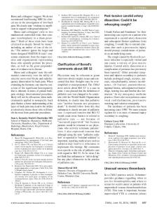
FIG 1. A 26-year-old woman with known ulcerative colitis who presented after a tonic-clonic seizure. A–D, MR images obtained 10 days after the onset of symptoms show a parenchymal hematoma with surrounding edema in the left posterior frontal lobe. The signal intensity of the mass is primarily hyperintense on sequential axial T1-weighted (500/15/1 [repetition time/echo time/excitations]) spin-echo images (A and B) as well as on proton density– and T2-weighted images (2000/20/2 and 2000/80/4, respectively) (C and D), consistent with extracellular methemoglobin. Central hypointensity becomes apparent within the mass on the T2-weighted image, consistent with intracellular methemoglobin. An overlying tubular structure (large arrows) with high signal intensity is seen on the T1- and proton density–weighted images. This hyperintense tubular structure lies on the cortical surface and extends directly to the hematoma (small arrow, A). The signal intensity of this structure is heterogeneous on the corresponding T2weighted image (D) and most likely represents a thrombosed cortical vein with both intracellular and extracellular methemoglobin. Focal hypointensity is present at the junction of this structure with the hematoma, possibly indicating flow void due to partial recanalization. In addition, the focus of low signal intensity adjacent to the high-signal-intensity thrombosed vein (large arrows) seen on all images may represent either flow void due to partial recanalization or an adjacent collateral vessel. Left lateral subtracted conventional angiogram in early (E) and late (F) venous phase. In the early phase, a relative paucity of contrast material is present in the frontal opercular region superior to the arrow, corresponding to the cortex superficial to the hematoma seen on the MR study. There is delayed filling of a sylvian vein, absent on the early image (arrow, E) and poorly opacified on the late image due to a filling defect (arrow, F).
490
DERDEYN
isointense on T1-weighted images and hypointense on T2-weighted images to hyperintense on both T1- and T2-weighted images. The signal change on T2weighted images occurred later than on T1-weighted images. The signal intensity of the thrombus observed in our patient, studied 10 days after the development of clinical symptoms, appears to match this subacute pattern (Fig 1A–D). The list of etiologic causes of intraparechymal hematoma is long and includes cortical venous thrombosis. Isolated cortical venous thrombosis is most often associated with pregnancy and puerperium and with use of oral contraceptives (5, 9, 10). Two cases of ulcerative colitis have also been reported (10). Owing to the often subtle findings in cortical venous thrombosis, it is important for neuroradiologists to be aware of this association. Several authors have reported an association between inflammatory bowel disease (IBD) and thrombotic events (1–3, 10, 11). Talbot and coworkers (11) retrospectively identified 92 thromboembolic complications in 7199 patients with IBD seen during an 11-year period. Nine patients (mean age, 38 years) without other possible etiologic factors had cerebrovascular accidents during active IBD. Ten additional patients (mean age, 58 years) also experienced cerebrovascular events but had other risk factors. In a review of the literature concerning an association between IBD and stroke, Johns (1) found that of the 42 reported patients with IBD and cerebrovascular events, 17 had arterial thromboembolism and 14 had cortical venous or dural sinus thrombosis. Criteria for the diagnosis of underlying arterial or venous abnormalities were variable in this review. Arteriographic confirmation was available in most patients. Some patients were diagnosed on the basis of history and clinical findings (12). Two of the 14 venous thromboses were in isolated cortical veins, and both these patients had ulcerative colitis. The frequency of both arterial and venous thromboembolic complications in patients with ulcerative colitis appears to be greater than in patients with Crohn disease. Twelve of the 17 arterial and 12 of the 14 venous thromboembolic events occurred in patients with ulcerative colitis. Events occurred more frequently in patients with active disease (70%) and in those receiving corticosteroids (59%). Although active disease and steroid use are associated with cerebrovascular events, a large minority of events occur in the absence of either. Yerby and Bailey (13) reported a case of a 28-year-old man presenting with sagittal sinus thrombosis 10 years after panproctocolectomy for ulcerative colitis. He had no other risk factors, such as dehydration or steroid use. Extensive laboratory studies of coagulation factors were all normal. The mechanism by which patients with ulcerative
AJNR: 19, March 1998
colitis may become hypercoagulable is not known. Abnormalities of the coagulation system during active IBD have been reported, including elevated platelet counts, elevated levels of factors V and VIII and fibrinogen, and decreased levels of antithrombin III (14). These levels return to normal, however, after successful treatment of the IBD exacerbation. Fibrinolytic and prothrombotic abnormalities have also been described (15, 16).
Conclusion An association exists between ulcerative colitis and isolated cortical venous thrombosis, in addition to dural sinus thrombosis. The presence of neurologic signs or symptoms or the identification of a parenchymal hematoma in a patient with known ulcerative colitis should prompt a careful search for underlying cortical venous or dural sinus thrombosis. The MR signal characteristics of cortical venous thrombosis are similar to those found in dural sinus thrombosis.
References 1. Johns DR. Cerebrovascular complications of inflammatory bowel disease. Am J Gastroenterol 1991;86:367–370 2. Harrison MJG, Truelove SC. Cerebral venous thrombosis as a complication of ulcerative colitis. Am J Dig Dis 1967;12:1025–1028 3. Schniederman JH, Sharpe JA, Sutton MC. Cerebral and retinal vascular complications of inflammatory bowel disease. Ann Neurol 1979;5:331–337 4. Bousser MG, Chiras J, Bories J, Castaigne P. Cerebral venous thrombosis: a review of 38 cases. Stroke 1985;16:199 –213 5. Buonanno FS, Moody DM, Ball MR, Laster DW. Computed cranial tomographic findings in cerebral sinovenous occlusion. J Comput Assist Tomogr 1978;2:281–290 6. Dorndorf D, Wessel K, Kessler C, Kompf D. Thrombosis of the right vein of Labbe: radiological and clinical findings. Neuroradiology 1993;35:202–204 7. Macchi PJ, Grossman RI, Gomori JM, Goldberg HI, Zimmerman RA, Bilaniuk LT. High field MR imaging of cerebral venous thrombosis. J Comput Assist Tomogr 1986;10:10 –15 8. Chang Y-J, Huang C-C, Wai Y-Y. Isolated cortical venous thrombosis, discrepancy between clinical features and neuroradiologic findings: a case report. Angiology 1995;46:1133–1138 9. Isensee C, Reul J, Thron A. Magnetic resonance imaging of thrombosed dural sinuses. Stroke 1994;25;29 –34 10. Averbach P. Primary cerebral venous thrombosis in young adults: the diverse manifestations of an underrecognized disease. Ann Neurol 1978;3:81– 86 11. Talbot RW, Heppell J, Dozois RR, Beart RW. Vascular complications of inflammatory bowel disease. Mayo Clin Proc 1986;61:140 – 145 12. Mayeux R, Fahn S. Strokes and ulcerative colitis. Neurology 1978; 28:571–574 13. Yerby MS, Bailey GM. Superior sagittal sinus thrombosis 10 years after surgery for ulcerative colitis. Stroke 1980;11:294 –296 14. Lam A, Borda IT, Inwood MJ, Thomson S. Coagulation studies in ulcerative colitis and Crohn’s disease. Gastroenterology 1975;68: 245–251 15. Dejong E, Porte RJ, Knot EAR, et al. Disturbed fibrinolysis in chronic inflammatory bowel disease: a study in blood, plasma, colon mucosa and faeces. Gut 1989;30:188 –194 16. Conlan MG, Haire WD, Burnett DA. Prothrombotic abnormalities in inflammatory bowel disease. Dig Dis Sci 1989;34:1089 –1093