Copyright 2000 by the Genetics Society of America
Isolation and Characterization of HRT1 Using a Genetic Screen for Mutants Unable to Degrade Gic2p in Saccharomyces cerevisiae Marc Blondel,1 Jean-Marc Galan1 and Matthias Peter Swiss Institute for Experimental Cancer Research (ISREC), 1066 Epalinges/VD, Switzerland Manuscript received December 23, 1999 Accepted for publication March 24, 2000 ABSTRACT Skp1p-cullin-F-box (SCF) protein complexes are ubiquitin ligases required for degradation of many regulatory proteins involved in cell cycle progression, morphogenesis, and signal transduction. Using a genetic screen, we have isolated a novel allele of the HRT1/RBX1 gene in budding yeast (hrt1-C81Y). hrt1C81Y mutant cells exhibited an aberrant morphology but were viable at all temperatures. The cells displayed multiple genetic interactions with mutations in known SCF components and were defective for the degradation of several SCF targets including Gic2p, Far1p, Sic1p, and Cln2p. In addition, they also failed to degrade the F-box proteins Grr1p, Cdc4p, and Met30p. Wild-type Hrt1p but not Hrt1p-C81Y was able to bind multiple F-box proteins in an F-box-dependent manner. Hrt1p-C81Y harbors a single mutation in its ring-finger domain, which is conserved in subunits of distinct E3 ligases. Finally, Hrt1p was localized in both nucleus and cytoplasm and despite a short half-life was expressed constitutively throughout the cell cycle. Taken together, these results suggest that Hrt1p is a core subunit of multiple SCF complexes.
P
OST-TRANSLATIONAL modification by ubiquitin targets many proteins for rapid degradation by the 26S proteasome (Hochstrasser 1996). Covalent attachment of ubiquitin to lysine residues of the substrate requires the coordinated action of three classes of enzymes: the E1 ubiquitin-activating enzymes, the E2 ubiquitin-conjugating enzymes, and the E3 ubiquitin ligases (Hershko et al. 1983; Peters 1998). The E1 and E2 enzymes are primarily involved in activating and transferring ubiquitin through high-energy thioester bonds, while E3 enzymes provide specificity of substrate recognition. To date four classes of E3 enzymes have been identified: the HECT (homology to E6-AP carboxyl terminus) family, the ring-finger ligase family, the SCF family, and the anaphase-promoting complex (APC) (Hochstrasser 1996). The HECT domain proteins are the only ligases whose mechanism of action is understood. Mammalian E6-AP (Nuber et al. 1996) and budding yeast Rsp5p (Wang et al. 1999) are members of this class and have been shown to accept activated ubiquitin from E2 enzymes through a transient thiolester formed on a conserved cysteine residue within their HECT domains. It is not known whether non-HECT E3s catalyze substrate ubiquitination via a similar thioester cascade. The members of the ring-finger family all share a small metal binding sequence rich in cysteine residues (Saurin et al. 1996).
Corresponding author: Matthias Peter, ISREC, Chemin des Boveresses 155, 1066 Epalinges/VD, Switzerland. E-mail:
[email protected] 1 These authors contributed equally to this work. Genetics 155: 1033–1044 ( July 2000)
Ring-finger ligases participate in a wide variety of unrelated processes: in budding yeast, Ubr1p, Rad18p, and Hrd1p/Der3p are involved in the N-end rule pathway (Varshavsky 1996), DNA repair (Bailly et al. 1997), and quality control in the ER, respectively (Bordallo and Wolf 1999). In mammals, Cbl is required for ubiquitination of various plasma membrane receptors (Waterman et al. 1999), whereas Mdm2 is involved in the regulation of the tumor suppressor protein p53 (Honda et al. 1997). The APC and SCF are multiprotein complexes discovered because of their roles in the regulation of the cell cycle (Peters 1998): the APC promotes entry into anaphase and exit from mitosis, whereas SCF complexes regulate the G1-S phase transition. In budding yeast, entry into S-phase requires ubiquitin-dependent degradation of the cyclin-dependent kinase inhibitor Sic1p (Schwob et al. 1994), and genetic and biochemical analysis revealed that a high molecular weight complex containing Cdc4p, Cdc34p, Cdc53p, and Skp1p is required for its ubiquitination (Feldman et al. 1997; Skowyra et al. 1997). CDC34 encodes an E2 ubiquitin-conjugating enzyme (Goebl et al. 1988) whereas Cdc4p contains a conserved motif called the F-box, which mediates the interaction with Skp1p (Bai et al. 1996). A large family of proteins contains an F-box motif, and it is thought that these proteins are substrate-specific adaptor subunits that recruit substrates to a core ubiquitination complex composed of Cdc53p, Cdc34p, and Skp1p (Bai et al. 1996; Patton et al. 1998). There is biochemical evidence for the existence of three SCF complexes in yeast, SCFCdc4, SCFGrr1, and SCFMet30. Ubiquitination of
1034
M. Blondel, J.-M. Galan and M. Peter TABLE 1
Genetic interactions between m27 and various SCF mutants
Cross cdc34-1 ⫻ m27 cdc53-1 ⫻ m27 skp1-11 ⫻ m27 skp1-12 ⫻ m27 cdc4-1 ⫻ m27 grr1⌬ ⫻ m27 a
No. of tetrads dissecteda
No. of double mutants expected
No. of double mutants obtained
43 38 35 38 33 41
43 38 35 38 33 41
0 0 5 3 1 0
All experiments were performed at 25⬚.
Cdc6p, Sic1p, and Far1p requires SCFCdc4 (Drury et al. 1997; Henchoz et al. 1997; Skowyra et al. 1997); ubiquitination of the G1-cyclins Cln1p and Cln2p (Barral et al. 1995) and the bud emergence protein Gic2p (Jaquenoud et al. 1998) requires SCFGrr1, whereas degradation of the Cdk-inhibitory kinase Swe1p is dependent on SCFMet30 (Kaiser et al. 1998). Recently, Hrt1p/Rbx1p/Roc1p has been identified as a component associated with SCF complexes in both yeast and mammalian cells (Kamura et al. 1999; Ohta et al. 1999a; Seol et al. 1999; Skowyra et al. 1999). Hrt1p was also identified as a stoichiometric subunit of the von Hippel Lindau (VHL) complex that possibly acts as an E3 ligase (Kamura et al. 1999; Stebbins et al. 1999). Hrt1p contains a ring-finger domain and interacts with Cdc53p, Cdc34p, and at least three different F-box proteins (Kamura et al. 1999; Skowyra et al. 1999). Recon-
stituted Hrt1p-SCF complex greatly stimulates the activity of Cdc34p (Skowyra et al. 1999) and together with an E1 enzyme is able to efficiently ubiquitinate phosphorylated Sic1p and Cln2p in vitro (Skowyra et al. 1999). The closest yeast homologue of Hrt1p is the APC component Apc11p, suggesting that Apc11p and Hrt1p may define a new subclass of ring-finger proteins engaged in various E3 multiprotein complexes. To understand the regulation of SCF complexes, we designed a genetic screen aimed at identifying subunits and regulators of SCFGrr1. At the time we initiated the screen, it was shown that in vitro reconstituted SCFGrr1 was able to specifically bind but not ubiquitinate its targets (Skowyra et al. 1997), suggesting that additional subunits or post-translational modifications were necessary. Gic2p was previously identified because of its role as an effector of the small GTPase Cdc42p in polarization of the actin cytoskeleton at bud emergence (Brown et al. 1997; Chen et al. 1997). After actin polarization has occurred, efficient bud emergence requires degradation of Gic2p by the SCFGrr1 complex (Jaquenoud et al. 1998). As with other SCF substrates, phosphorylation of Gic2p is a prerequisite for its ubiquitin-dependent degradation in vivo (Jaquenoud et al. 1998) most likely by regulating its interaction with the F-box protein Grr1p (Deshaies 1997). Cells with a reduced ability to degrade Gic2p, either because they are defective for SCFGrr1 function or because they fail to phosphorylate Gic2p, are inviable when Gic2p is overexpressed ( Jaquenoud et al. 1998). On the basis of these observations, we isolated mutants unable to grow in the presence of high levels of Gic2p. Here we report the identification and molecular characterization of HRT1.
TABLE 2 Yeast strains Strain K699 JMG11 JMG136 JMG227 YBM200 YBM201 YBM202 JMG230 JMG231 YMT670 YMT740 YMT668 Y552 Y554 YM2957 JMG101 JMG68 JMG205 JMG206 JMG190
Relevant genotype
Source/reference
Wild type gic2::LEU2 his3-1::GAL-GIC2-HIS3 m27 gic2::LEU2 his3-1::GAL-GIC2-HIS3 m27 m27 skp1-11 m27 skp1-12 m27 cdc4-1 cln2::CLN2-HA m27 cln2::CLN2-HA cdc34-2 cdc53-1 cdc4-1 skp1-11 skp1-12 grr1⌬ grr1::GRR1-MYC-TRP1 met30::MET30-HA-His3-MX6 m27 grr1::GRR1-MYC-TRP1 m27 met30::MET30-HA-His3-MX6 hrt1::HRT1-9myc
K. Nasmyth This study This study This study This study This study This study This study This study M. Tyers M. Tyers M. Tyers S. Elledge S. Elledge C. Mann Galan and Peter (1999) Galan and Peter (1999) This study This study Seol et al. (1999)
SCF Components in Budding Yeast MATERIALS AND METHODS Yeast strains, genetic manipulations, and database searches: Genetic interactions between m27 and SCF mutants are shown in Table 1. Yeast strains are described in Table 2. All strains derived from K699 strain: MATa ade2-1 trp1-1 can1-100 leu2-3,112 his311,15 ura3 GAL⫹ psi⫹ ssd1-d2 (W303 background). Standard yeast growth conditions and genetic manipulations were used as described (Guthrie and Fink 1991). Yeast transformations were performed by the lithium acetate procedure (Gietz et al. 1995). Database searches were performed using the SGD (Stanford University) and the NCBI BLAST program (National Institutes of Health). Integration of TRP1 at the HRT1 locus was obtained by transforming m27 ( JMG136) with the plasmid pBM110 linearized with BsmI. EMS mutagenesis and screening: JMG11 cells (1.0 ml) grown to early log phase at 30⬚ were pelleted, washed twice with 0.001 m KPO4 (pH 7.0), and incubated 90 min with 20 l of ethyl methanesulfonate (EMS, Sigma, St. Louis). The reaction was stopped by adding 1 ml of 5% sodium thiosulfate; the cells were then washed three times with YPD medium and plated with a density of 400–500 colonies per plate on selective medium containing 2% raffinose. This treatment usually leads to 20–25% survival. Cells were replica plated on media containing 2% galactose to identify those that fail to grow on galactose as a carbon source. Among ⵑ80,000 colonies tested, 42 showed a reproducible growth defect on galactose plates. Each Gal⫺ mutant clone (numbered m1 to m42) was tested for the inability to grow when Gic2p was overexpressed from the TET-promoter and subsequently backcrossed to a K699 parental strain. Segregation of the Gal⫺ phenotype with the
1035
GAL-GIC2 construct (marked with HIS3) was determined in ⵑ20 complete tetrads for each cross. DNA manipulations: Plasmids are listed in Table 3. Standard procedures were used for recombinant DNA manipulations (Ausubel et al. 1991). To identify the mutation in m27, we isolated genomic DNA from wild-type or m27 cells and amplified the HRT1 locus by PCR. Genomic DNA was prepared as described (Hoffman and Winston 1987). PCR reactions were performed with the Expand polymerase kit as recommended by the manufacturer (Boehringer Mannheim, Indianapolis). Oligonucleotides were synthesized by Genset (Paris). The PCR fragments were subcloned and sequenced on both strands by the ISREC sequencing facility. To express Gic2p from the TET-promoter, GIC2 was amplified by PCR using the primers oTP538 (5⬘-CCTGACAGAATACGCGTTTAAACGAACG-3⬘) and oTP540 (5⬘-GAAAAAAGAATGCGGCCGCGAATGAATG AGAT-3⬘), and the PmeI-NotI fragment was ligated into the vector pCM190 (Gari et al. 1997) to yield plasmid MJ489. Two-hybrid and green fluorescent protein (GFP)-tagged constructs of HRT1 were constructed by PCR using the oligo pairs oTP737 (5⬘-TAGGAATTCATGAGCAACGAAGTTGACAGGA TGG-3⬘) and oTP738 (5⬘-GGGTGCATACAACTCGAGTATGC TATGTATATAAC-3⬘), and oTP739 (5⬘-AAAGGATCCAACAT GAGCAACGAAGTTGACAGGATGG-3⬘) and oTP740 (5⬘-CAT TTTTGAATTCCCTACCGCATCTTGCTAAC-3⬘), respectively. The amplified fragments were digested with EcoRI/XhoI and BamHI/EcoRI, respectively, and cloned into the vectors pEG203 or pJG4-6 for two-hybrid analysis (Gyuris et al. 1993), or pRS314-GFP ( Jaquenoud et al. 1998) for the GFP fusions. The coding sequence of CDC4 was amplified using oligos oTP758 (5⬘-GGCAAAAATTACGCGGGCCCATGGGGTCG TTT-3⬘) and oTP759 (5⬘-GCTTATTCTCTGGCTCGAGTCAT
TABLE 3 Plasmids Plasmid MJ 230 MJ489 JMG14 pYES2-GST-SKP1 pMT1577 pMT1733 pMT914 pDK101 pHY314-3HA-RBX1 pTP613 pTP62 MJ193 pMT184 pUB23-Leu JMG73 JMG74 JMG73 JMG74 JMG32 JMG33 pBM116 JMG85 pMT1297 JMG31 JMG75 JMG76 pBM110
Relevant characteristics GAL-GIC2 HIS3 integrative TET-GIC2 URA3 2 GAL-GRR1-myc URA3 CEN GAL-SKP1-GST URA3 2 CDC53 URA3 2 GAL-CDC34 URA3 2 CDC4 URA3 CEN RBX1 TRP1 CEN GAL-RBX1-HA TRP1 CEN GAL-CLN2 URA3 CEN GAL-FAR1 URA3 CEN GAL-GIC2 URA3 CEN CLN2-HA LEU2 integrative GAL-Ub-leu-GAL URA3 2 pEG203 HRT1 pEG203 HRT1-C81Y pJG4-6 HRT1 pJG4-6 HRT1-C81Y pJG4-6 GRR1 pJG4-6 GRR1-3A pJG4-6 CDC4 pJG4-6 F-Box CDC4 pEG202 CDC53 pJG4-6 SKP1 GAL-HRT1-GFP TRP1 CEN GAL-HRT1-C81Y-GFP TRP1 CEN pRS304 RBX1 integrative
Source/reference M. Jaquenoud This study Galan and Peter (1999) Kishi and Yamao (1998) M. Tyers M. Tyers M. Tyers S. Elledge S. Elledge F. Chang Henchoz et al. (1997) Jaquenoud et al. (1998) M. Tyers Bachmair et al. (1986) This study This study This study This study Galan and Peter (1999) Galan and Peter (1999) This study This study M. Tyers Galan and Peter (1999) This study This study This study
1036
M. Blondel, J.-M. Galan and M. Peter
GGTATTATA-3⬘) and cloned as an XhoI-ApaI fragment into pJG4-6 to yield plasmid pBM116. The F-box of CDC4 (amino acids 268–330) was amplified using oligos oTP766 (5⬘-CAAGG ATGAATTCAATTTAAAGAGGGACCTAATAACGTC-3⬘) and oTP767 (5⬘-GAATTAAAACCCCTCGAGTTTGGGCTCACAA AATTTTCCGATATCAG-3⬘) and cloned as an XhoI-EcoRI fragment into pJG4-6. Two-hybrid vectors for SKP1 and GRR1 have been described previously (Galan and Peter 1999). To integrate TRP1 at the HRT1 locus, the HRT1 coding sequence was cloned into the integrating vector pRS304 (Sikorsky and Hieter 1989) to yield plasmid pBM110. Antibodies, Western blots, and microscopy: Standard procedures were used for yeast cell extract preparation and immunoblotting. Antibodies against actin (Boehringer Mannheim), the HA-epitope (HA11, Babco, Berkeley), and -GAL (Sigma) were used as recommended by the manufacturers. 9E10 antibodies were produced by the ISREC antibody facility, and polyclonal antibodies specific for Cdc34p (Patton et al. 1998), Gic2p ( Jaquenoud et al. 1998), Clb2p and GFP were kindly provided by M. Tyers (Toronto), M. Jaquenoud (ISREC), C. Mann (CEA), and P. Nurse (ICRF), respectively. For microscopy, cells were grown to early log phase in rich medium at 30⬚, fixed with 3.7% formaldehyde, and photographed on a Zeiss axiophot fluorescence microscope with a Photometrics CCD camera. Hrt1p-GFP was visualized using a Chroma GFPII filter (excitation 440–470 nm), photographed with a Photometrics CCD camera, and analyzed with Photoshop 4.0 software (Adobe). Photographs shown are both phase-contrast and fluorescence images of the same cells. Cells expressing Hrt1p-GFP from the inducible GAL promoter were grown to early log phase at 25⬚ in selective media containing raffinose (2% final concentration), at which time galactose was added (2% final concentration) for 6 hr. For 4⬘,6-diamidino-2-phenylindole (DAPI) staining cells were fixed 5 min in EtOH 70% containing DAPI to a final concentration of 2.5 g/ml and then washed twice with PBS. Determination of half-lives: Cultures were grown to early log phase in rich medium at 30⬚ (25⬚ for temperature-sensitive mutants), at which time cycloheximide (CHX; Sigma) was added to a final concentration of 50 g/ml (stock solution: 10 mg/ml). Aliquots were collected at the times indicated, and protein levels were analyzed by immunoblotting with specific antibodies. The half-lives of Hrt1p-HA, Ub-Leu-Gal, and Far1p were determined in strains carrying plasmids allowing regulated expression of these proteins under the control of the inducible GAL1 promoter as described previously (Henchoz et al. 1997). Two-hybrid assays: Two-hybrid assays were performed as described (Ausubel et al. 1991) in the yeast strain W303 containing the LacZ reporter plasmid pSH18.34, using pEG202based plasmids expressing LexA DNA-binding domain fusions and pJG4-5-based plasmids containing fusions to the B42 transcriptional activation domain (Gyuris et al. 1993). Miller Units are averages from three independent experiments with standard deviations. Cell cycle arrest and ␣-factor release experiments: Cells carrying HRT1-myc ( JMG190) were grown to early log phase in rich medium at 25⬚, at which time they were arrested for 5 hr by addition of hydroxyurea (200 mm; Sigma), nocadazole (15 g/ml; Sigma), or ␣-factor (50 g/ml final concentration). Arrest at the appropriate cell cycle stage was monitored microscopically. Cell synchronization experiments by release from ␣-factor arrest were performed as described (Breeden 1997); the synchrony was monitored by fluorescence-activated cell sorting analysis (FACS) and microscopic determination of the budding index. FACS analysis was carried out as described in Epstein and Cross (1992). Briefly, cells were fixed in 70% ethanol, washed, and digested with RNase A for 5 hr.
The DNA was stained with propidium iodide and analyzed on a FACScan (Becton-Dickinson).
RESULTS
Isolation and characterization of the m27 mutant: Wild-type cells overexpressing SCF substrates including Far1p, Sic1p, Cln2p, and Gic2p are viable probably because the ubiquitin-proteasome system maintains low levels of these proteins. In contrast overproduction of these proteins is toxic in cells defective for SCF-dependent ubiquitination (Bai et al. 1996; Deshaies et al. 1997; Henchoz et al. 1997; Jaquenoud et al. 1998). We took advantage of this observation to identify new elements of the machinery required to degrade Gic2p. A strain was constructed that contains GIC2 under control of the strong inducible galactose promoter (GALGIC2) integrated in the genome at the HIS3 locus; the strain also harbors a plasmid expressing Gic2p from the tetracyclin (TET) promoter (MJ489; Gari et al. 1997). This strain was submitted to EMS mutagenesis and screened for colonies (80,000 colonies total) unable to grow on plates containing galactose (GAL promoter on). Each Gal⫺ mutant was subsequently tested for its inability to grow when Gic2p was overexpressed from the TET promoter. All selected mutants (m1 to m42) were backcrossed with a wild-type strain; in most cases the Gal⫺ phenotype did not require the GAL-GIC2 construct, indicating that these mutants fail to use galactose as a carbon source. In contrast, all Gal⫺ spores from
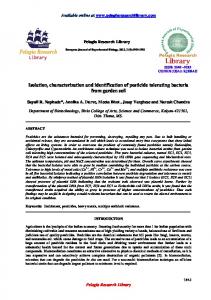
Figure 1.—m27 cells exhibit an aberrant morphology and are unable to grow when Gic2p is overexpressed. (A) JMG11 (wt) and JMG136 (m27) were grown at 30⬚ for 2 days on media containing glucose (GLU; left) or galactose (GAL; right). (B) The morphology of JMG11 (wt) and JMG227 (m27) grown at 30⬚ in rich media containing glucose (GAL-GIC2 not expressed) was analyzed by phase-contrast microscopy after fixation with formaldehyde and shown at 2000⫻ actual size. Note the aberrant morphology of m27 cells.
SCF Components in Budding Yeast
the m27 backcross carried GAL-GIC2, suggesting that m27 cells were unable to grow because of Gic2p overproduction (Figure 1A). Moreover, segregation of the Gal⫺ phenotype strongly suggested that it was caused by mutation(s) of a single locus unlinked to GAL-GIC2 integrated at HIS3 (23 tetrads with 3:1 Gal⫹:Gal⫺ ratio; 3 tetrads with a 2:2 ratio and 4 tetrads with a 4:0 ratio). Heterozygous m27/wt diploid cells were able to grow on galactose, indicating that the m27 mutation is recessive.
1037
Interestingly, m27 cells exhibit an elongated shape (Figure 1B) and a slight growth defect on glucose plates at all temperatures (Figure 1A, data not shown). FACS analysis revealed no major alteration in the DNA content of m27 as compared to wild-type cells (data not shown). These results suggest that m27 cells share some characteristics with mutants defective for the SCFGrr1 function. Genetic interactions between m27 and SCF encoding
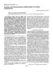
Figure 2.—Genetic interactions between m27 and SCF genes. (A) Overexpression of Cdc34p restores growth of m27 cells overproducing Gic2p. JMG136 cells (m27, GAL-GIC2) were transformed with pGALGRR1myc (GRR1), pYES2GST-SKP1 (SKP1), pMT1577 (CDC53), pMT1733 (CDC34), pMT 914 (CDC4), or a control vector (pRS426; empty vector) and tested for their ability to grow on plates containing glucose (GLU; left) or galactose (GAL; right). JMG11 (wt, GALGIC2) cells transformed with an empty pRS426 vector (Sikorski and Hieter 1989) are included as a positive control. (B and C) JMG227 (m27) was crossed with YMT 670 (cdc34-2), YMT 740 (cdc53-1), YMT 668 (cdc4-1), Y552 (skp1-11), Y554 (skp1-12), and YM2957 (grr1⌬), and ⵑ40 tetrads were analyzed for each cross. We never obtained spores harboring m27 in combination with cdc34-2, cdc53-1, and grr1⌬ (Table 1). Some spores containing m27 in combination with skp1-11, skp112, or cdc4-1 were viable (Table 1) but showed both enhanced thermosensitivity (C and data not shown) and a strongly aberrant morphology (B and data not shown). The genotype of the analyzed mutants is indicated. (D) Overexpression of Cln2p, Gic2p, and Far1p is toxic in m27 cells. Wild-type (K699) or m27 ( JMG227) cells transformed with either pTP613 (GAL-CLN2), pTP62 (GALFAR1), MJ193 (GAL-GIC2), or the empty control vector pRS316 (Sikorski and Hieter 1989) were compared for their ability to grow at 30⬚ on plates containing glucose (GLU, left) or galactose (GAL, right).
1038
M. Blondel, J.-M. Galan and M. Peter
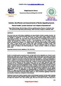
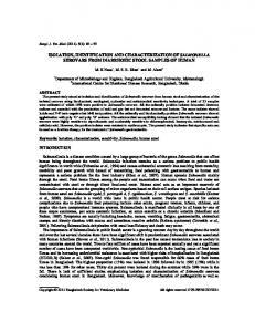
Figure 3.—Determination of the half-lives of multiple SCF substrates in m27 cells. (A) The half-lives of Gic2p (left) or Cln2p-HA (right) were determined by CHX chase experiments (see materials and methods) in either wild-type (left; JMG11 and JMG230, respectively) or m27 cells (right; JMG136 and JMG231, respectively). Cells were grown to mid-log phase at which time CHX was added (time 0) to 50 g/ml final concentration. Aliquots were removed at the times indicated (in minutes) and analyzed by immunoblotting with Gic2p polyclonal antibodies (left) or HA-monoclonal antibodies to detect Cln2p-HA (right). Note that phosphorylated forms of Gic2p (bracket) are accumulating in m27 cells. (B) The half-life of Far1p expressed from the GAL promoter (pTP62) in either wild-type (K699; upper) or m27 cells ( JMG227; lower) was determined by Gal shut-off experiments (see materials and methods). Cells were grown in medium containing galactose (2% final concentration) to mid-log phase at which time glucose was added (2% final concentration) to repress the GAL promoter (time 0). Aliquots were removed at the times indicated (in minutes) and analyzed for the presence of Far1p by immunoblotting. Note that phosphorylated forms of Far1p accumulate in m27 cells. The steady-state levels of Sic1p in K699 (WT), JMG227 (m27), YMT670 (cdc34-2), YMT740 (cdc531), and Y552 (skp1-11) growing in log phase were compared by immunoblotting using polyclonal antibodies against Sic1p. An unspecific protein (*) detected by the Sic1p antibody was used as a loading control. (C) The half-lives of Grr1p-myc (left) and Met30p-HA (right) were determined in wild-type ( JMG101; left) or m27 ( JMG205, right) cells using CHX chase experiments as described above. (D) The half-life of the N-end rule substrate Ub-Leu-Galp (Bachmair et al. 1986) expressed from the GAL promoter (pUB23-Leu) in wild-type (K699) or m27 ( JMG227) cells was determined by Gal shut-off experiments as described above. The lower molecular mass species (*) corresponds to a partially degraded form of Ub-Leu-Galp. Note that m27 cells degraded Ub-Leu-Galp (arrowhead) as efficiently as wild-type cells.
genes: To examine the relationship between m27 and SCF complexes, we explored genetic interactions between m27 and genes encoding known subunits of SCF complexes. Interestingly, overproduction of Cdc34p restored growth of m27 cells overproducing Gic2p on plates containing galactose, whereas overproduction of Skp1p, Cdc4p, Grr1p, and Cdc53p had no major effect (Figure 2A). However, overproduction of Cdc34p did not restore wild-type morphology to m27 cells (data not shown), suggesting that the m27 mutation(s) did not alter the CDC34 locus (see also below). We tested for synthetic lethal interactions between m27 and mutant alleles of SCF genes including cdc34-2, cdc53-1, skp1-11, skp1-12, cdc4-1, and grr1⌬. Among ⵑ40 tetrads dissected for each cross, we never obtained spores containing m27
in combination with cdc34-2, cdc53-1, and grr1⌬, strongly suggesting that those alleles are synthetic lethal (Table 1). Some spores containing m27 in combination with skp1-11, skp1-12, or cdc4-1 were viable, but showed both enhanced thermosensitivity and a grossly aberrant morphology (Figure 2, B and C; data not shown). Thus, m27 exhibits strong genetic interactions with mutations in several genes encoding SCF, suggesting that the protein defective in m27 cells is part of multiple SCF complexes and/or regulates the activity or expression of a common SCF subunit. Several SCF substrates are stabilized in m27 cells: To test the specificity of the m27 mutation we examined whether overexpression of SCF targets other than Gic2p would inhibit its growth. Both Far1p and Cln2p were
SCF Components in Budding Yeast
1039
Figure 4.—Both growth and morphology defects of m27 cells are fully complemented by HRT1. JMG136 (m27, GALGIC2) cells transformed with a low-copy-number plasmid carrying HRT1 (pDK101), or for control an empty vector (pRS314), were plated on media containing glucose (GLU, left) or galactose (GAL, right); JMG11 (wt, GAL-GIC2) cells transformed with an empty vector (pRS314) were included as a positive control. The morphology of the cells was examined by phase-contrast microscopy (bottom). Note that HRT1 on a low-copy plasmid and expressed from its own promoter restores both growth and wild-type morphology of m27 cells.
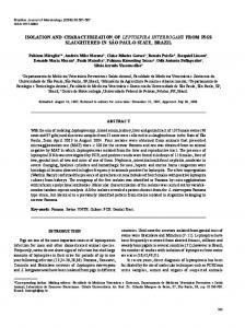
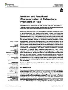
Figure 5.—m27 harbors a single cysteine-to-tyrosine mutation in HRT1 that prevents its interaction with F-box proteins. (A) The sequence of the HRT1 locus from m27 cells harbors a single A-to-T substitution resulting in a mutation of cysteine 81 to a tyrosine (C81Y). C81 is located within the conserved ring-finger domain of Hrt1p but is not predicted to be involved in binding of Zn2⫹. (B) Two-hybrid analysis with wild type (Hrt1p-wt) or the C81Y mutant (Hrt1p-C81Y). Hrt1p-wt or Hrt1p-C81Y fused to an activation- or DNA-binding domain was tested for their ability to interact with Skp1p, Grr1p-wt, Grr1p-3A, Cdc4p-wt, and its F-box motif (amino acids 268– 330). Expression of the -Gal reporter was quantified as described in materials and methods and shown as Miller Units with standard deviation (SD). Note that the C81Y mutation of Hrt1p and the 3A mutation in the F-box of Grr1p both abolish the interaction between these two proteins.
toxic in m27 cells when overproduced from the galactose-inducible promoter (Figure 2D). Because Far1p is targeted for degradation by SCFCdc4, and Gic2p and Cln2p by SCFGrr1, these results suggest that the m27 mutation affects multiple SCF complexes and is thus likely to alter a general factor involved in SCF activity. As expected, Gic2p was stabilized in m27 cells (Figure 3A, left). Since Gic2p accumulated in the hyperphosphorylated form (Jaquenoud et al. 1998), m27 cells are not defective for Gic2p phosphorylation. In addition, we observed a total or partial stabilization of Cln2p and Far1p, respectively, with accumulation of hyperphosphorylated Far1p (Figure 3A, right; Figure 3B, left). Finally, quantitative Western-blot analysis revealed that the steady-state level of Sic1p was moderately increased in m27 cells (Figure 3B, right), suggesting that degradation of Sic1p also depends on m27. We were unable to determine the half-life of Sic1p by cycloheximide chase (data not shown), probably because Cln2p and Cdc4p are both rapidly degraded during these experiments (Figure 3A, Galan and Peter 1999). F-box proteins themselves are degraded in a SCF-dependent manner (Zhou and Howley 1998; Galan and Peter 1999; Rouillon et al. 2000). Indeed, Grr1p is stable in m27 cells (Figure 3C, left) and Met30p is partly stabilized (Figure 3C, right). These results indicate that m27 cells are defective in degradation of multiple SCF targets, raising the possibility that m27 cells are deficient for proteoso-
mal degradation. To exclude this hypothesis we measured the turnover of the fusion protein Ub-Leu-Gal that is degraded by the 26S proteasome by the N-end rule pathway (Bachmair et al. 1986). As shown in Figure 3D, both the kinetics of disappearance of Leu-Gal (arrow head) as well as the appearance of a characteristic degradation product (asterisk) were unaffected in m27 cells, indicating that these cells are not defective for proteosomal degradation. m27 alters the HRT1 locus: To clone the gene defective in m27 cells, we transformed m27 cells harboring GAL-GIC2 with a multicopy library and plated the cells on media containing galactose. One of the plasmids that was able to restore growth contained HRT1. HRT1 under control of its own promoter cloned on a lowcopy plasmid was also able to restore both growth on galactose and wild-type morphology to m27 cells overexpressing Gic2p (Figure 4). To confirm that m27 harbors a mutation in the HRT1 gene, HRT1 marked with TRP1 was integrated at the HRT1 locus of wild-type cells, which were then crossed to m27 cells. Out of 30 complete tetrads, all m27 cells were not able to grow on plates lacking tryptophan (data not shown), demonstrating that m27 is indeed an allele of HRT1. We found that HRT1 from m27 cells bore a single A-to-T substitution changing cysteine 81 to tyrosine (C81Y; Figure 5A). Cysteine 81 is located within the ring-finger domain of Hrt1p (Saurin et al. 1996), but is not part of the con-
1040
M. Blondel, J.-M. Galan and M. Peter
Figure 6.—Hrt1p is unstable but constitutively expressed throughout the cell cycle. (A) The half-lives of Hrt1p-HA (left) and Hrt1p-myc (right) were determined in wild-type cells (K699) by Gal shut-off or by CHX chase experiments as described in materials and methods. Cdc34p was used as a loading control (bottom). Wild-type cells (K699) transformed with pHY314-3HA-RBX1 were grown until early log phase in selective medium containing 2% raffinose as a carbon source. At time 0 the culture was divided and treated with glucose (Gal-shut off) or with CHX (CHX chase); aliquots were removed at the times indicated (in minutes) and analyzed by immunoblotting with HAmonoclonal antibodies (left). JMG190 (hrt1::HRT1-9myc) cells were grown at 25⬚ to early log phase and treated with CHX at time 0; samples were analyzed by immunoblotting with 9E10 antibodies after the times indicated (in minutes) as described above. Note that regardless of the experimental protocol or epitope tag, Hrt1p is unstable with a half-life of ⵑ30 min. (B) JMG190 (hrt1:: HRT1-9myc) cells were released from an ␣-factor block (time 0). Aliquots were removed at the times indicated (minutes) and analyzed by immunoblotting for the levels of Hrt1p-myc (top), Clb2p (middle), and Cdc34p (bottom). An extract prepared from exponentially growing JMG190 cells is shown in the left lane (“as”). BE marks the time of bud emergence as determined microscopically. The synchrony of the cells was monitored by FACS analysis (bottom). The steady-state levels of Hrt1p-myc were determined in JMG190 cells arrested in G1 by ␣-factor (␣F), in S-phase by HU, and in mitosis by Noc. An extract prepared from exponentially growing JMG190 cells is shown in the left lane (“as”). Note that the level of Hrt1p-myc remains constant throughout the cell cycle.
served cysteine residues predicted to bind Zn2⫹ (Figure 5A). One of the previously identified temperature-sensitive HRT1 mutations (rbx1-1, Skowyra et al. 1999) also affects this residue (cysteine 81 and lysine 72 are both substituted by arginine residues). Hrt1p-C81Y is defective for binding to the F-box proteins Grr1p and Cdc4p: We compared the ability of Hrt1p-WT and Hrt1p-C81Y to interact with SCF components by two-hybrid assay. Hrt1p-WT interacted significantly with the F-box proteins Grr1p and Cdc4p (Figure 5B), consistent with previous reports (Kamura et al. 1999; Skowyra et al. 1999). The C81Y mutation severely decreased binding of Hrt1p to Grr1p, suggesting that an intact ring-finger domain is required for binding to F-box proteins. Although Hrt1p-C81Y was able to degrade Sic1p in vivo, its interaction with Cdc4p was similarly impaired. These results suggest that Hrt1pC81Y is defective for interaction with multiple F-box proteins. As the F-box partners of Hrt1p share only the F-box motif, it is tempting to speculate that Hrt1p binds to the F-box. In fact, we found that a GRR1 mutant
harboring three substitutions in conserved residues in the F-box motif (Grr1p-3A) was unable to interact with Hrt1p. As Grr1p-3A was already shown to be defective in Skp1p binding (Galan and Peter 1999), our data support a model in which Hrt1p competes with Skp1p for binding to F-box proteins. Indeed, overexpression of Skp1p significantly decreased the interaction between Hrt1p and Cdc4p in two-hybrid assays (data not shown). Furthermore, we were unable to detect an interaction between Skp1p and Hrt1p (data not shown), and these two proteins also fail to bind in direct assays (Seol et al. 1999; Skowyra et al. 1999). However, the F-box motif alone (amino acids 268–330 of Cdc4p) was unable to interact with Hrt1p, although it was able to bind Skp1p efficiently (Figure 5B). Thus, binding of Hrt1p to F-box proteins depends on an intact F-box but may also require additional sequences. Hrt1p is unstable but its steady-state levels do not change during the cell cycle: Recent studies showed that the core components of SCF complexes and Cdc34p are stable whereas F-box proteins are constitutively de-
SCF Components in Budding Yeast
graded by an SCF-dependent mechanism (Zhou and Howley 1998; Galan and Peter 1999; Mathias et al. 1999; Rouillon et al. 2000). To determine the stability of Hrt1p, we measured the turnover of epitope-tagged Hrt1p either by galactose shutoff experiments or CHX chase (Figure 6A). Regardless of the experimental protocol, Hrt1p was degraded with a half-life of ⵑ30 min (Figure 6A). To determine the steady-state levels of Hrt1p through the cell cycle, we used a strain with the endogenous HRT1 locus replaced with a myc-tagged version. Cells were synchronized by an ␣-factor block/ release protocol (Figure 6B) and the levels of Hrt1pmyc, Clb2p, and Cdc34p were analyzed by immunoblotting (Figure 6, top). The synchrony of the cells was monitored by the expression of endogenous Clb2p and FACS analysis (bottom). We found that the protein levels of Hrt1p were constant throughout the cell cycle. Consistent with these results, Hrt1p-myc levels remained unchanged in cells arrested in G1 by ␣-factor (␣F), in S-phase by hydroxyurea (HU), or in mitosis by nocodazole (Noc; Figure 6B, right). These data support the notion that the expression and degradation of Hrt1p is not cell cycle regulated. Hrt1p is localized in the nucleus and the cytoplasm: To determine the subcellular localization of Hrt1p, we fused wild type and the C81Y mutant of Hrt1p to the GFP. Although both proteins were expressed at comparable levels (Figure 7B), expression of wild-type Hrt1pGFP but not Hrt1p-C81Y-GFP was able to complement the growth and morphology defects of m27 cells overexpressing Gic2p (Figure 7A), suggesting that the fusion protein is functional in vivo. Hrt1p-GFP was predominantly nuclear with a diffuse cytoplasmic staining (Figure 7C), but was excluded from large round structures that likely correspond to the vacuole (Figure 7D). This localization was not influenced by the cell cycle stage or by treatment with pheromones (data not shown). Finally, the localization of Hrt1p-C81Y-GFP was identical to wild-type Hrt1p-GFP (data not shown), suggesting that assembly of Hrt1p into functional SCF complexes is not required for its proper subcellular localization. DISCUSSION
Hrt1p is an essential subunit of multiple SCF complexes: We have identified Hrt1p as an additional subunit of multiple SCF complexes. HRT1 is an essential gene and the hrt1-C81Y allele exhibited strong genetic interactions with several SCF components. It has been suggested that Hrt1p may be required to recruit Cdc34p into the SCF complex by bridging or stabilizing the interaction between Cdc34p and Cdc53p (Skowyra et al. 1999). Supporting this idea, we observed the strongest genetic interactions with the E2 enzymes Cdc34p and Cdc53p: hrt1-C81Y was synthetic lethal with cdc34-2 and cdc53-1, and conversely, overexpression of Cdc34p was able to suppress the growth defect of hrt1-C81Y cells
1041
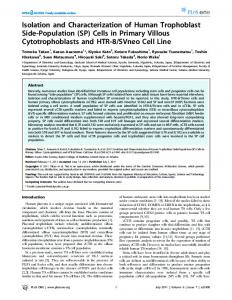
Figure 7.—Hrt1p-GFP is localized in the nucleus and the cytoplasm. (A) JMG136 (m27, GAL-GIC2) cells transformed with pDK101 (HRT1), JMG75 (HRT1-GFP), JMG76 (HRT1C81Y-GFP), or the pRS314-GFP control vector were plated on medium containing glucose (GLU, left) or galactose (GAL, right). Note that expression of Hrt1p-GFP but not Hrt1pC81Y-GFP restores growth of JMG136 cells on galactose plates. (B) The expression of Hrt1p-GFP and Hrt1p-C81Y-GFP was analyzed by immunoblotting with polyclonal antibodies against GFP; cells with an empty vector were used as a control. Note that wild type and the C81Y mutant form of Hrt1p are present at similar levels. (C and D) Wild-type cells (K699) transformed with JMG75 (GAL-HRT1-GFP) were grown to early log phase at 25⬚ in selective media containing raffinose (2% final concentration), at which time galactose was added (2% final concentration) for 6 hr. Cells were fixed, incubated with DAPI as described in materials and methods, and analyzed by fluorescence microscopy (C). Cells in D were neither fixed nor incubated with DAPI. Note that Hrt1p-GFP is localized in both the nucleus and the cytoplasm.
overexpressing Gic2p. Finally, purified Cdc34p is able to bind Hrt1p in vitro and copurifies in stoichiometric amounts in Cdc53p immunoprecipiates (Seol et al. 1999). This latter interaction requires the conserved C-terminal cullin box (W. Krek, personal communication). Finally, as reported here and elsewhere, Hrt1p interacts with at least three different F-box proteins (Kamura et al. 1999; Skowyra et al. 1999). The Hrt1p-
1042
M. Blondel, J.-M. Galan and M. Peter
C81Y mutant protein was defective for interaction with both Cdc4p and Grr1p, although hrt1-C81Y cells are viable and thus must be able to assemble sufficient SCFCdc4 to degrade Sic1p in vivo. An intact F-box is required but not sufficient for interacting with Hrt1p, suggesting that sequences outside the motif are also involved in binding. Alternatively, it is possible that efficient binding of Hrt1p requires an assembled core complex, although Hrt1p makes independent contacts with several subunits. Hrt1p is expressed at constitutive levels throughout the cell cycle and is localized in both nucleus and cytoplasm. The localization of SCF components is poorly documented. The F-box proteins Cdc4p and Met30p have been shown to be nuclear (Choi et al. 1990; Rouillon et al. 2000), while Grr1p is found in both the nucleus and the cytoplasm (M. Blondel, J.-M. Galan and M. Peter, unpublished results). To date nothing is known about the localization of Cdc53p and Skp1p in yeast, but it was recently reported that mammalian Skp1p and CUL1 are localized in the nucleus and the cytoplasm. In addition, Skp1p is associated with centrosomes (Freed et al. 1999; Gstaiger et al. 1999). SCF targets have various localizations in yeast: Gcn4p is nuclear, Gic2p is localized at the site of bud emergence, and Far1p shuttles between nucleus and cytoplasm (Ma and Ptashne 1987; Brown et al. 1997; Chen et al. 1997; Blondel et al. 1999). Hrt1p is present in both cytoplasm and nucleus throughout the cell cycle, consistent with the notion that it is required to degrade both targets of Cdc4p and Grr1p. It is thus tempting to speculate that core subunits that function in several SCF complexes (i.e., Hrt1p, Skp1p, and cullins) are localized throughout the cell, whereas F-box proteins are found in subcompartments where they are specifically required to degrade their substrates. Supporting this model we have recently found that ectopic expression of Cdc4p in the cytoplasm is sufficient to destabilize a cytoplasmic mutant Far1p that is stable in wild-type cells (M. Blondel, J.-M. Galan and M. Peter, unpublished results). Thus, the localization of F-box proteins may be an important determinant to spatially control degradation of substrates. What is the role of ring-finger proteins in E3 ligases? A large number of ring-finger-containing proteins promote E2-dependent ubiquitination in vitro (Lorick et al. 1999), suggesting that many E3 ligases may contain a subunit with a ring-finger domain (Saurin et al. 1996). For example, ring-finger-containing subunits are present in the APC (APC11, Zachariae et al. 1998), as well as the VHL-complex that functions as a ubiquitin ligase to ubiquitinate still unknown substrates (Kamura et al. 1999; Lisztwan et al. 1999). Hrt1p-m27 contained a single substitution of cysteine 81 to tyrosine, which is located within the ring-finger domain (Figure 5A), demonstrating that the ring-finger motif is essential for the function of Hrt1p in vivo. Similarly, Skowyra et al. (1999) have generated a thermosensitive allele of HRT1
(rbx1-1) in which cysteine 81 and lysine 72 are both substituted by arginine residues (Skowyra et al. 1999). Cysteine 81 of Hrt1p is conserved in several, but not all, ring-finger E3 proteins including Apc11p and the mammalian kf-1 (Lorick et al. 1999). Hrt1p-C81Y was defective for its interaction with F-box proteins, suggesting that the ring-finger domain could function as a protein-protein interaction motif. C81 may directly bind Zn2⫹, although it is not one of the conserved residues within the ring-finger consensus (Saurin et al. 1996), or alternatively, C81 could be important for the correct folding of the ring-finger motif. How do these ring-finger-containing subunits function together with E2s to catalyze ubiquitination? The mammalian E6-AP and Rsp5 proteins accept activated ubiquitin from the E2 enzyme through a transient thioester formed on a cysteine residue within their HECT domains (Huibregtse et al. 1995; Wang et al. 1999). Similarly, the ring-finger oncoprotein Mdm2 forms a thioester bond with ubiquitin on a conserved cysteine residue (Honda et al. 1997), and several ring-finger proteins have recently been shown to be modified by ubiquitin in vitro (Joazeiro et al. 1999; Lorick et al. 1999). Finally, the ring finger of Ubr1p is required for the ubiquitination of N-end rule substrates, but is not involved in binding of the E2 enzyme or the target protein (Xie and Varshavsky 1999). The formation of a thioester between an E2 and a ring-finger protein may therefore be required for many ubiquitination reactions. However, Hrt1p and several mammalian ring-finger proteins are resistant to incubation with high concentration of alkylating agents that fully inactivate HECT E3s by modifying cysteine residues, suggesting that these proteins promote ubiquitination of substrates by a mechanism that does not involve an E3-ubiquitin thioester intermediate (Lorick et al. 1999; Seol et al. 1999). Alternatively, ubiquitin may be ligated onto the ring-finger proteins in a substrate reaction. We found that Hrt1p is unstable (t1/2 ⵑ 30 min), suggesting that the turnover of Hrt1p may be regulated by ubiquitindependent degradation. Indeed, mammalian Roc proteins are degraded by a proteasome-dependent pathway and are stabilized by association with cullin partners (Ohta et al. 1999b). Are there additional subunits of SCF complexes? While our genetic screen was successful in isolating Hrt1p, we did not find mutations in all known SCF subunits or other proteins involved in degradation of Gic2p. For example, we did not identify mutations in SGT1, an essential gene required to degrade Sic1p in vivo and also for ubiquitination of Cln1p in vitro (Kitagawa et al. 1999). Because Sgt1p is involved in degrading substrates of both SCFCdc4 and SCFGrr1, we expect that Gic2p is similarly stabilized in sgt1 mutant cells. In addition, we failed to identify the protein kinase that phosphorylates Gic2p, thereby targeting it for degradation. Gel filtration experiments demonstrated that
SCF Components in Budding Yeast
Grr1p migrates in complexes of ⵑ350 kD (data not shown), suggesting that SCFGrr1 may contain additional subunits. Our screen was based on the identification of cells unable to grow on media containing galactose, which makes it difficult to screen large numbers of colonies. In addition, it may be difficult to isolate SCF components that could be involved in sugar metabolism such as GRR1 (Flick and Johnston 1991). It is also possible that some of the missing SCF subunits may not be essential for Gic2p degradation or that they may be functionally redundant with other proteins. The identification of such components will thus most likely require biochemical methods such as purification of the SCFGrr1 complex or two-hybrid interaction screens using known SCF subunits as baits. We thank M. Tyers, T. Kishi, M. Jaquenoud, E. Herrero, S. Elledge, R. Deshaies, C. Marchal, P. Nurse, and C. Mann for providing antibodies, plasmids, and strains. We are grateful to W. Krek for helpful suggestions, N. Perrinjaquet and P. Page´ for excellent technical assistance, Marco Vazquez for help during early stages of this work, members of the laboratory for discussion, and R. Iggo and V. Simanis for critical reading of the manuscript. M.B. and J.-M.G. are supported by European Molecular Biology Organization postdoctoral fellowships; M.P. is supported by the Swiss National Science Foundation, the Swiss Cancer League, and a Helmut Horten Incentive Award.
LITERATURE CITED Ausubel, F. M., R. Brent, R. E. Kingston, D. D. Moore, J. G. Seidman et al., 1991 Current Protocols in Molecular Biology. Greene Publishing Associates and Wiley-Interscience, New York. Bachmair, A., D. Finley and A. Varshavsky, 1986 In vivo half-life of a protein is a function of its amino-terminal residue. Science 234: 179–186. Bai, C., P. Sen, K. Hofmann, L. Ma, M. Goebl et al., 1996 SKP1 connects cell cycle regulators to the ubiquitin proteolysis machinery through a novel motif, the F-box. Cell 86: 263–274. Bailly, V., S. Lauder, S. Prakash and L. Prakash, 1997 Yeast DNA repair proteins Rad6 and Rad18 form a heterodimer that has ubiquitin conjugating, DNA binding, and ATP hydrolytic activities. J. Biol. Chem. 272: 23360–23365. Barral, Y., S. Jentsch and C. Mann, 1995 G1 cyclin turnover and nutrient uptake are controlled by a common pathway in yeast. Genes Dev. 9: 399–409. Blondel, M., P. M. Alepuz, L. S. Huang, S. Shaham, G. Ammerer et al., 1999 Nuclear export of Far1p in response to pheromones requires the export receptor Msn5p/Ste21p. Genes Dev. 13: 2284–2300. Bordallo, J., and D. H. Wolf, 1999 A RING-H2 finger motif is essential for the function of Der3/Hrd1 in endoplasmic reticulum associated protein degradation in the yeast Saccharomyces cerevisiae. FEBS Lett. 448: 244–248. Breeden, L. L., 1997 Alpha-factor synchronization of budding yeast. Methods Enzymol. 283: 332–341. Brown, J. L., M. Jaquenoud, M. P. Gulli, J. Chant and M. Peter, 1997 Novel Cdc42-binding proteins Gic1 and Gic2 control cell polarity in yeast. Genes Dev. 11: 2972–2982. Chen, G. C., Y. J. Kim and C. S. Chan, 1997 The Cdc42 GTPaseassociated proteins Gic1 and Gic2 are required for polarized cell growth in Saccharomyces cerevisiae. Genes Dev. 11: 2958–2971. Choi, W. J., M. W. Clark, J. X. Chen and A. Y. Jong, 1990 The CDC4 gene product is associated with the yeast nuclear skeleton. Biochem. Biophys. Res. Commun. 172: 1324–1330. Deshaies, R. J., 1997 Phosphorylation and proteolysis: partners in the regulation of cell division in budding yeast. Curr. Opin. Genet. Dev. 7: 7–16. Deshaies, R. J., V. Chau and M. Kirschner, 1997 Ubiquitination
1043
of the G1 cyclin Cln2p by a Cdc34p-dependent pathway. EMBO J. 14: 303–312. Drury, L. S., G. Perkins and J. F. Diffley, 1997 The Cdc4/34/53 pathway targets Cdc6p for proteolysis in budding yeast. EMBO J. 16: 5966–5976. Epstein, C. B., and F. R. Cross, 1992 CLB5: a novel B cyclin from budding yeast with a role in S phase. Genes Dev. 6: 1695–1706. Feldman, R. M., C. C. Correll, K. B. Kaplan and R. J. Deshaies, 1997 A complex of Cdc4p, Skp1p, and Cdc53p/cullin catalyzes ubiquitination of the phosphorylated CDK inhibitor Sic1p. Cell 91: 221–230. Flick, J. S., and M. Johnston, 1991 GRR1 of Saccharomyces cerevisiae is required for glucose repression and encodes a protein with leucine-rich repeats. Mol. Cell. Biol. 11: 5101–5112. Freed, E., K. R. Lacey, P. Huie, S. A. Lyapina, R. J. Deshaies et al., 1999 Components of an SCF ubiquitin ligase localize to the centrosome and regulate the centrosome duplication cycle. Genes Dev. 13: 2242–2257. Galan, J. M., and M. Peter, 1999 Ubiquitin-dependent degradation of multiple F-box proteins by an autocatalytic mechanism. Proc. Natl. Acad. Sci. USA 96: 9124–9129. Gari, E., L. Piedrafita, M. Aldea and E. Herrero, 1997 A set of vectors with a tetracyclin-regulatable promoter system for modulated gene expression in Saccharomyces cerevisiae. Yeast 13: 837–848. Gietz, R. D., R. H. Schiestl, A. R. Willems and R. A. Woods, 1995 Studies on the transformation of intact yeast cells by the LiAc/ SS-DNA/PEG procedure. Yeast 11: 355–360. Goebl, M. G., J. Yochem, S. Jentsch, J. P. McGrath, A. Varshavsky et al., 1988 The yeast cell cycle gene CDC34 encodes a ubiquitinconjugating enzyme. Science 241: 1331–1335. Gstaiger, M., A. Marti and W. Krek, 1999 Association of human SCF(SKP2) subunit p19(SKP1) with interphase centrosomes and mitotic spindle poles. Exp. Cell Res. 247: 554–562. Guthrie, C., and G. R. Fink, 1991 Guide to Yeast Genetics and Molecular Biology. Academic Press Inc., San Diego. Gyuris, J., E. Golemis, H. Chertkov and R. Brent, 1993 Cdi1, a human G1 and S phase protein phosphatase that associates with Cdk2. Cell 75: 791–803. Henchoz, S., Y. Chi, B. Catarin, I. Herskowitz, R. J. Deshaies et al., 1997 Phosphorylation- and ubiquitin-dependent degradation of the cyclin-dependent kinase inhibitor Far1p in budding yeast. Genes Dev. 11: 3046–3060. Hershko, A., H. Heller, S. Elias and A. Ciechanover, 1983 Components of ubiquitin-protein ligase system. Resolution, affinity purification, and role in protein breakdown. J. Biol. Chem. 258: 8206–8214. Hochstrasser, M., 1996 Ubiquitin-dependent protein degradation, pp. 405–439 in Annual Reviews of Genetics, edited by Annual Reviews Inc., Palo Alto, CA. Hoffman, C. S., and F. Winston, 1987 A ten-minute DNA preparation from yeast efficiently releases autonomous plasmids for transformation of Escherichia coli. Gene 57: 267–272. Honda, R., H. Tanaka and H. Yasuda, 1997 Oncoprotein MDM2 is a ubiquitin ligase E3 for tumor suppressor p53. FEBS Lett. 420: 25–27. Huibregtse, J. M., M. Scheffner, S. Beaudenon and P. M. Howley, 1995 A family of proteins structurally and functionally related to the E6-AP ubiquitin-protein ligase. Proc. Natl. Acad. Sci. USA 92: 2563–2567. Jaquenoud, M., M. P. Gulli, K. Peter and M. Peter, 1998 The Cdc42p effector Gic2p is targeted for ubiquitin-dependent degradation by the SCFGrr1 complex. EMBO J. 17: 5360–5373. Joazeiro, C. A., S. S. Wing, H. Huang, J. D. Leverson, T. Hunter et al., 1999 The tyrosine kinase negative regulator c-Cbl as a RING-type, E2-dependent ubiquitin-protein ligase. Science 286: 309–312. Kaiser, P., R. A. Sia, E. G. Bardes, D. J. Lew and S. I. Reed, 1998 Cdc34 and the F-box protein Met30 are required for degradation of the Cdk-inhibitory kinase Swe1. Genes Dev. 12: 2587–2597. Kamura, T., D. M. Koepp, M. N. Conrad, D. Skowyra, R. J. Moreland et al., 1999 Rbx1, a component of the VHL tumor suppressor complex and SCF ubiquitin ligase. Science 284: 657– 661. Kishi, T., and F. Yamao, 1998 An essential function of Grr1 for the degradation of Cln2 is to act as a binding core that links Cln2 to Skp1. J. Cell Sci. 111: 3655–3661.
1044
M. Blondel, J.-M. Galan and M. Peter
Kitagawa, K., D. Skowyra, S. J. Elledge, J. W. Harper and P. Hieter, 1999 SGT1 encodes an essential component of the yeast kinetochore assembly pathway and a novel subunit of the SCF ubiquitin ligase complex. Mol. Cell 4: 21–33. Lisztwan, J., G. Imbert, C. Wirbelauer, C. M. Gstaiger and W. Krek, 1999 The von Hippel-Lindau tumor suppressor protein is a component of an E3 ubiquitin-protein ligase activity. Genes Dev. 13: 1822–1833. Lorick, K. L., J. P. Jensen, S. Fang, A. M. Ong, S. Hatakeyama et al., 1999 RING fingers mediate ubiquitin-conjugating enzyme (E2)-dependent ubiquitination. Proc. Natl. Acad. Sci. USA 96: 11364–11369. Ma, J., and M. Ptashne, 1987 A new class of yeast transcriptional activators. Cell 51: 113–119. Mathias, N., S. Johnson, B. Byers and M. Goebl, 1999 The abundance of cell cycle regulatory protein Cdc4p is controlled by interactions between its F box and Skp1p. Mol. Cell. Biol. 19: 1759–1767. Nuber, U., S. Schwarz, P. Kaiser, R. Schneider and M. Scheffner, 1996 Cloning of human ubiquitin-conjugating enzymes UbcH6 and UbcH7 (E2-F1) and characterization of their interaction with E6-AP and RSP5. J. Biol. Chem. 271: 2795–2800. Ohta, T., J. J. Michel, A. J. Schottelius and Y. Xiong, 1999a ROC1, a homolog of APC11, represents a family of cullin partners with an associated ubiquitin ligase activity. Mol. Cell 3: 535–541. Ohta, T., J. J. Michel and Y. Xiong, 1999b Association with cullin partners protects ROC proteins from proteasome-dependent degradation. Oncogene 18: 6758–6766. Patton, E. E., A. R. Willems, D. Sa, L. Kuras, D. Thomas et al., 1998 Cdc53 is a scaffold protein for multiple Cdc34/Skp1/ F-box protein complexes that regulate cell division and methionine biosynthesis in yeast. Genes Dev. 12: 692–705. Peters, J. M., 1998 SCF and APC: the yin and yang of cell cycle regulated proteolysis. Curr. Opin. Cell Biol. 10: 759–768. Rouillon, A., R. Barbery, E. E. Patton, M. Tyers and D. Thomas, 2000 Feedback-regulated degradation of the transcriptional activator Met4 is triggered by the SCFMet30 complex. EMBO J. 19: 282–294. Saurin, A. J., K. L. Borden, M. N. Boddy and P. S. Freemont,
1996 Does this have a familiar RING? Trends Biochem. Sci. 21: 208–214. Schwob, E., T. Bohm, M. D. Mendenhall and K. Nasmyth, 1994 The B-type cyclin kinase inhibitor p40SIC1 controls the G1 to S transition in S. cerevisiae. Cell 79: 233–244. Seol, J. H., R. M. Feldman, W. Zachariae, A. Shevchenko, C. C. Correll et al., 1999 Cdc53/cullin and the essential Hrt1 RINGH2 subunit of SCF define a ubiquitin ligase module that activates the E2 enzyme Cdc34. Genes Dev. 13: 1614–1626. Sikorski, R. S., and P. Hieter, 1989 A system of shuttle vectors and yeast host strains designed for efficient manipulation of DNA in Saccharomyces cerevisiae. Genetics 122: 19–27. Skowyra, D., K. L. Craig, M. Tyers, S. J. Elledge and J. W. Harper, 1997 F-box proteins are receptors that recruit phosphorylated substrates to the SCF ubiquitin-ligase complex. Cell 91: 209–219. Skowyra, D., D. M. Koepp, T. Kamura, M. N. Conrad, R. C. Conaway et al., 1999 Reconstitution of G1 cyclin ubiquitination with complexes containing SCFGrr1 and Rbx1. Science 284: 662–665. Stebbins, C. E., W. G. Kaelin, Jr. and N. P. Pavletich, 1999 Structure of the VHL-ElonginC-ElonginB complex: implications for VHL tumor suppressor function. Science 284: 455–461. Varshavsky, A., 1996 The N-end rule: functions, mysteries, uses. Proc. Natl. Acad. Sci. USA 93: 12142–12149. Wang, G., J. Yang and J. M. Huibregtse, 1999 Functional domains of the Rsp5 ubiquitin-protein ligase. Mol. Cell. Biol. 19: 342–352. Waterman, H., G. Levkowitz, I. Alroy and Y. Yarden, 1999 The RING finger of c-Cbl mediates desensitization of the epidermal growth factor receptor. J. Biol. Chem. 274: 22151–22154. Xie, Y., and A. Varshavsky, 1999 The E2-E3 interaction in the N-end rule pathway: the RING-H2 finger of E3 is required for the synthesis of multiubiquitin chain. EMBO J. 18: 6832–6844. Zachariae, W., A. Shevchenko, P. D. Anrews, R. Ciosk, M. J. Stark et al., 1998 Mass spectrometric analysis of the anaphase-promoting complex from yeast: identification of a subunit related to cullins. Science 279: 1216–1219. Zhou, P., and P. M. Howley, 1998 Ubiquitination and degradation of the substrate recognition subunits of SCF ubiquitin-protein ligases. Mol. Cell 2: 571–580. Communicating editor: M. Johnston