hybridization studies indicate thatcertain sequences of the human. MHC genes have been conserved through evolution while other portions have diverged ...
Proc. Nati Acad. Sci. USA Vol. 80, pp. 3716-3720, June 1983 Cell Biology
Isolation and characterization of human myosin heavy chain genes (multigene family/repetifive sequences/polymorphism) LESLIE A. LEINWAND*, LINO SAEZ*, ELIZABETH MCNALLY*, AND BERNARDO NADAL-GINARDt *Department of Microbiology and Immunology, Albert Einstein College of Medicine, Bronx, New York 10461; and tDepartment of Pediatrics, Harvard Medical
School, and Department of Cardiology, Children's Hospital Medical Center, 300 Longwood Avenue, Boston, Massachusetts 02115 Communicated by Harry Eagle, March 7, 1983
clones have been compared to those in analogous rat MHC genomic clones and found to be vastly different. It has been shown that MHC genes are polymorphic in rats (5). Preliminary evidence is presented that shows that a sequence directly flanking one of the MHC genes is polymorphic in human populations. These polymorphisms should prove useful in the genetic analysis of this multigene family. MATERALS AND METHODS Bacteria, Bacteriophage, and Plasmids. Clones were obtained by screening a human genomic DNA library containing human DNA partially cleaved with EcoRI and cloned into bacteriophage ACharon 4A. The library was generously provided by A. Banks (Columbia University). Bacteriophage were grown in Escherichia coli strain LE392 in NZY.T broth (14). Screenings were done on five 20-cm2 plates, each containing approximately 105 plaque-forming units of recombinant bacteriophage. Selection of positive clones was performed according to Benton and Davis (15). Bacteriophage DNA was isolated according to Maniatis et al. (16). EcoRI fragments were subsequently subcloned into pBR322. MHC cDNA and Repetitive DNA Clones. Rat MHC cDNA clones were constructed as described from L6E9 fused myotubes (1), adult skeletal (unpublished data) and adult cardiac muscle (6). A clone containing DNA corresponding to the human Alu family of interspersed repetitive DNA was generously provided by Carl Schmid (University of California at Davis) and has been described in Houck et al. (11). A rodent Alu family clone has been described (17). These clones were named pBLUR8 and p49, respectively. Nick-translations were done according to Rigby et al. (18). RNA Analysis. RNA from various rat muscle and nonmuscle tissues was isolated, denatured, and electrophoresed on formaldehyde/agarose gels according to Derman et al. (19) and transferred to Gene Screen (New England Nuclear). Hybridizations were carried out in 0.75 M NaCI/0.075 M Na citrate/ Denhardt's solution/0.2% NaDodSO4/0.01 M Na2HPO4, pH 7.4/100 Ag of denatured salmon sperm DNA per ml/10% dextran sulfate at 650C for 16 hr. Filters were always prehybridized in the above mixture without dextran sulfate for at least 4 hr. Radioactive DNA probes were used at a final concentration of 2 x 10⁵ cpm/ml. The specific activity of the probes was between 5 X 10⁷ and 2 X 10⁸ cpm/pug. DNA Analysis. Bacteriophage, plasmid, and human genomic DNAs were cleaved with restriction enzymes obtained from Bethesda Research Laboratories, New England BioLabs, and Boehringer Mannheim and were used according to suppliers' instructions. Transfer of DNA from agarose gel to filter was performed according to Southern (20), except that Gene Screen was used in place of nitrocellulose for binding of cloned DNA
ABSTRACT We have isolated four unique human sarcomeric myosin heavy chain (MHC) genomic clones using rat MHC cDNA clones as probes. Three of these clones contain adult skeletal muscle-specific DNA sequences, whereas one clone contains embryonic skeletal muscle-specific sequences. This developmental and tissue specificity was determined by hybridization of each of the human clones to MHC mRNA from different muscle tissues. Crosshybridization studies indicate that certain sequences of the human MHC genes have been conserved through evolution while other portions have diverged considerably. Preliminary evidence demonstrates that the MHC gene family is polymorphic in human populations. Each of the human MHC genes was shown to have repetitive sequences in multiple positions, both within the gene and in adjacent flanking DNA sequences. We have shown that, in contrast, four rat MHC genes have far fewer repetitive sequences even though two of the four genes contain the same muscle specificity as the human genes. Therefore, these genes may be useful to study gene evolution and repetitive sequence transposition.
Myogenesis is a process whereby mononucleated myoblasts fuse to form multinucleated myotubes. Myotube formation is accompanied by the coordinate production of many muscle-specific proteins. Myosin heavy chain (MHC) is a major protein of the contractile apparatus whose synthesis in myotubes is accomplished by transcriptional activation of the MHC gene(s) encoding it (1). In the rat, a minimum of 13 sarcomeric MHC genes has been suggested (2, 3) as well as a large number in the chicken (4). The rat sarcomeric MHC gene family has diverged enough from smooth muscle and nonmuscle MHC that there is no cross-hybridization between them (5-7). Studies of the genes coding for the various MHCs may elucidate the molecular basis for this differential gene expression as well as provide valuable insight into the evolution of multigene families. In this report we describe the isolation and characterization of four human MHC genomic clones that are unique and nonoverlapping. We have determined that three of these clones contain adult skeletal muscle-specific sequences and one contains embryonic skeletal muscle-specific sequences. We have also identified the position of repetitive sequences in the four clones. Repetitive DNA sequences have been localized both in transcribed regions of genes as well in their flanking sequences (refs. 8-10; this report). Most of these repetitive sequences have been identified as the Alu family of interspersed repetitive DNA (11, 12). Many roles, including regulation of gene expression and genetic transposition, have been suggested for interspersed repetitive DNA sequences (12, 13). Therefore, we determined the location of Alu family and non-Alu family interspersed repetitive DNA sequences in the four human MHC clones. The positions and abundance of repetitive sequences in the human The publication costs of this article were defrayed in part by page charge payment. This article must therefore be hereby marked "advertisement" in accordance with 18 U.S.C. §1734 solely to indicate this fact.
Abbreviations: MHC, myosin heavy chain; kbp, kilobase pair(s); kb, kilobase(s); IVS, intervening sequence.
3716
Cell Biology: Leinwand et al. fragments. Human genomic DNA was isolated from peripheral blood leukocytes as described (21). To obtain uniform cleavage with restriction enzymes we found it necessary to centrifuge the genomic DNA samples to equilibrium in CsCl, followed by extensive dialysis against 0.01 M Tris HCI, pH 7.4/1 mM EDTA. Hybridization of filters containing bacteriophage DNA was carried out as described above, but without dextran sulfate, whereas hybridization of filters with digested genomic DNAs was carried out in the presence of dextran sulfate.
Proc. Natl. Acad. Sci. USA 80 (1983)
3' A--
AA
A
5*
AA
XHMHC2
A
p2-4 Lim p2-5
I
-
-1
I
p2-2
p2-3
p2-I
XHMHC8
A
RESULTS Isolation of Clones Containing Human MHC Sequences. The isolation of human MHC genomic sequences was carried out by screening recombinant bacteriophage plaques with a mixture of three previously described rat MHC cDNA probes: pMHC25, pCMHC5, and pMHC62. pMHC25 corresponds to the 3' 650 nucleotides from an embryonic rat MHC mRNA isolated from the differentiated rat myogenic cell line L6E9 (1). pMHC25 shows substantial hybridization to all sarcomeric MHC mRNAs tested but does not hybridize to nonsarcomeric MHC mRNA (5). pCMGC5 and pMHC62 are cDNA clones corresponding to the light meromyosin region of cardiac and adult skeletal MHC mRNAs, respectively (ref. 6; unpublished data). Four human MHC-hybridizing genomic clones that are unique and nonoverlapping were isolated and chosen for further study. Because all of the cDNA clones used in the screening contain sequences close to and including the 3' end of the MHC mRNA, our clones, by definition, should contain sequences coding for the rod portion of the MHC molecule. Restriction Mapping. The sizes of the total cloned human DNA segments in these four clones are: 18.1, 15.8, 17.6, and 18.4 kilobase pairs (kbp). Three of the four clones contain two EcoRI fragments and one clone contains seven EcoRI fragments (see Figs. 1 and 3A). The four A clones were mapped with respect to EcoRI, BamHI, Xba I, HindIII, and Bgl II restriction endonuclease sites (Fig. 1). Clone AHMHC9 had no BamHI and too many HindIII, Bgl II, and Xba I sites to be ordered. This major difference in the number of specific restriction endonuclease sites implies that this clone (AHMHC9) is divergent in its sequence composition from the other three clones. Each of the EcoRI fragments and other restriction fragments from the four clones were subcloned into pBR322, as indicated in Fig. 1, and used as hybridization probes (see below) and for restriction mapping purposes. It is clear from the restriction maps that each of the clones is unique and nonoverlapping. Specificity of the Human MHC Clones. To demonstrate that these clones code for MHC and to determine their tissue and developmental specificity, RNA from various rat muscle and nonmuscle tissues was hybridized to each cloned human DNA probe (Fig. 2). [Rat rather than human tissue was used because of the difficulty of obtaining fresh human tissue. It was not necessary to use human tissue due to the previously observed conservation of the MHC multigene family (5).] All four human genomic clones showed specific hybridization to a single 7-kilobase (kb) RNA species- that was previously shown to correspond to MHC mRNA (1). Further, the four clones showed developmental as well as muscle tissue specificity. AHMHC2 DNA hybridized most strongly to embryonic skeletal muscle RNA (Fig. 2A), whereas AHMHC8, AHMHC9, and AHMHC10 DNA probes showed adult skeletal muscle specificity (Fig. 2 B-D). There was no hybridization of these probes to smooth muscle and nonmuscle RNA (data not shown) and very little to cardiac mRNA (Fig. 2 A and B, lanes 1 and 2). The weak, but reproducible, hybridization of the human probes to MHC mRNA from more than one muscle type is due to the conservation of
3717
p-H p8-2 p8-3
p8-1
an tt
t
XHMHC9
i p9-1
t tat t ? I
VIO
at T II
p1O-3 pIO-6j i 1-4 Ikb t Hind m A XbaI
A BglII
pII
i
.plO-5 i I j
4 ist? pIO-4\ plO-7
it
X HMHC 10
p1O-2
p1O-8 p1O-9 p10-10 ? Bom HI t EcoRi
s77 Repetitive -
Repetitive + pMHC 62 pMHC 62
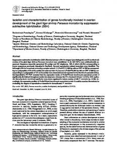
FIG. 1. Restriction maps of the four AHMHC recombinant phage. Restriction sites and MHC-encoding regions were determined as described. Shaded boxes indicate restriction fragments that hybridize to rat MHC cDNA clones, pMHC62, and total genomic DNA. Open boxes indicate restriction fragments that hybridize only to rat MHC cDNA clones. Hatched boxes indicate restriction fiagments that hybridize only to total genomic DNA. Subclones generated from the A clones are indicated below each restriction map and are designated with "p" as a prefix. The direction of transcription for clone AHMHC 2 is indicated above the map.
the mRNA sequences in the region coding for the rod portion of the protein (5-7). MHC-Encoding Regions. MHC-encoding regions within each clone were identified by hybridizing rat cDNA probes to restriction digests of the human clones. The pattern of one such hybridization is shown in Fig. 3B. The sizes of the EcoRI fragments that hybridized to the MHC cDNA clone were 13.0, 5. 1, 3.8, 12.0, 6.3, and 2.0 kbp. Because the cDNA probes represent only a fraction of the total mRNA molecule (i.e., pMHC62 contains 2,200/7,100 nucleotides), other regions of the clones could also contain protein-encoding sequences. The tentative direction of transcription of one of the clones (AHMHC2) was determined by examining the patterns of hybridization of the human MHC clones to two rat MHC cDNA probes of varying lengths. This is denoted by the arrow in Fig. 1. pMHC62 has a 2,200-nucleotide insert and extends further in the 5' direction of the full-length mRNA than does pMHC25, which has a 650-nucleotide insert. A restriction fragment of clone HMHC2 was identified that hybridized to pMHC62 but not to pMHC25. p2-2 in HMHC2 hybridized to pMHC62 but not to pMHC25, tentatively defining its orientation as 5' to p2-1, which hybridized to both cDNA probes (see Fig. 1). The other three clones showed the same pattern of hybridization with both cDNA probes. This demonstrates that these clones contain sequences encoding the 3' end of the MHC mRNA molecule.
Proc. Natl. Acad. Sci. USA 80 (1983)
Cell Biology: Leinwand et al.
3718
1
3
2
4 :..
:
......
28 S
.....
-
clones (data not shown). This DNA fragment also contained a repetitive element that corresponded to a non-Alu family interspersed repetitive DNA sequence (see below). Repetitive Sequences in Human MHC Clones. The same
.Q7. .
.. A
S-
28
B
0
28 S
-
Co
28 S-
D
FIG. 2. MHC specificity of the human MHC clones. Ten micrograms of RNA from each rat muscle tissue was denatured and electrophoresed on 1% formaldehyde/agarose gels. The RNA was transferred
to filters and hybridized to. radioactively labeled human MHC genomic cloned fragments: (A) AHMHC2; (B) AHMHC9; (C) AHMHC8; and (D) AHMHC10. Lane 1, embryonic cardiac muscle; lane 2, adult cardiac muscle; lane 3, embryonic skeletal muscle; and lane 4, adult skeletal muscle. Migration of 28S ribosomal RNA is indicated by 28S.
Hybridization patterns and the mapping of protein-encoding regions in the human MHC genomic clones resulted in the localization and subcloning of one intervening sequence (IVS) in the AHMHC10 clone that was at least 1.54 kb in length (plO6 in Fig. 1). This IVS was unique to the clone of its origin, as indicated by the total lack of hybridization to the other three 2 )1
kh Au~p 23.6
it
1
2
3 4
1 2
3 4
1
2
:3
4
1 2 3 4
-
9. i>
6.6 4.3 2.2
-
-
-
1.9 -
:1>
A
B
C7
D
FIG. 3. Hybridization patterns of genomic clones. HMHC clones were digested with EcoRl and electrophoresed, and the DNA was visualized by staining with ethidium bromide (A). Lane 1, AHMHC2; lane 2, AHMHC8; lane 3, AHMHC9; and lane 4, AHMHC10. DNA was transferred to Gene Screen and hybridized to (B) pMHC62; (C) pBLUR 8, and "Alu family" probe; (D) p104; and (E) plO-7. EcoRI fragments in the four human clones, as visualized by staining, are indicated by squares. HindIEI markers are indicated-along the vertical axis of A.
filter obtained from transfer of the gel shown in Fig. 3A was erased and hybridized to pBLUR8, a clone containing the Alu family of interspersed repetitive DNA sequences (11). The results shown in Fig. 3C indicate that the Alu family of repeated sequences is quite prevalent in the human MHC clones. Three of the four clones have Alu sequences in both EcoRI fragments, whereas the fourth has Alu sequences in two of the seven EcoRI fragments. The individual members of the Alu family of interspersed repetitive DNA sequences are highly conserved (12). Therefore, any fragment containing an Alu repeat hybridizes to all other fragments also containing an Alu repeat (data not shown). In addition to the Alu family, clones AHMHC2 and AHMHC10 contain a sequence corresponding to a non-Alu family interspersed repetitive DNA. sequence (data not shown). This sequence family is less abundant than the Alu family (22). It is of interest to note that the IVS in clone AHMHC10 contains such a repetitive element. Attempts to determine the interrelatedness of the four clones by electron microscopic analysis of heteroduplex molecules proved futile due to the extremely complicated structures formed by inter- and intramolecular hybridization between these repetitive sequences. Sequence Relatedness of Four Human MHC Clones. To determine the extent of the sequence relatedness among the four human MHC clones, restriction fragments that did not contain repetitive DNA sequences were- isolated from each clone and hybridized to restricted DNA from each of the other clones. An example of such an experiment is shown in Fig. 3D. The 2.0-kb RI fragment from AHMHC10, which is known to contain protein-encoding sequences close to the 3' end of the gene (Fig. 3B, lane 4), was subcloned into pBR322 (p1O-3, Fig. 1) and hybridized to the same filter shown in Fig. 3B and C. As would be expected, that fragment hybridized strongly back to itself. In addition, it also hybridized weakly to one fragment each in AHMHC2 and AHMHC9, but not at all to AHMHC8. The intensity difference between the hybridization of the probe back to itself and to the two other clones suggests that, in that region of the MHC gene, the three human MHC clones have diverged considerably, probably in their IVS. The lack of hybridization to the AHMHC8 clone most probably indicates that the region of the gene represented by p10-3 is not contained within clone AHMHC8. p10-1 (Fig. 1) from HMHC10, which also contained coding sequences, was hybridized to the same filter and hybridized back to itself as well as weakly to the following fragments in the other three clones: p2-1 in AHMHC2, p8-1 in AHMHC8, and the larger RI fragment in AHMHC9 (data not shown). No cross-hybridization among the four clones was detected when a DNA fragment corresponding to a sequence flanking the transcribed region of AHMHC10 was used in a hybridization (Fig. 3E). Similar results were obtained when fragments p2-3 and p8-2 were used as probes (data not shown). This type of analysis demonstrated that the adult (AHMHC8, -9, and -10) and embryonic (AHMHC2) skeletal MHC genes are as related to each other as are two different adult genes. It should be pointed out that these analyses are limited to small numbers of restriction fragments by the prevalence of repetitive sequences in the human clones. We have isolated restriction fragments that are unique to each clone and also fragments that contain sequences homologous to other clones. These results indicate that the four clones show limited relatedness to each other and are obviously quite divergent even though they were selected with the same closely related cDNA probes. The results presented here further confirm the data in Fig. 3B that indicate a significant degree of conservation among the coding
Cell Biology: Leinwand et al.
sequences and a high degree of divergence between the intragenic and intergenic sequences. Human MHC Genes Are Encoded by a Multigene Family. It has been shown that in the rat, there is a minimum of 13 sarcomeric MHC genes (2, 3). To estimate whether the number of MHC genes in the human genome is similar and to determine what proportion of the total genes we had isolated in our screening, we hybridized several human probes containing MHC-encoding or flanking, but not repetitive, sequences to human genomic DNA. Each of the subclones hybridized to a restriction fragment corresponding in size to itself and showed very little, if any, detectable cross-hybridization with other fragments (see Fig. 4). This result was obtained with both coding and flanking probes (data not shown). The weak cross-hybridization among the subeloned probes containing coding sequences is most likely due to the mixture of exon and intron sequences because it has been shown that the ratio of exon/intron sequences in MHC genes is close to 1:3 (23). The sizes of the EcoBl fragments that are found in our four human clones and contain sequences that hybridize to pMHC62 are 13.0, 12.0, 6.3, 5.1, 3.8, and 2.0 kbp (see Fig. 3). We were not able to use many regions of the genomic clones as probes because of repetitive DNA sequences. As mentioned earlier, we did not isolate any cardiac MHC genes. Because we have isolated four unique clones that are skeletal muscle-specific, there must be more than four genes in the human genome that contain MHC-encoding sequences. Also, it should be kept in mind that the size of a MHC gene is around 20-30 kb (unpublished data). Because the total insert size of any one of our clones does not exceed 18 kb, we are detecting only a subset of the myosin-encoding restriction endonuclease fragments in a genomic digest. Therefore, to test this possibility the rat cDNA clone pMHC25 was hybridized to a blot containing human and mouse genomic DNA digested with HindIII, as shown in Fig. 4A. The number of bands detected in both mouse and human is very similar and less than half of them are represented in the four genomic clones thus far isolated. These results suggest, although do not prove, that humans and rodents have a similar number of sarcomeric MHC genes. Repeated DNA Sequences in Rat and Human MHC Genomic Clones. To gain some insight into the evolution of MHC genes, the frequency and position of repetitive DNA sequences in the four human clones described in this report and four rat MHC genomic clones described elsewhere (23) were compared. (These four rat MHC genomic clones were isolated with the same cDNA clones used in this report; therefore, they contain the same region of the MHC genes as do the human clones.) This was accomplished by hybridizing restriction digests of the clones to two radioactively labeled probes-total genomic DNA and a clone containing the Alu family of interspersed repetitive DNA sequences (a human or rodent probe, depending on which genomic clones were being studied) (11, 12). Total mammalian genomic DNA that is labeled to a specific activity of 108 cpm/,ug hybridizes to sequences that are repeated at least 50 times in the genome (see ref. 8). As shown in Fig. 5A, only one rat MHC gene [one which contains embryonic MHC-encoding sequences (23)] hybridized to total rat genomic DNA. These same fragments hybridized to a cloned rodent Alu family sequence (data not shown). This pattern is in strking contrast to that seen in the human genes (Fig. 5B and Fig. 3C), in which most of the BamHI and EcoRI fragments hybridize to total human genomic DNA and to pBLUR8, an Alu family probe. These results indicate that repetitive sequences are a recent acquisition in MHC gene evolution. It is possible that the abundance of repetitive sequences in the human MHC genes has been accomplished by transposition of Alu family members into MHC genes during evolution. However, we have
3719
Proc. NatL Acad. Sci. USA 80 (1983) 1 2 3 4 5 6 7 8 9
12 3
kbp
kbp 23.6
23.6-
6.6 4.3 -
4
_*
*pro
0
-
.a"
-
.-
an
- 9.7 - 6.6
A.* ft-
4-
- 4.3 6.
.6
-
2.2 -
1.9
2.2 1.9
B
-
A
FIG. 4. Hybridization of MHC clones to genomic DNA. (A) Lanes 1 and 3, human DNA, and lane 2, mouse DNA. Genomic DNA was digested with HindM, electrophoresed, blotted, and hybridized to radioactively labeled DNA from pMHC25, a rat cDNA clone. (B) DNA from nine healthy individuals of various genetic backgrounds was isolated, digested with Hindu, electrophoresed, transferred to a filter, and hybridized to radioactively labeled p10-3 and p2-3 (see Fig. 1). AHindIl size markers are indicated along the vertical axis. Each lane represents DNA from a different individual.
no evidence that the four rat MHC genomic clones represent the same alleles as any of the human MHC clones. At this time the only data available indicate that they share sequence ho-
mology and muscle type specificity. Human MHC Genes Are Polymorphic. To determine whether the sequence organization of the human MHC genes differed among individuals, we isolated genomic DNA from a number of healthy individuals of various genetic backgrounds. DNA was digested with HindIII and the fragments were separated by electrophoresis and transferred to nitrocellulose. The filter was hybridized to two human MHC probes, p10-3 and p2-3. The former probe contains MHC-encoding sequences from the AHMHC10 clone, whereas the latter represents a unique sequence directly flanking the embryonic skeletal MHC clone, AHMHC2. The autoradiogram shown in Fig. 4B demonstrates one very distinct polymorphic band in the DNA from individual no. 4. This new HindIII band and the 7.3-kb band are the result of hybridization to the flanking region probe (p2-3) isolated from the embryonic skeletal MHC genomic clone and not the coding 1
2 3
4
1
2
-9_
3
4
kbp - -23.6 - 99.7 -
0.b
-
4.3
-
2.2
- 1.9
A
B
FIG. 5. Repetitive sequences in human and rat MHC genes. DNA from four rat (A) and four human (B) MHC genomic clones was digested with BamHI, electrophoresed, transferred to filters, and hybridized to radioactively labeled total rat genomic DNA (A) or total human genomic DNA (B). Each lane contains a different unique and nonoverlapping MHC clone. The four genomic clones are described elsewhere (23). (A) Lane 1, A287A1; lane 2, A287A4; lane 3, A287A3; and lane 4, A287A6. (B) Lane 1, AHMHC2; lane 2, AHMHC8; lane 3, AHMHC9; and lane 4, AHIMHC10.
3720
Cell Biology: Leinwand et al.
sequence probe (plo-3) (data not shown). Both probes were used in the same hybridization for this figure to provide an internal control, showing that other DNA fragments from individual no. 4 are the same size as bands from the other eight individuals. The fainter, higher molecular weight band seen in lane 1 of Fig. 4B is a partial digestion product, as we have not detected this band in other digestions. The additional HindIII band is probably an allelic form of the 7.3-kb "wild-type" band. This new band cannot be due to partial restriction by genomic DNA because it is of lower molecular weight than the wild-type allele. Also, individual no. 4 shows an additional band when digested with BamHI (data not shown). A more detailed analysis of this polymorphism in members of the family of individual no. 4 is necessary. It will be of particular interest to determine how extensive the polymorphism is and whether it is generated by Alu family repetitive DNA sequence transposition.
DISCUSSION It has recently been shown that vertebrate sarcomeric MHC is encoded by a conserved, multigene family (5). In this report, we have demonstrated that human MHC genes are encoded by a multigene family showing sequence conservation among skeletal MHC genes. The four clones isolated are unique and nonoverlapping. Each of these clones contains sequences that are highly homologous to 3' portions of rat MHC mRNAs. However, the human sequences have diverged both from the rat and from each other over most of their length during the evolution of this family. At least part of that sequence divergence must be due to the nonhomology of IVSs (24). Only skeletal muscle-specific human genomic clones were obtained despite the fact that a mixture of rat skeletal and cardiac MHC cDNA clones was used as probes. This result could be due to a high degree of divergence between rat and human cardiac MHC genes. A more trivial explanation for this result is that the genomic library did not contain the clones representing any of the human cardiac genes. The abundance of repetitive sequences in the human genes and the heterologous nature of the cDNA probes used have made it impossible to accurately determine the exact number of human sarcomeric MHC genes. Because the rat MHC gene family has been estimated to contain 13 members (3) and human DNA probed with the rat MHC cDNA clone gives a similar hybridization pattern, it is reasonable to assume that the human genome contains a similar gene number. Because the MHC gene family is composed of a number of members, it is of interest to determine whether they are clustered or dispersed to several chromosomes. We have recently shown that the former is the case because the genomic clones described here map to the short arm of human chromosome 17 (unpublished data). However, it should be kept in mind that without further analysis, the data presented here do not allow the discrimination of pseudogenes from functional genes. The most surprising finding in this study was the vast difference between the prevalence of repetitive sequences in the human and rat MHC clones. The lack of hybridization of the total genomic and Alu family probes to the rat MHC genes might suggest a different gross genome organization between the two organisms. That this was not the case was previously shown by screening a portion of a rat genomic library with a hamster Alu family probe. Ninety-nine percent of the clones (with an average insert size of 15 kb) showed positive hybridization, indicating a general repetition frequency for the Alu family of >105 copies per genome (17). In addition, studies of the repetitive DNA sequence organization of the rat genome suggest that it is similar to that in the human genome (25). It has been suggested, both by virtue of flanking direct repeats (12) and a
Proc. Natd Acad. Sci. USA 80 (1983)
different position for an Alu family member in two growth hormone genes (9), that the Alu family is a class of transposable elements. Our results raise the possibility that at least some human Alu family members might have such a function. The polymorphism shown in Fig. 4 occurred in one of nine surveyed individuals. The fragments that were used to screen for polymorphisms represented a coding segment from AHMHC10 and a flanking segment from AHMHC2. The polymorphism that occurred was in the flanking sequence probe rather than within the coding portion of the gene. It will be of interest to determine whether this polymorphism is generated by transposition of Alu family repetitive DNA sequences. We thank B. O'Brien and E. Bekesi for excellent technical assistance, Dr. Geoffrey Childs for critically reading the manuscript, and B. Donovan and A. Stockhausen for secretarial assistance. These studies were supported by grants from the American Cancer Society (NP-348) and National Institutes of Health (GM 29090) awarded to L.A.L. and from National Institutes of Health (GM 31281) awarded to B.N.-G.
Medford, R., Wydro, R., Nguyen, H. & Nadal-Ginard, B. (1980) Proc. Nati Acad. Sci. USA 77, 5749-5753. 2. Whalen, R., Schwartz, K., Bouveret, P., Sell, S. & Gros, F. (1979) Proc. Natl Acad. Sci. USA 76, 5197-5201. 3. Nadal-Ginard, B., Medford, R., Nguyen, H., Periasamy, M., 1.
4.
5. 6.
7. 8. 9. 10. 11. 12.
13. 14.
15. 16. 17. 18. 19.
20. 21.
22.
23. 24.
25.
Wydro, R., Hornig, D., Gubits, R., Garfinkel, L., Weiczorek, D., Bekesi, E. & Mahdavi, V. (1982) in Muscle Development: Molecular and Cellular Control, ed. Pearson, M. & Epstein, H. H. (Cold Spring Harbor Laboratory, Cold Spring Harbor, NY), pp. 143-168. Robbins, J., Freyer, G., Chisholm, D. & Gilliam, T. C. (1981)J. Biol, Chem. 256, 550-556. Nguyen, H., Gubits, R., Wydro, R. & Nadal-Ginard, B. (1982) Proc. Natli Acad. Sci. USA 79, 5230-5234. Mahdavi, V., Periasamy, M. & Nadal-Ginard, B. (1982) Nature (London) 297, 659-664. Sinha, A. M., Umeda, P. K., Kavinsky, C. J., Rajamanickam, C., Hsu, H. J., Jakovcic, S. & Rabinowitz, M. (1982) Proc. Natl, Acad. Sri. USA 79, 5847-5851. Shen, C. & Maniatis, T. (1980) Cell 19, 345-391. Page, G., Smith, S. & Goodman, H. (1981) Nucleic Acids Res. 9, 2067-2104. Della Favera, R., Gelman, E., Gallo, R. & Wong-Staal, F. (1981) Nature (London) 292, 31-35. Houck, C., Rinehart, F. & Schmid, C. (1979)J. Mol BioL 132, 289306. Haynes, S., Toomey, T., Leinwand, L. & Jelinek, W. (1981) Mol. Cell Biol 1, 573-583. Davidson, E. & Britten, R. (1979) Science 204, 1052-1059. Blattner, I., Williams, B., Blechl, A., Denniston-Thompson, K., Faber, H., Furlong, L., Grunwald, D., Kieter, D., Moore, D., Schuman, J., Sheldon, E. & Smithies, 0. (1977) Science 196, 161169. Benton, W. & Davis, R. (1977) Science 196, 180-182. Maniatis, T., Fritsch, E. & Sambrook, J. (1982) Molecular cloning: A laboratory manual (Cold Spring Harbor Laboratory, Cold Spring Harbor, NY). Leinwand, L., Wydro, R. M. & Nadal-Ginard, B. (1982) MoL Cell, Biol 2, 1320-1330. Rigby, P., Dieckmann, M., Rhodes, C. & Berg, P. (1977) J. MoL Biol 113, 237-251. Derman, E., Krauter, K., Walling, L., Weinberger, C., Ray, M. & Darnell, J. E., Jr. (1981) Cell 23, 731-739. Southern, E. M. (1975) J. Mol Biol. 98, 503-518. Bell, G., Karam, J. & Rutter, W (1981) Proc. Nati Acad. Sci. USA 78, 5759-5763. Schmid, C. W., Fox, G., Dowds, B., Lowensteiner, D., Paulson, E., Shen, C. J. & Leinwand, L. (1983) in Perspectives on Genes and the Molecular Biology of Cancer, eds. Robertson, D. & Saunders, G. (Raven, New York), pp. 35-41. Wydro, R., Nguyen, H., Gubits, R. & Nadal-Ginard, B. (1983)J. Biol Chem. 258, 670-678. Van Den Berg, J., Van Ooyen, A., Mantei, N., Schambock, A., Grosveld, G., Flavell, R. & Weissmann, C. (1978) Nature (London) 276, W.37-44. Pearson, R., Wu, J. R. & Bonner, J. (1978) Biochemistry 17, 51-59.