genes in the nif regulon and adjacentdeoxyribonucleic acid of K. .... The nifmutations in the K. pneumoniae strains ...... Shanmugam, K. T., and C. Morandi. 1976.
OF BACTRIOLOGY, Mar. 1980, p. 1264-1271 0021-9193/80/03-1264/08$02.00/0
Vol. 141, No. 3
JOURNAL
Isolation and Characterization of Lambda Specialized Transducing Bacteriophages Carrying Klebsiella pneumoniae nif Genes DOUGLAS MAcNEIL,t MARTHA M. HOWE, AND WINSTON J. BRILL Department of Bacteriology and Center for Studies of Nitrogen Fixation, University of Wisconsin, Madison, Wisconsin 53706
Seven Xdnif specialized tansducing bacteriophages were isolated from Escherichia coli strains containing plasnids carrying the his-nif region of Kiebsiella pneumoniae. These phages collectively carry deoxyribonucleic acid for all of the genes in the nif regulon and adjacent deoxyribonucleic acid of K. pneumoniae. The phages were isolated by using Mu insertions in the nif region to direct the integration of XpMu phages in nif via formation of XpMu-Mu dilysogens which, upon induction, yielded Xdnifphages. This procedure should be generally applicable for isolating A specialized transducing phages carrying genes from E. coli or other bacteria. Although nitrogenase, the enzyme which reduces N2 to NH4', contains only three different polypeptides, 17 contiguous nif genes arranged in seven operons have been identified in Klebsiella pneunoniae by characterizing the complementation pattern (8,9, 13,16), fine-structure map (13), and polarity of over 500 Nif point, insertion, and deletion mutants (8, 9, 13, 16, 21; D. MacNeil G. P. Roberts, and W. J. Brill, manuscript in preparation). The function of most of the nif gene products has been identified (22). Regulation of the nif genes is complex, and the molecular mechanism of this regulation are largely unknown. Synthesis ofactive nitrogenase does not occur in high levels of NH4' (27), high levels of amino acids (24), the presence of 02 (23), or the absence of MoO42- (3). Studies of mutants with defective glutamine synthetase suggest that glutamine synthetase, or factors which regulate glutamine synthetase, regulate several N-metabolizing enzymes (reviewed in reference 26), and may mediate NHE4 repression of Nif (25); however, other factors also may be involved in NH4' repression (2) and amino acid repression of Nif (24). The product of nifA may be a positive regulatory protein, because it is required for synthesis of the other known nifspecific proteins (22). One approach to studying the regulation of Nif is to isolate purified nif DNA and use it as a probe to assay nif-specific mRNA synthesis. DNA-RNA hybridization studies could be used to provide an independent estimate of the numt Present address: Merck Sharp & Dohme Research Lab-
oratories, Rahway, NJ 07065.
ber and size of transcripts previously deduced from polarity studies, and to determine the amount of nif mRNA's made under various repressing conditions and in specific mutants. Ideally, isolated nif DNA should be free of non-nif DNA and should be easy to isolate in significant quantities. Previously, a large ("108-dalton), low-copy-number plasmid, pRD1, containing the his-nif genes of K. pneumoniae, was constructed in vivo (7). Attempts to clone nif DNA from this plasrid onto small, amplifiable plasmids have been only parially successful (2, 4, 5). Several fragments resulting from restriction enzyme cleavage of the pRD1 his-nif region have been cloned and used 1 physically map the nif region (21), but these clones, collectively, do not contain all the nif DNA (2, 4, 5). In this paper we describe the construction of Adnif specialzed transducing phages. Together, two of these phages carry all of the nif genes and adjoining K. pneumoniae DNA. To isolate the Xdnif phages, we have used a technique first described by Casadaban (6) and further developed by M. M. Howe, B. R. Paul, and L. Parada (manuscript in preparation). This technique takes advantage of the random integration properties of phage Mu (10) to allow the isolation of strains containing a Mu prophage inserted in or near the gene(s) of interest. The Mu prophage then directs the integration of a ApMu by homologous recombination between Mu DNA sequences. When the A-Mu dilysogen is induced for A, it yields a lysate containing a small proportion of specialized trnsducing phages carrying host DNA sequences originally adjacent to the Mu prophage.
1264
LAMBDA NIF SPECIALIZED TRANSDUCING PHAGES
VOL. 141, 1980
1265
MATERIALS AND METHODS
5 kilobases of the c immunity end of Mu located between the J and red genes of A (18). The A phages Media. Recipes for liquid and solid media including (ApMu522, ApMu526, AcI857S7, Acb2, and Xvir) and LC, soft agar, SM buffer, K (N-free medium), and KN Mu phages (Mucts6l and Mucts62hP15, a Mu phage (K with 0.2% ammonium acetate) have been previ- with P1 host range) were from the collection of ously described (12, 17). A Broth contains 20 g of M.M.H. Most P1 transductions were performed as maltose, 10 g of tryptone, 2.5 g of NaCl, 1 g of yeast previously described utilizing Plkchl (13), an extended extract, and 0.6 g of MgSO947H20 per liter. Filter- host-range mutant of Plkc. The transduction of strain sterilized spectinomycin (gift from G. B. Whitfield, UQ6 to RecA- was performed with PlCMclrlOO grown The Upjohn Co., Kalamazoo, Mich.) at 200 ,ug/ml and by induction on strain UQ2 because Plkc grows poorly tetracycline (Lederle Laboratories, Pearl River, N.Y.) on RecA- hosts. at 20 ,g/ml were added when required. Bacterial matings. Transfer of pTM plasmids Bacterial strains and plasmids. The K. pneu- from K. pneumoniae to E. coli or between E. coli moniae and E. coli strains used are listed in Tables 1 strains was accomplished by spotting 10 to 30 #1 of and 2. The nifmutations in the K. pneumoniae strains overnight LC cultures of the donor onto LC plates spread with 0.1 ml of an overnight culture of LC-grown are designated as previously described (13), with three exceptions. Further biochemical and genetic studies recipient. The mating mixture was incubated at 30°C (D. MacNeil, G. P. Roberts, and W. J. Brill, manu- overnight and then replicated or streaked onto selecscript in preparation) have subdivided three previ- tive minimal media. Construction of E. coli strains with nif plasously described nif genes. nifL has been divided into nifL and nifR (a regulatory site), nifF has been divided mids. E. coli K-12 is naturally Nif- but becomes Nif+ into nifF and nifW, and nifS has been divided into when it contains the nif genes of K. pneumoniae (7). nifS and nifU. The recombinant plasmid pRD1 con- E. coli strains with nif point mutations were isolated tains the transfer, replication, and kan tet amp genes by mating K. pneumoniae strains with the pTM plasof RP4 (7), contains the K. pneumoniae his-nif region mids listed in column 3 of Table 2 with E. coli strains (7), and confers resistance to phages Mu and P1 (12). UQ6 or UQ20 and selecting for transfer of the pTM Plasmids pTM4010 (his+ nif+), pTM4041 (hisD4209 plasmid by selecting His' Spcr exconjugants. Because nif+), and other pTM plasmids (his+ nif) are deriva- the pTM plasmids segregate markers (12), the excontives of pRD1 which do not confer resistance to phages jugants were tested by back mating to the K. pneuMu or P1 (12). Various nif mutations were transferred moniae Nif- mutants listed in column 2 of Table 2, to pTM4041 by transducing its host, strain UN1516 or selecting for Nif+ on K medium. UQ6 or UQ20 derivstrain UQ67, to His+ with P1 grown on a Nif- mutant atives with pTM plasmids, which gave Nif+ recombinants with all K. pneumoniae Nif- strains except the and scoring for cotransduction of the nif mutation. Bacteriophage strains. XpMu522 and ApMu526 strain with the same nif allele, were saved. E. coli strains containing pTM plasmids with Mu were isolated as plaque-forming A phages arising after induction of a AcI857S7plac5 prophage containing a insertions in nif were constructed by P1 transduction Mucts62 prophage inserted in the lac5 DNA (14, 18). of the Mu insertion from K. pneumoniae into They are int and presumably att and contain less than pTM4041 in E. coli strain UQ67. This strain is restric-
TABLE 1. Bacterial strains Genotype
Strain
Source/reference
E. coli AB2880
CSH26 JC10240 MH812 MH867
QD5003 SB1801
UQ2 UQ4 UQ5 UQ6 UQ8 UQ19
UQ20 UQ67 K. pneumoniae UN976 UN1179 UN1516
his pro arg thi araD shiA A(pro-lac) ara thi Hfr P045 srl-300-.:Tn1O recA56 thr-300 ilv-318 thi-1 rpsE300 hsdM hsdR thr leu met lac'supE hsdM hsdR thr leu met lac supE nalA (Muc+) pro mel supF
Ahis-750 ara-14galK2 malAl xyl-5 mtl-I rpsL JSC10240 (PlCMclrlOO) A(pro-lac) ara thi rpsE Al(pro-lac) ara thi rpsE hisD A(pro-lac) ara thi rpsE hisD Mur pro mel supF Mur hsdM hsdR thr leu met lac supE his nalA (Muc+) A(pro-lac) ara thi rpsE hisD Mur srl::TnlO recA56
T. Pittard (19) Cold Spring Harbor Collection L. Csonka 921 of W. Arber Nalr Muc+, derivative of MH812
(18)
UQ19(pTM4041)
P. Hartman Lysogeny of JC10240 Spontaneous of CSH26 Diethyl sulfate induced of UQ4 Spontaneous of UQ5 Spontaneous of QD5003 P-2 eductant of MH867 Transduction UQ2 -+ UQ6 Mate UN1516 -+ UQ19
A(his-nif)4646pro-4001 (pTM4010) A(his-nif)4498 A(his-nif)4646pro-4001 (pTM4041)
(13) (13) (13)
1266
MAcNEIL, HOWE, AND BRILL
J. BACTERIOL.
TABLE 2. Pedigree of Nif strains nif allele Point mutations nifQ4969 nifB4398 nifB4106 , nifA4683 nifA4176 nifL4373 nifF4066 nifM4027 nifV4944 nifS4389 nifS4618
nifN4740 nifN4687 nifN4724 nifE4675 nifE4701 nifK4729 nifK4115 nifD4716 nifD4685 nifD4697 nifH4060 nifH4083 nifJ4727 nifJ4702 nifJ4057
K. pneumoniae strain with allele
Derivative of pTM4041 with allele
nifplasmid transferred to: UQ6 (Rec+)
UQ20 (Rec-)
UN2138 UN1074 UN106 UN1647 UN176 UN1030 UN66 UN27 UN1990 UN1046 UN1488 UN1771 UN1651 UN1688 UN1639 UN1665 UN1693 UN115 UN1680 UN1649 UN1661 UN60 UN83 UN1691 UN1666 UN57
pTM4194 pTM4185 pTM4042 pTM4164 pTM4044 pTM4209 pTM4046 pTM4048 pTM4198 pTM4162 pTM4197 pTM4182 pTM4149 pTM4150 pTM4176 pTM4175 pTM4161 pTM4052 pTM4155 pTM4154 pTM4153 pTM4057 pTM4055 pTM4180 pTM4181 pTM4062
UQ70 UQ71 UQ72 UQ73 UQ74 UQ75 UQ76 UQ77 UQ78 UQ79 UQ80 UQ81 UQ82 UQ83 UQ84 UQ85 UQ86 UQ87 UQ88 UQ89 UQ9O UQ9L UQ92 UQ93 UQ94 UQ95
UQ96
UN1125 UN1088 UN1102 UN2608 UN2609 UN1126 UN1119 UN1091 UN1100 UN1133 UN1162 UN1122 UN1114
pTM4082 pTM4071 pTM4080 pTM4222 pTM4223 pTM4083 pTM4173 pTM4070 pTM4073 pTM4065 pTM4067 pTM4066 pTM4081
UQ109 UQ110
UQ97
UQ98
UQ99 UQ100 UQIOI UQ102 UQ103 UQ104
nifW4445 nifU4438 nifN4411 nifE4420 nifK4452 nifD4481 nifJ4441 nifJ4434
X p)
a
KKS
UQ105 nit
his
UQ107
S
E
AC
L
H
K
J
X i ngratoion
nit
his
UQ108
nif
Mu
m
O
UQ106
Mu insertions
nifB4444 nifB4408 nifA4422 nifL5181 nifL5182
prophage as shown in Fig. 1. Lysates of ApMu522 and ApMu526 were grown by infection as described previously (18), except that QD5003 was grown in A broth. Lysates had titers between 109 and 10'0 plaque-forming units (PFU) per ml. One drop from each lysate was spotted on soft agar lawns of the E. coli Nif- Mu lysogens listed in column 4 of Table 2, and the plates were incubated overnight at 300C. From the area of the spot, survivors were streaked onto LC plates spread with -107 PFU of Acb2, and these plates were incubated at 30°C overnight. Surviving colonies were cross-streaked against lines containing 107 PFU of Acb2 or Avir on an LC plate at 30°C. A Lysogens, identified as Acb2 resistant, but.Avir sensitive, were mated to strain SB1801. His' exconjugants were selected, purified, and tested for A release into a lawn of Mu-resistant strain UQ8 at 42°C. Strains which could transfer A were presumed to be dilysogens, with the A integrated adjacent to Mu as indicated in Fig. 1. Preparation and assay of low-frequency-
XpMu
Mbu
S
0
E S
J
Ac
L
nif AK K H
Acm R N
J
A excision
UQ1ll UQ112 UQ113 UQ114 UQ115 UQ116 UQ117 UQ118
Xdnif
UQ122 UQ123 UQ124 UQ125
UQ119 UQ120 UQ121
tion deficient and contains a Muc+ prophage which prevents zygotic induction during the transduction. His' transductants were tested for Mu release into a soft agar lawn of strain MH812 at 42'C. His' (Mucts61)+ transductants gave a large clear zone of lysis. MH867 or His' (Mucts6l)- transductants gave only a few single, turbid plaques. His' Mucts6l-releasing clones were mated into strain UQ6, and His' exconjugants were isolated and back mated to Nif- K. pneumoniae strains as described above. Clones were saved if they gave Nif+ recombinants with all strains tested except the parental Mu lysogen. Construction of A-Mu dilysogens. Because ApMu522 and ApMu526 are deficient in A-mediated integration at the E. coli attA site, lysogens of these phages form only at low frequencies. One mechanism of lysogeny is homologous recombination between the Mu DNA of the ApMu's and a Mu prophage, resulting in the integration of the ApMu at the c end of the Mu
MA A KA5
0
m
b nit
his
nit
M
uA
X integration
X pmu
nit
his 0
X excision
5
E
cA
N
R
coK
A
mu J
cA
L
S
K
nif K
J
j
Xdnif C os
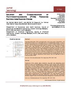
FIG. 1. Formation of Adnif phages carrying DNA adjacent to Mu prophages. The Mu prophage directs the integration of a ApMu by recombination between homologous Mu DNA sequences. The resulting ApMu-Mu dilysogens are induced to yield a proportion of Adnif phages in which nif DNA has replaced some A DNA during A excision. (a) A Mu prophage in a + orientation generates a Anif carrying his-distal nif genes. (b) A Adnif derived from a Mu prophage in the - orientation carries the his-proximal nif genes.
VOL. 141, 1980
LAMBDA NIF SPECIALIZED TRANSDUCING PHAGES
transducing Anif lysates. Cultures (10 ml each) of the A-Mu dilysogens of Su-UQ6 were heat induced as described by Miller for AcI857S7 lysogens (17). This procedure induced both Mu and A. The lysates were titrated on Mu-resistant strain UQ8 and used to transduce Mu-resistant derivatives of strain UQ8 to prevent infection by Mu. A Transductions for Nif+ were initially performed by mixing 0.2 ml of the Nif recipient cells (column 4 of Table 2) grown to 2.5 x 10⁸ cells/ml in broth with 5 x 10⁸ PFU of the low-frequency-transducing lysate and 10' PFU of AcI857S7 helper. The presence of the A helper results in preferential integration of both the helper and the integration-defective Anif at the bacterial attA site. This mixture was incubated at 30°C for 20 min, plated on N-free K medium, and incubated anaerobically for 7 days at 30°C. These plates contained few if any Nift clones and only about 103 tiny Nif- clones. This suggested that anaerobic, N-starving conditions greatly favored lytic development of A. In subsequent transductions, the absorption mixture was plated on LC plates and incubated overnight aerobically at 30°C and was then replicated to K medium. This increased the number ofsurvivors and Nif+ clones by approximately 40-fold. Preparation of high-frequency-transducing (HFT) lysates. Nif+ clones isolated as described above could arise by reversion of the nif mutation, recombination between the nifplasmid and an incoming Anifphage, complementation between the nifplasmid and a Anif integrated along with the A helper at the attA site, or integration of a Adnif into the nif plasmid and integration of the A helper into the att site. Only the latter two should release plaque-forming A and, upon induction, yield a significant number of Anif-transducing phages. Thus, Nift transductants which also released A were heat induced, and 10 pl of the lysate was spotted on LC lawns of the Nif UQ6 strains (Table 2, column 4), which were then incubated overnight and replicated to K medium to select for Nift transductants. After anaerobic incubation for 5 days at 300C, HFT lysates produced confluent patches of growth with at least some of the Nif- recipients. Mapping nif DNA on Anif phages. E. coli Nifstrains (columns 4 and 5 of Table 2) were grown overnight in A broth, and 0.1 ml of each was spread on LC agar. A 10-pl amount of HFT Anif lysates with titers of 108 to 1012 PFU/ml was spotted on the Nif lawns, incubated at 30°C overnight, and replicated to N-free medium. Growth was scored after 5 to 7 days of anaerobic incubation at 360C for NifQ-, NifV-, and NifS- strains or 30°C for the other Nif- strains. Mapping experiments used RecA+ strains, and complementation experiments used RecA- strains (columns 4 and 5, respectively, of Table 2). Purification of Anifphages. Anifpurification was accomplished by using the procedures described by Miller (17). Cultures (400 ml each) of the Aniflysogens were induced at 42°C for 20 min, then shifted to 37°C for 80 min and centrifuged at 10,000 x g for 15 min, and the pellet was suspended in 10 ml of SM buffer to which 0.2 ml of CHC16 was added to lyse the cells (17). Alternatively, after the 37°C incubation, 1 ml of CHC13 was added, and after 10 min, the cell debris was removed by centrifugation at 10,000 x g for 15 min. A
1267
40-g amount of polyethylene glycol 6000 and 11.6 g of NaCl were added to 400 ml of supernatant (1). After 18 h of incubation at 4°C, the phages were collected by centrifugation and suspended in 10 ml of SM buffer (1). Anifphages were purified by ultracentrifugation of the lysates on block gradients of 1.7, 1.5, and 1.4 g of CsCl per cm3 and, subsequently, on an equilibrium gradient of 1.5 g of CsCl per cm3 (17). Phage bands were withdrawn through the side of the tube with a syringe, and after the equilibrium gradient they were dialyzed three times against 300 volumes of SM buffer
(17).
Testing Anif phages for plaque-forming ability. The inclusion of A helper phage in the procedure described above for isolating HFT Anif lysates allows the isolation of both plaque-forming and defective Anif specialized transducing phages. Anif lysates were tested for plaque-forming Anifphages by two methods. Because the Anif phages contain the S7am mutation they do not form plaques on UQ6 or its derivatives, but do plaque on UQ8, a SupF strain. Two hundred plaques from each Anif lysate plated on a lawn of strain UQ8 were picked into 0.5 ml of SM buffer (5 plaques per tube) mixed with CHC13 and blended in a Vortex mixer for 10 s. These lysates were then tested for their ability to transduce E. coli Nif strains as described above for mapping Anif phages. Also, the phage isolated after CsCl equilibrium gradient centrifugation of Anif lysates were titrated on strain UQ8 for plaque-forming ability and on Nif UQ6 strains for Nif-transducing ability.
RESULTS To isolate A specialized transducing phages, it is advantageous to use a bacterial strain that is sensitive to A infection, and to have a procedure which allows the isolation of lysoge&s carrying a X prophage integrated close to the genes of interest. Since K. pneumoniae is naturally resistant to X, we used an E. coli strain containing derivatives of pRD1 (12). These plasmids carry the his-nif region from K. pneumoniae (7, 12). To obtain lysogens in which A integrated in nif, we infected strains containing Mu prophages in nif with XpMu-transducing phages and then selected for A lysogens. These X phages integrate by homologous recombination with the Mu prophage. Induction of the dilysogens resulted in the production of potential low-frequency A transducing lysates. The specific steps involved in isolating the Anif phages were as follows: (i) the isolation of strains with Mu insertions in the K. pneumoniae nif genes, (ii) the transfer of these insertions and a collection of well characterized nif point mutations to E. coli, (iii) the construction of A-Mu dilysogens utilizing ApMu's, (iv) the induction of the dilysogens for X, and (v) the screening of Nift transductants for ability to yield HFT Anif lysates. Isolation of nifmutations and their trans-
1268
MACNEIL, HOWE, AND BRILL
J. BACTERIOL.
fer to E. coli Previously, during genetic analysis of nif, over 100 Mu insertions in nif in K. pneumoniae were isolated and characterized (13). Thirteen of these, inserted in 11 different nif genes, were transferred onto the his nift pTM4041 plasmid in E. coli strain UQ67 by P1 cotransduction of the Nif His' properties. The resulting plasmids were then transferred by mating into strain UQ6 for subsequent analysis. Also, several hundred point mutations were previously isolated in K. pneumoniae, and many of these were transduced onto plasmid pTM4041 (13). Of these plasmids, 26 were transferred by conjugation to E. coli. Table 2 summarizes the K. pneumoniae and E. coli strains with Mu insertions and point mutations in nif used in this
Construction of A-Mu dilysogens in nif. A-Mu dilysogens were constructed by lysogenizing derivatives of strain UQ6 containing nif::Mu plasmids (Table 2, column 4) with ApMu's. These ApMu's lack the A phage att site and the A int gene and can integrate only by Mu-Mu homologous recombination at the c end of a Mu prophage as shown in Fig. 1. The A-Mu dilysogens were found at approximately the same frequency as spontaneous Ar clones (10-6). The low frequency of A lysogeny is probably due to the limited target for homologous recombination. The Mu prophage on the plasmid may be in either of two orientations (Fig. 1). The orientations of the eight Mu prophages in the strains lysogenized by the ApMu's were determined durpaper. ing isolation of nif-lacZ fusions (6; D. MacNeil The phenotypes of the E. coli strains with nif and W. J. Brill, manuscript in preparation), and mutations were similar to the phenotypes of K. are indicated in Table 3. ApMu lysogeny of 3 pneumoniae strains carrying the same muta- strains with a Mu prophage in the "+" orientations. E. coli strains with point mutations in tion formed ApMu-Mu dilysogens with the nifQ (UQ70), nifV (UQ78), and nifS (UQ79) are ApMu integrated on the his distal side of Mu. In leaky Nif mutants, showing some growth on N- five strains containing a Mu prophage in the free medium. E. coli strains containing muta- "-" orientation, the ApMu's integrated on the tions in the other nif genes showed no evidence his-proximal side of Mu. of growth on N-free medium. This growth patIsolation of Anif specialized transducing tern is the same as that observed in K. pneu- phages. All dilysogens were heat induced to moniae (13). The nif mutations reverted at sim- yield A lysates with titers between 10i and 109 ilar frequencies in both E. coli and K. pneumoPFU/ml. Each lysate was tested for its ability niae strains. Strains with Mu insertions in nifQ, to transduce four E. coli Nif- strains to Nif+. B, A, F, M, V, S, N, E, K, D, H, or J did not The Nif- strains used for each transduction conrevert, but strains with Mu insertions in nifL, tained mutations which were located adjacent W, or U did revert. When E. coli Nif' revertants to and on either side of the dilysogen (13). Nif+ of Mu inswtions in nifL, W, or U were crossed transductants appeared infrequently (O to 10 with strain UN1179 (total his-nif deletion) the Nift colonies per 2 x i05 infected cells). ApproxNift phenotype was cotransferred with His', imately 60% of the Nif+ colonies were apparently indicating that the reversion mutation was on lysogenized by the Ak helper phage since they the plasmid and not the E. coli chromosome. released plaque-forming A. The Nift colonies Together these results suggest that there are no which failed to release X may have arisen by reversion of the nif mutation in the recipient or cryptic nif genes in E. coli K-12. TABLE 3. A-Mu dilysogens used to isolate Anifphages Mu lysogen
Mu insertion
Munon-
A-Mu dilysogen
UQ109 UQllO UQ116
nifB4444 nifB4408 nifN4411
+ +
UQ139 (ApMu526) UQ140 (ApMu526) UQ141 (ApMu526)
UQ117
nifE4420 nifK4452
-
UQ142 (ApMu526) UQ143 (XpMu522)
UQ118
UQ119
nifD4481
UQ144 (ApMu522)
Aniflysogensb UQ148 (Anif24) UQ149 (Anifl8) UQ150 (Anif3l) UJQ151 (Anif19) UQ152 (Anif15) UQ153 (Anifl6) UQ154 (Xnifl7)
+ UQ120 nifJ4441 UQ145 (ApMu522) UQ121 nifJ4434 UQ146 (ApMu526) a +, Mu genes c through S are in a clockwise orientation relative to the order of genes on the standard E. coli map (K. pneumoniae has a similar gene order [15]); -, counterclockwise orientation. Thus, in a + lysogen the c immunity end of Mu is distal to his. ' The Anif lysogens also contain a A helper prophage.
LAMBDA NIF SPECIALIZED TRANSDUCING PHAGES
VOL. 141, 1980
1269
tation pattern of the Xnifphages is in agreement with previous polarity studies (13) and indicates that nifN-nifE and nifK-nifD-nifH are operons transcribed right to left. Furthermore, nifJ and nifU are not part of either of these operons. In agreement with the mechanisms shown in Fig. 1, Xnifphages isolated from strains with Mu insertions in the + orientation carry nif DNA distal to his, whereas Xnif phages derived from strains with Mu in the - orientation carry nif DNA proximal to his. The Anif phages carry nif DNA beginning at the site of the Mu insertion and extending to one side, except Xnif24 (see Fig. 2). Anif24, derived from Mu insertion nifB4408, may have resulted from a deletion of nifB and nifA DNA to give the mapping pattern indicated in Fig. 2. The Anifphages are expected to contain the Mu immunity gene, gene c (Fig. 1). This was tested by spotting a lysate of Mucts62hP15 on lysogens of each Xnif. A Mu
by recombination between a nif-transducing phage and the nif plasmid. HFT Anif lysates were obtained from 8% (17 of 213) of the Xreleasing Nif+ transductants. Anif phages obtained from induction of independent transductants arising from the same low-frequency-transducing lysate often mapped identically and were presumed to be siblings. All X-Mu dilysogens were induced at least twice and tested for their ability to yield Nif+ transductants; however, not all dilysogens yielded Anif phages. The dilysogens which did yield Xnif phages are indicated in Table 3. Genetic characterization of Anif phages. The extent of the nif DNA carried on each Anif was determined by assaying in spot tests the ability of HFT lysates of the Xnif phages to transduce to Nift a series of E. coli strains, each containing a mutation in one of the nif genes. Seven Anif phages were mapped by assaying their ability to recombine with point mutations and Mu insertions in RecA+ strains (column 4 of Table 2) and to complement point mutations in RecA- strains (column 5 of Table 2). In general, the Xnifphages which recombined with all point mutations in a particular nif gene gave a confluent patch when tested for complementation with mutations in that gene. However, if a phage recombined with only some of the mutations in a gene, for example Anifl7 with nifA mutations, only a few single colonies were observed in the complementation test with point mutations in that gene. These Nift colonies probably arose due to Xred-mediated recombination. Figure 2 illustrates the mapping results, and Table 4 summarizes the complementation data. It is not known if Anifl6 carries nifl. The complemen-
TABLE 4. Mapping and complementation of Anif phages with nif mutations Phage
nif genes detected by
nif genes detected by
complementation mappinga QBALFWMVSUNEKD`C QBALFWMVSUNE
Anifl5b QBALFWMSVU Anifl9 QBALFWMVSUNE' FWMVSU Anif24 'ALFWMVSUN FWMVSUNE Anifl7 'ALFWMVSUNEKD' FWMVSUNE Anifl6 FWMVSUNEKD' EKDH Anif31 'NEKDHJ' Anifl8 'NEKDHJ EKDHJ I The site for NH4' regulation, nifR (G. Roberts and W. J. Brill, manuscipt in preparation), is located between nifL and nifF. No mutations in nifR were used to characterize the Xnif phages. bAnifl5 also transduced mutations in hisD. Indicates that the phage recombined with some but not all mutations tested in that gene. "
MUTATION 4209 D
6
4106 4396 4373 4400 4176 5161 4969 4444 4663 4422 5162 A a L
4667 4115 4665 4369 4724 4675 4452 4697 4066 4445 4027 '4944 4618 4438 4411 4740 4701 4420 47259 4716 F
w
M
V
S
u
N
E
K
4057
4060 4441 4434
4461 64063 4727 4702 D
H
J
his fWf
wn PHAGE Init 18 )Xnlf 31
unit
16
Xnif 17 Mit 24
NiW 19
Xnif I5 FIG. 2. Mapping of Xnifphages. The nif DNA carried on each Anifphage is represented by the lower lines. Anifl8 extends beyond nifJ, and Xnifl5 and Anifl9 extend beyond nifQ as indicated by the arrowheads. The allele of each mutation used in mapping the Anifphages is indicated above the line. Phages Xnifp5, Xnifl6, and Xnifl7 are derived from Mu insertion nifD4481, Xnifl8 and Xnif3l from Mu insertion nifN4411, Xnifl9 from Mu insertion nifE4420, and Xnif24 from Mu insertion nifB4408. The arrows immediately below the top line indicate the extent and direction of transcription of the 7 nif operons.
1270
MAcNEIL, HOWE, AND BRILL
with P1 host range was used because the Anif lysogens are derivatives of Mu-resistant, Pl-sensitive UQ6. All Anif lysogens are Mu immune. The Anifphages were also tested for their ability to transduce the his and shiA genes near nif (7, 19). Only Anifl5 lysates transduced strains AB2800, UQ6, and UQ67 to His+; none transduced strain AB2880 to ShiA+. The seven Anif phages all appear to be defective for plaque formation; the Aniflysogens that release phage also contain a A helper phage. Single plaque lysates of the Xnif phages picked from lawns of strain UQ8 were unable to transduce Nif derivatives of strain UQ6 to Nif. These plaques were probably X helper phage. Also, when the Xnif lysates were purified on CsCl gradients, two bands were observed. In each case, the band at the density of the helper phage had a 10 to 10i higher (dependent on band separation) ratio of PFU to Nif-transducing ability than the other band. Xnifl5, Anifl7, and Anifl8 were more dense than the helper, whereas the other 4 Anif phages were less dense. The helper and XpMu phages were chosen as S7am phages to allow for induction of a large lysogenic culture, centrifugation of the cells, and suspension of the pellet in a small volume of A buffer in which the cells could be lysed by addition of CHC13 (17). However, three of the Anif lysogens did not show this phenotype. UQ152 (Xnifl5) lysed before centrifugation. UQ149 (Anifl8) and UQ150 (Anif3l) both lysed only poorly both before or after concentration. For CsCl equilibrium banding of these phages, the above three strains were heat induced and lysed without concentration, and the phages were precipitated with polyethylene glycol (1).
DISCUSSION This paper describes the recovery of all the K. pneumoniae nifgenes on specialized transducing phages. Seven different Xnif phages, all defective, collectively contain DNA for all nif genes of the 7 nif operons (9, 13, 16). These phages were isolated from four different ApMu-Mu dilysogens of E. coli containing plasmids with the K. pneumoniae his-nif region. The extent of the nif DNA on these phages was determined by genetic mapping. Among the seven Adnifphages are phages which have acquired only nif DNA and phages which carry DNA beyond nifQ toward his. Included in this latter class is one phage, Xdnifl5, which contains hisD. Another phage, Adnifl8, which recombines with and complements all tested mutations in the most his distal nif gene, nifJ, probably also extends beyond nifJ into adjoining DNA. Higher-titer lysates ofthese Xdnifphages were
J. BACTERIOL.
prepared by heat induction of Adnif-A helper lysogens. Large quantities of nif DNA from these phages were easily prepared (J. Collins, personal communication). nif DNA can also be prepared from recombinant plasmids constructed in vitro (2, 5, 6). However, not all of the nif genes have been cloned. Thus, when DNA containing many contiguous genes is desired, it may be easier to use the method utilized here than in vitro recombinant techniques. The DNA isolated from these Adnif phages will be useful for physically mapping the many insertions and deletions in nif (9, 13, 16). Some of these have been mapped using clones derived in vitro (21). DNA isolated from the Xdnif phages can also be used to assay for nif-specific mRNA synthesis and to provide nif DNA for in vitro transcription studies, investigations of protein-nif DNA binding interactions, and DNA sequencing. Purified nif DNA may also be useful to identify nif DNA and to physically map nif genes in other nitrogen-fixing organisms where genetic techniques are more limited than in K.
pneumoniae. We have used a method to obtain Anif specialized transducing phages which is generally applicable to isolating A phages carrying any gene(s) in E. coli. As demonstrated in this report, this includes genes from other organisms which can be transferred to E. coli. This technique was.first described by M. Casadaban (6), and has been further developed by Howe et al. (in preparation). The method requires a Mu insertion within or near the gene(s) of interest which subsequently directs the integration of a ApMu phage. When the dilysogen is induced for A, the lysate may contain specialized transducing phages for nearby genes and these can be detected by transducing an appropriately marked strain. We have also shown that the method will work when both A and Mu are thermoinducible. As an altemative to Mu, other insertion elements can be used to provide homology and direct the integration of phages. Reyes et al. (20) have shown that the insertion element IS2 can direct the integration of a A containing IS2 into an IS2 insertion in gal. In Salmonella typhimurium, the antibiotic resistance transposon, TnlO, was used to direct a P22 carrying TnlO DNA to integrate near his. Excision of the P22 led to isolation of P22 his specialized transducing phages (11). ACKNOWLEDGMENTS This research was supported by the College of Agricultural and Life Sciences, University of Wisconsin, Madison, by National Science Foundation grants PCM76-24271 (to W.J.B.) and PCM75-02465 (to M.M.H.), and by Public Health Service grant GM22130 from the National Institutes of Health (to
VOL. 141, 1980
LAMBDA NIF SPECIALIZED TRANSDUCING PHAGES
W.J.B.). D.M. was supported by Public Health Service training grant GM07133 from the National Institute of General Medical Sciences. M.M.H. is the recipient of Public Health Service Research Career Development award AI00274. We thank John Collins for purifying Anifphages and Richard Goldberg for helpful suggestions.
1. 2.
3.
4.
5.
6.
7.
8.
9.
10. 11.
12.
LITERATURE CITED Allet, B., K. J. Katogiri, and R. F. Gesteland. 1973. Characterization of polypeptides made in vitro from bacteriophage lambda DNA. J. Mol. Biol. 78:589-606. Ausubel, F., G. Riedel, F. Cannon, A. Peskin, and R. Margolskee. 1977. Cloning nitrogen fixing genes from KlebsieUa pneumoniae in vitro and the isolation of Nif promoter mutants affecting glutamine synthetase regulation. In A. Hollaender (ed.), Genetic engineering for nitrogen fixation. Plenum Press, New York. Brill, W. J., A. L Steiner, and V. K. Shah. 1974. Effect of molybdenum starvation and tungsten on the synthesis of nitrogenase components in Klebsiella pneumoniae. J. Bacteriol. 118:986-989. Cannon, F. C., G. E. Riedel, and F. M. Ausubel. 1977. Recombinant plasmid that carries part of the nitrogen fixation (nif) gene cluster of Kiebsiella pneumoniae. Proc. Natl. Acad. Sci. U.SA. 74:2963-2967. Cannon, F. C., G. E. Riedel, and F. ML Ausubel. 1979. Overlapping sequences of Klebsiella pneumoniae nif DNA cloned and characterized. Mol. Gen. Genet. 174: 59-66. Casadaban, M. 1976. Transposition and fusions of the lac genes to selected promoters in E. coli using bacteriophage X and Mu. J. Mol. Biol: 104:541-555. Dixon, R., F. Cannon, and A. Kondorosi. 1976. Construction of a P plasmid carrying nitrogen fixation genes from Klebsiella pneumoniae. Nature (London) 260: 269-271. Dixon, R., C. Kennedy, A. Kondorosi, V. Krishnapilai, and M. Merrick. 1977. Complementation analysis of Klebsiella pneumoniae mutants defective in nitrogen fixation. Mol. Gen. Genet. 157:189-198. Elmerich, C., J. Houmard, L Sibold, I. Manheimer, and N. Charpin. 1978. Genetic and biochemical analysis of mutants induced by bacteriophage Mu DNA. Mol. Gen. Genet. 165:181-189. Howe, M. M., and E. G. Bade. 1975. Molecular biology of bacteriophage Mu. Science 190:624-632. Kleckner, N., J. Roth, and D. Botatein. 1977. Genetic engineering in vivo using translocatable drug-resistance elements: new methods in bacterial genetics. J. Mol. Biol. 116:125-159. MacNeil, T., W. J. Brill, and M. M. Howe. 1978. Bacteriophage Mu-induced deletions in a plasmid containing the nif (N2 fixation) genes of Klebsiellapneumoniae.
1271
J. Bacteriol. 134:821-829. 13. MacNeil, T., D. MacNeil, G. P. Roberts, M. A. Supiano, and W. J. Brill. 1978. Fine-structure mapping and complementation analysis of nif (nitrogen fixation) genes in Kkebsiella pneumoniae. J. Bacteriol. 136:253266. 14. Magazin, M., M. Howe, and B. Allet. 1977. Partial correlation of the genetic and physical maps of bacteriophage Mu. Virology 77:677-688. 15. Mat8umoto, H., and T. Tazakl. 1971. Genetic mapping of aro, pyr and pur markers in Kkebsiella pneumoniae. Jpn. J. Microbiol. 15:11-20. 16. Merrick, M., M. Filser, C. Kennedy, -nd R. Dixon. 1978. Polarity of mutations induced by insertion of transposons Tn5, Tn7, and TnlO into the nif gene cluster of Kkbsiella pneumoniae. Mol. Gen. Genet. 165:103-111. 17. Miller, J. IL 1972. Experiments in molecular genetics. Cold Spring Harbor Laboratory, Cold Spring Harbor, N.Y. 18. O'Day, K., D. Shultz, W. Ericesen, L. Rawluk, and M. Howe. 1979. Correction and refinement of the genetic map of bacteriophage Mu. Virology 93:320-328. 19. Pittard, T., and B. J. Wallace. 1966. Gene controlling the uptake of shikimic acid by Eacherichia coli. J. Bacteriol. 92:1070-1076. 20. Reyes, O., M. Gottesman, and S. Adhya. 1979. Formation of lambda lysogens by IS2 recombination: gal operon-laknbda PR promoter fusions. Virology 94:400408. 21. Reidel, G. E., F. M. Ausubel, and F. C. Cannon. 1979. Physical map of chromosomal nitrogen fixation (nif) genes of Klebsiella pneumoniae. Proc. Natl. Acad. Sci. U.S.A. 76:2866-2870. 22. Roberts, G. P., T. MacNeil, D. MacNeil, and W. J. Brill. 1978. Regulation and characterization of protein products coded by the nif (nitrogen fixation) genes of Klebsiella pneumoniae. J. Bacteriol. 136:267-279. 23. St. John, R. T., V. K. Shah, and W. J. Brill. 1974. Regulation of nitrogenase synthesis by oxygen in KkebsieUa pneumoniae. J. Bacteriol. 119:266-269. 24. Shanmugam, K. T., and C. Morandi. 1976. Amino acids as repressors of nitrogenase biosynthesis in Kkbsiella pneumoniae. Biochim. Biophys. Acta 437:322-332. 25. Streicher, S. L., K. T. Shanmugam, F. Ausubel, C. Morandi, and R. S. Goldberg. 1974. Regulation of nitrogen fixation in Kkebsiella pneunmniae: evidence for a role of glutamine synthetase as a regulator of nitrogenase synthesis. J. Bacteriol. 120:815-821. 26. Tyler, B. 1978. Regulation of the assimilation of nitrogen compounds. Annu. Rev. Biochem. 47:1127-1162. 27. Yoch, P. C., and R. M. Pengra. 1966. Effect of amino acids on the nitrogenase system of Kkebsiella pneumoniae. J. Bacteriol. 92:618-622.