Mar 25, 2017 - clotting (3), plasmin has been implicated in complement ac- tivation (4) ... crude urokinase from Leo Pharmaceuticals (Ballerup, Denmark);.
THE JOURNALOF BMILOGICAL CHEMISTRY Vol. 257, No. 6 , Issue of March 25, pp. 3276-3283. 1982 Printed in U.S.A.
Isolation and Characterizationof Urokinase from Human Plasma* (Received for publication, August 17, 1981)
T.-C. Wun#, W.-D.Schleuning, and E. Reich From the Rockefeller University, New York, New York 10021
kinase is lo4- to 105-fold more active as a plasminogen activator.* The high catalytic efficiency suggested that if urokiPlasminogen activators catalyze the formation of plasmin nase, or its proenzyme, circulated as normal constituents of by hydrolyzing a single peptide bond in the precursor, plas- plasma, trace concentrations might suffice to meet the funcminogen, a proenzyme that is present at high concentrations tional needs for plasminogen activation. This consideration in the blood and body fluids (1, 2). These enzymes are of prompted us to undertake a search for urokinase in human interest because agrowing body of evidence suggeststhat they plasma. In radioimmunoassays of plasma, Astedt (23) found participate in a wide variety of biological functions. Apart occasional low levels of material thatcross-reacted withrabbit from its assumed fibrinolytic role in the regulation of blood antisera raised against urinary urokinase, and while he reclotting (3), plasmin has been implicated in complement ac- garded these asbeing of questionable significance, his results tivation (4), kinin formation ( 5 ) , prohormone conversion (6), added encouragement to the searchfor plasma urokinase. In and the generation of localized extracellular proteolysisduring what follows, we describe the isolation of a urokinase-like tissue remodeling, cell migration, carcinogenesis, and neopla- molecule from human plasmafractions, and theidentification of the corresponding enzymatic activity in plasma. sia (7). Plasminogen activators haveusually been named according EXPERIMENTALPROCEDURES to the starting material from which they were isolated (e.g. urinary, ortissue, or cell culture plasminogen activators, etc.) Materials (see Refs. 3, 8, and 9 for reviews), and the separatemolecular CNBr-activated Sepharose 4B-CL, protein A-Sepharose, DEAEidentities of these enzymes has been in dispute (10). Immu- Sephacel were obtained from Pharmacia, Inc., Piscataway, NJ; benznological data, catalytic efficiency, and studies of specificity amidine hydrochloride,p-aminobenzamidine,4-methylumbelliferylpwith small peptide substrates suggest that, in man, there are guanidinobenzoate,4-methylumbelliferone, aprotinin fromSigma; two distinct enzymes that are physiologically plausible plas- crude urokinasefrom Leo Pharmaceuticals (Ballerup, Denmark); * This work was supported by the American Cancer Society(Grant ACS PDTI) and The National Cancer Institute (Grant CA08290). T h e costs of publication of this article were defrayed in part by the payment of page charges. This article must thereforebe hereby marked “advertisement” in accordance with 18 U.S.C. Section 1734 solely to indicate this fact. $ Recipient of a fellowship from The Damon Runyon-Walter Winchell Cancer Fund.
BioRex 70 from Bio-Rad fluorescarnine from Hoffman-LaRoche; Lpyroglutamyl-glycyl-L-arginine-p-nitroanilide(5-2444). D-Valy1-hcyl-lysyl-p-nitroaniide (S-2251) from Ortho Diagnostics; ’*‘I-sodium iodide from Amersham Corp.; Spectrapore 2 from VWR Scientific Inc., San Francisco. Aquacide 11-A from Calbiochem-Behring: fresh human plasma from the New York Blood Bank; Cohn fractions 111, IV, IV-1, and PPF plasma fractions were generously provided by Dr.
3276
’ P. Aiyappa, A. Guha, and E. Reich, unpublished results.
Downloaded from www.jbc.org by guest, on July 12, 2011
The presence of activators of the fibrinolyticsystem minogen activators: the firstis the urinaryenzyme, urokinase in blood plasma has been assumed for a long time but (ll),and thesecond is an activator isolated fromuterine tissue never convincinglydocumentedbythe isolation of (121, generally designated as tissue activator, which resembles characterized and physiologically plausible enzymes. an enzyme originally purified from pig heart (13). The enof previouslyidentified ThelowCatalyticefficiency zymes variously isolated from cell culture (14), vascular enplasma plasminogen activators, which has made their dothelium (15), or from blood following venous occlusion (16) physiological significance uncertain, prompted us to are probablyall identical with one or anotherof the above. search for other plasma enzymes, resembling especially The presence of a fibrinolytic pathway intrinsic to plasma, the potent urinary activator,urokinase. We report here a plasminogen activator, and their identity,if they exist, are thedetection of aurokinase-like activity in human also unsettled questions. Coleman (17) reported that plasma plasma, and the isolation of the enzyme from whole plasma protein fractions.The purified enzymeis indis- kallikrein could activate plasminogen. Subsequently, Kaplan tinguishable from the 53,000-dalton components of hu-and Austen (18) described a partially purified plasminogen man urinary urokinase in the following respects: ap- proactivator, ultimatelyshown to be inseparable from plasma kallikrein (19, 20). More recently, coagulation factor XIa has parent M, on sodium dodecyl sulfate-polyacrylamide been proposed as a plasminogen activator (21), and plasmingel electrophoresis, doubleimmunodiffusion,amino ogen activation by factor XIIa has also been observed (22). acid analysis, two-dimensional tryptic peptide maps, catalytic efficiency with synthetic peptide substrates, All three of these enzymes are of doubtful significance as and inhibitor spectrum. The results suggest (a)that the physiologically relevant plasminogen activators, fwstly, beplasminogen as substrate) is very enzyme is present in plasma in a latent form whose cause catalytic activity (with nature remains to be defined, and (b) that the circulat- sluggish, and, secondly, because genetically determined defiing concentration is at least 5 to 10 pg/liter, and suffi- ciency states give no indication of functional limitations that cient to generate substantial levels of plasmin, partic- could reasonably be attributed to reductions in plasminogen ularly if activation were somehow confined by locali- activation or fibrinolysis. zation at specificsites. In comparison with plasma kallikrein, human urinary uro-
Urokinase
Plasma
Human
Duane Schroeder of Cutter Laboratories (Berkeley, CA); 6-aminocaproyl-p-aminobenzamidineSepharose was prepared as described elsewhere (24). Erythrina inhibitor was supplied by Dr. Eugene B. Dowdle, University of Cape Town, Cape Town, South Africa. Trasylol was a gift of Dr. E. Truscheit, Bayer A. G., West Germany. Methods
-
The abbreviations used are: SDS, sodium dodecyl sulfate; PAGE, polyacrylamide gel electrophoresis; MUGB, 4-methylumbelliferyl-pguanidinobenzoate; iPrZP-F, diisopropylfluorophosphate; PBS, phosphate-buffered saline; MES, 4-morpholinoethanesulfonic acid.
cellular debris and the supernatant was frozen and stored in 200-ml aliquots at -70 "C. The frozen plasma samples (200 m l ) were thawed in 37 "C water bath anddialyzed against 2 liters of 10mM benzamidine in double distilled water at 4 "C for 3 h using Spectrapore No. 2 dialysis membrane. This preparation was cleared by centrifugation at 16,000 x g for 60 min and subsequently mixed with 50 ml of BioRex 70 (100 to 200 mesh) which previously had been equilibrated in 10 mM NaP04, 10 mM benzamidine, pH 8.0. After 10 min, the mixture was packed intoa plastic column and drained. The column was washed with 100 ml of starting buffer and subsequently eluted with a solution containing 50 mM NaP04, 50 mM benzamidine, and 0.4 M NaCI, pH 6.9, The eluate was pooled (40 ml), adjusted to pH 5.2 by adding 4 ml 1 M Na acetate, pH 4.5, and concentrated to 20 ml in a dialysis bag covered with Aquacide IIA. Immunoaffinity Purification of Plasma Urokinase from Cohn Fraction IV-I-Human plasma Cohn fraction IV-1 (500 g) was mixed with 2300 ml of glass-distilled water and homogenized in a Waring blendor for 3 min. The mixture was titrated to pH 2.0 by dropwise addition of 6 N HCl, stirred at room temperature for 30 min, neutralized to pH 7.4 by addition of 6 N NaOH, and stirred further at room temperature for 2 h. Benzamidine/HCl (11.8 g), NaCl (87.7 g), and aprotinin (20,700 kallikrein inhibitor units) were added. The total volume was adjusted to 3,000 ml and the solution centrifuged at 1O , OO X g for 15 min. The supernatant was then passed through a preimmune rabbit Ig-Sepharose 4B column (2.6 X 3.5 cm) connected in tandem with a second column (1.5 X 2.0 cm) of affmity purified antiurokinase-Ig-Sepharose 4B, at a pressure of 70 cm of water and flow rate of 40 to 50 d / h . When this process was complete another 500 g of Cohn fraction IV-1 was processed in the same way and loaded onto the same columns. The preimmune rabbit Ig-Sepharose 4B column was then detached from the anti-urokinase Ig-Sepharose column, the latter was washed with 200 ml of PBS supplemented with 1%Triton X-100,0.4 M NaCl, and 0.1 M benzamidine, pH 7.4 (buffer A), and then 50 mI of the same buffer containing no Triton X-I00 (buffer B). Elution of urokinase was accomplished with 160 ml of 0.1 M glycine/ HCl, pH 2.2. The preimmune rabbit Ig-Sepharose 4B column was washed in the same way, and no urokinase-related plasminogen activator was observed in the eluate asassayed by zymography. Three preparations of the eluate of anti-urokinase-Ig-Sepharose4 8 column were combined, adjusted to 0.1 M in benzamidine and neutralized to pH 7.4 by dropwise addition of I M Tris/OH. The solution was subjected to a second cycle of affinity chromatography identical with the fist except that the anti-urokinase-Ig-Sepharose4B-CL column was smaller ( I X 1.1 cm), and was washed with smaller volumes, 30 ml of buffer A and 10 ml of buffer B. The urokinase was eluted in about 20 ml of 0.1 M glycine/HCl, pH 2.2, and concentrated to 1.5 ml in a dialysis bag covered with Aquacide I1 A. Preparative SDS-PAGE-Preparative SDS-PAGE was carried out in a Savant disc electrophoresis apparatus at room temperature. The Laemmli gel system (25) was used except that Tris/borate, pH 8.3, was substituted for Tris/glycine, pH 8.3, as the running buffer. The column containing a separating gel (10% polyacrylamide, 1.5 X 3.5 cm), overlaid with a stacking gel (4%polyacrylamide, 1.5 X 1.2 cm). The gel was pre-electrophoresed at 60 V for 30 min. SDS was added to thesample (1 to 2 m l ) to a final concentration of 0.5%. The sample was adjusted to 10%glycerol and 0.002% bromphenol blue and loaded onto the gel. A potential of 75 V was applied until the dye front had entered the separating gel, and was then increased to 150 V. The eluates were collected in I-ml fractions at IO-min intervals. Plasminogen Activator Activity of Urokinase and Plasma Activator-Plasminogen activator activity was determined either directly by hydrolysis of the chromogenic substrate S-2444, or indirectly by using 5-2251 as substrate for plasmin generated under conditions of plasminogen activation. The S-2444 assay was performed as follows: 0.23 ml of 0.1 M Tris/HCl, pH 8.8,0.5%Triton X-100 were mixed with 0.015 mi of sample containing 0.1%SDS and incubated a t 37 "C for 10 min. After the addition of 5 pl of IO" M S-2444, the absorbance increase at 405 nm was recorded in a Gilford 150 spectrophotometer. Urokinase reference standard (Leo) dissolved in 0.1% SDS was used to construct a standard curve. We found that SDSdoes not interfere with the assay of urokinase when it is trapped in mixed micelles by addition of Triton X-100. A linear relationship was found between the rate of absorbance change and the amount of activator over the range 1 to 8 Ploug units. The S-2251 assay was performed as follows: 0.4 ml of 0.1 M barbital, pH 8.3, 0.5% Triton X-100 were mixed with 0.05 ml of sample in 0.1% SDS and incubated at 37 "C for 10 min. Then, 40pgof plasminogen was added and the incubation was continued. Addition 10 of pl S-2251 M) and recording of
Downloaded from www.jbc.org by guest, on July 12, 2011
Electrophoretic Procedures and Zymography-Analytical SDSPAGE2 was performed according to the method of Laemmli (25), using a slabgel plate (1 X 95 X 110 mm). The separating and stacking gels contained 10% and 4% acrylamide, respectively. Plasminogen activator activity of the protein bands separated on the gelwas determined by the zymographic technique of Granelli-Piperno and Reich (26). Purification of Urokinase-32 mg of commercially available urokinase (Leo Lot No. SE 760230 and Lot UDT A 93) containing about 200,000 Ploug units were dissolved in 2 ml of a buffer containing 0.1 M KPOo, pH 7.0,0.4 M NaCl, and applied to a benzamidine-Sepharose 4B column (1 X 15 cm). The column was washed with 100 ml of starting buffer, and urokinase was then eluted using either 2% acetic acid or 0.1 M glycine/HCl. pH 2.2. A typical preparation yielded 2.84 mgof urokinase (assuming E;& = 13.4). The purified urokinase consisted of two major Coomassie blue-stainable bands (apparent M, = 53,000 and 33,000) inSDS-polyacrylamide gel electrophoresis under nonreducing conditions. Numerous weakly stained minor bands were also visible in the regions corresponding to M, 47,000 and 53,000 to 120,000. The corresponding proteins could also activate plasminogen when assayed by the zymographic technique of Piperno and Reich (26). Immunization Procedures-Healthy New Zealand white rabbits were treated with an initial injection into the popliteal lymph nodes of 250 pg of purified urokinase (inactivated by iProP-F as previously described (27) suspended in 100 p1of complete Freund's adjuvant. Booster injections of 100 pg in 100 p1 were administered 6, 7, and 8 weeks thereafter. Rabbits were bled weeklybeginning at 9 weeks and the titerof the anti-urokinase serum was determined by Ouchterlony double immunodiffusion analysis. Purification of Immunoglobulin G-Sera from unimmunized or urokinase-immunized rabbits were adjusted to 40% saturation in (NH&SO4. The precipitate was dissolved in PBS supplemented with 0.4 M NaCl and dialyzed uersus the same buffer. This solution containing 250mg of protein in 5 mlwas loaded onto a protein ASepharose column (4.7 ml bed volume). The column was washed with 5 column volumes of starting buffer and the IgG fraction was eluted with 0.1 M glycine/HCl buffer, pH 2.2, directly into collecting tubes that contained enough 1 M Tris/Cl, pH 9, to bring the eluate to pH 7.8. The yield was approximately 65 mg of protein. Alternatively, the immunoglobulin was purified by DEAE-Sephacel chromatography: the protein precipitated by (NH&S04 was dialyzed against 10 mM Tris/HCl, 10 mM benzamidine, pH 8.6, and applied to a column of DEAE-Sephacel (2.5 X 32 cm). The column was developed with a gradient from 0.0 to 0.5 M NaC1, total volume 2 liters. Fractions containing immunoglobulin G were identified by SDS-PAGE and pooled. Synthesis of Urokinase-Sepharose 4B-CL-0.5 mg of urokinase in 0.1 mM Na/MES, pH 6.5 (coupling buffer), was mixed with 2 ml of swollen cyanogen bromide-activated Sepharose 4B-CL previously equilibrated in coupling buffer. The mixture was shaken for 12 to 15 h a t 4 "C. Residual activated groups were subsequently quenched by incubating the resin for 2 h in 1M Tris/HCl, pH 9.0. The immobilized urokinase was stored in PBS, 0.02% NaN3 at 4 "C. Zmmunoaffinity Purification of Anti-Urokinase-IgG-The IgGcontaining fractions (20 mg/rnl) obtained after protein A-Sepharose 4B affinity chromatography or DEAE-Sephacel ion exchange chromatography were pooled and passed through a Urokinase-Sepharose 4B column (2 ml bed volume) equilibrated in PBS supplemented with 0.4 M NaCl. After washing the column with 10 ml of starting buffer, anti-urokinase-Ig was eluted employing 0.1 M glycine/HC1, pH 2.2, and immediately neutralized with 1M Tris/OH. The protein recovery in the eluate was approximately 5% of the initial Ig. Adsorptwn/Desorption of Plasma on BioRex 7O"Fresh human plasma was centrifuged at 10,ooO X g to sediment plateletsand
3277
3278
Human Plasma Urokinase rabbit IgG (170pg/ml) were added as a carrier, and precipitation was achieved by addition of 10 p1of goat anti-rabbit IgG and incubation at room temperature for 4 h. The precipitates were recovered by centrifugation and the radioactivity determined in a Packard y scintillation spectrometer. The radioactivity of the supernatants was determined the same way. To define a standard curve dilutions of urokinase in the range 5 to 300 n g / d were prepared and assayed in the same way. The ratios of bound to total antigen (B/T)were calculated and transformed to their logit functions according to the method of Robard et al. (35) and logit BT values were plotted as a function of the logarithm of urokinase concentration (log c). The concentrations of urokinase in plasma were then derived from the position of the respective logit values on the standard curve. All dilutions were made with PBS containing 0.1 mg/ml of bovine serum albumin. RESULTS
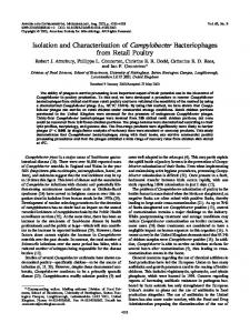
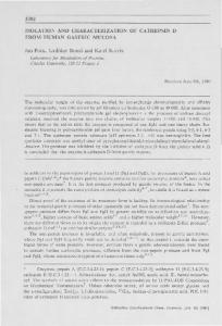
Presence of Urokinase-like Activity in Plasma-Analysis of fresh plasma by a zymographic gel procedure (26), for the presence of plasminogen activators shows twobands of activity: these are entirely accounted for by prekallikrein and factor XII, and no activity is seen near the molecular weight regions, 53,000 or 33,000, which are characteristic of urinary urokinase. A similar analysis of plasma aged for 3 to 5 days in the cold showed a much richer spectrum of plasminogen-dependent fibrinolytic bands corresponding especially to molecular weights above 100,OOO. When such aged samples were incubated for long periods (>24 h) to permit the development of activity bands, a weak zone of lysis appeared at a position corresponding to the migration of the 53,000 component of urinary urokinase. Stronger evidence for the presence of a urokinase-like molecule was obtained by passing fresh citrated or heparinized plasma over a column of washed glassbeads (CPG-10,ElectroNucleonics Inc.). After washing extensively with PBS, the column eluate obtained with 1 M KSCN contained an enzyme with the following properties. 1) It hydrolyzed the urokinase substrate S-2444. This reaction was inhibited by rabbit antiurokinase-Ig, obtained from antisera raised against highly purified human urinary urokinase, and isolated by affinity chromatography on columns of purified antigen. The hydrolM soybean trypsin ysis of S-2444 wasfully resistant to inhibitor, a characteristic property of urokinase. 2) When analyzed by the zymographic gel procedure (26), the KSCN fraction contained a plasminogen activator whose electrophoretic mobility was identical with the 53,000 component of urinary urokinase. While these observations suggested that plasma contained either a urokinase-like molecule or its precursor, the concentration in plasma was too low, and the adsorptive capacity of glass bead columns wastoo limited, to permit the isolation of useful amounts by this procedure. Since the “contact activation” and adsorptive properties of glass are considered to result in large part from the surface distribution of negative charge, we explored the use of the cation exchanger BioRex 70 for fractionating fresh plasma with the aim searching for urokinase-like enzymes.When fresh human plasma was adsorbed to BioRex 70 and eluted in the presence of benzamidine (as described under “Methods”) the eluate contained plasminogen-dependent fibrinolytic activity in the 80,000 to 90,000 region that was visualized in the fibrin-agar overlay (Fig. 1, slot 1);no fibrinolytic activity was present in the unadsorbed fractions. In work to be described elsewhere it hasbeen shown that the 80,000 to 90,000 activity is due largely, and perhaps entirely to kallikrein, factor XIIa, or a combination of the two. If this eluate was dialyzed to remove benzamidine, a new plasminogen-dependent fibrinolytic zone appeared in the 53,000 region (slot 2). Further development of activity and the appearance of lytic zones at lower molecular weight were achieved by incubating the di-
Downloaded from www.jbc.org by guest, on July 12, 2011
absorbance change a t 405 nm were made 13 min and 15 min, respectively, after plasminogen addition. A linear relationship between the rate of absorbance change and amount of activator was obtained in the range 0.1 to 1Ploug unit. Active Site Titration of Urinary Urokinase and Plasma Urokinase-The molarity of enzymatically active urokinase in solution was determined employing the conditions of Jameson et al. (28). A stock solution containing 2.4 ml of Na-barbital, pH 8.3, 0.5% Triton X-100, and 0.006 ml of 0.01 mM MUGB in 1 mM HCl was prepared. Aliquots (0.35 ml) of this solution were mixedwith 50 pl of 0.1%SDS or sample in 0.1% SDS. The increase in fluorescence intensities was recorded in a Hitachi MPF 2 spectrofluorimeter. The excitation and emission wavelengths were set a t 365 and 445 nm, respectively. The fluorescence increase reached a plateau after about 20 min indicating that the turnover of the titrant was negligible. The amount of the plasminogen activator was calculated from the fluorescence increase in 30 min in comparison with a reference standard containing a known amount of 4-methylumbelliferone and corrected for spontaneous hydrolysis of a blank incubated under identical conditions. This procedure allowed determination of as little as 0.1 to 1.0 X 10”’ mol of activator. Determination of Protein Concentration-Protein concentrations were determined by absorbance measurement at 280 nm assuming a specific absorbance of E% of 10 unless more accurate figures were available from the literature, as in the case of plasminogen (17), urokinase purified by benzamidine-Sepharose 4B (13), and Ig (11.8). Proteins purified by preparative SDS-PAGE were measured by the fluorescamine assay (29). Before protein determinations, the samples were dialyzed extensively against 0.1% SDS. A 50-pl aliquot of the dialyzed sample was mixed with 0.25 ml of 0.1 M Na-borate, pH 9.2, fluorescamine (0.025 mg/ml in acetone, 0.1 ml) was added and the sample was mixed on a Vortex shaker. A standard curve was constructed using varying concentrations of bovine y-globulin. Tryptic Peptide Mapping-The radioiodination of proteins in polyacrylamide gel slices, tryptic digestion, and peptide mapping were carried out as described by Elder et al. (30). Amino Acid Analysis-The protein solution (approximately 5 to 10 pg in 0.1% SDS) in Corning No. 9820 tube (10 X 75 m m ) was adjusted to 20% in trichloroacetic acid by adding 100% (w/v) trichloroacetic acid and incubated on ice overnight. The precipitate was recovered by centrifugation at 12,000 X g for 1 h. The precipitate and the wall of the tube were washed twice with 0.1 ml of dry methanol without stirring up the precipitate. Methanol was evaporated in a Savant Speed Vac concentrator. Constant boiling 6 N HCl(75 pl) was added, the tubes were sealed under vacuum ( 4 5 pm),and the samples were hydrolyzed a t 110 “C for 24 to 72 h. The amino acid composition was determined with a DurrumSpinco D-500 amino acid analyzer according to the method of Moore and Stein (31). Cysteine was determined as cysteic acid after performic acid oxidation according to the method of Hm (32). Immunodiffusion Analysis-Double immunodiffusion analysis according to the method of Ouchterlony and Nilsson (33) was carried out in 1%agar gels in 0.01 M sodium barbital, 1%Triton X-100,50 mM benzamidine. The samples obtained from preparative SDS-PAGE were adjusted to 2% Triton X-100, then added to the punched wells and allowed to diffuse overnight at 37 “C. Radioimmunoassay for Urokinase-A competitive radioimmunoassay for urokinase was developed by the following procedure. Commercial urokinase was purified by p-aminobenzamidine-Sepharose chromatography as described above, catalytically inactivated by treatment with iPr,P-F and then iodinated with ‘25iodineusing a modification of the chloramine-?‘ method (34). To free 1251-urokinase from material that had lost immunoreactivity during iodination the product was purified by immunoaffinity chromatography on acolumn of anti-urokinase-Sepharose.Antibodies were raised against highly purified urokinase as described above, goat anti-rabbit serum was obtained from Behring Calbiochem, and therespective IgG fractions were isolated by protein A-Sepharose chromatography. The tracer antigen (lz5I-iPr2P-urokinase)was stored in PBS supplemented with bovine serum albumin (0.1 mg/ml) and stored at 0 “C. Radioactivity of the tracer mixture was 0.4 pCi/ml. The binding capacity of the IgG fraction was estimated by determining equivalence points with defined amounts of antigen using the Ouchterlony double immunodiffusion technique: for anti-urokinase IgG, it was found to be 150 pg/ ml, and for goat anti-rabbit IgG, 250 pg/ml. To 830 pl of fresh citrated plasma (obtained from the New York Blood Center) were added 50 p1 of tracer antigen and 100 pl of1:lOOOdilution of anti-urokinase IgG, and the samples were kept on ice overnight. Then, lop1of nonimmune
Human Plasma Urokinase
100- :.
80 53 47 -
z
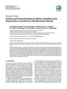
unacceptably low. 3) Columns based on affinity-purified, specific anti-urokinase Ig proved effective. Even so, the Cohn fraction IV-1 extracts required two cycles of immunoaffinity chromatography before achieving a degree of enrichment that permitted the isolation of highly purified plasma urokinase. Under these conditions, optimal results were obtained only when the amounts of unspecifically bound protein were reduced, firstly by washing the column with buffers containing Triton X-100 (1%) before elution of enzymes and, secondly, by passing the extracts over a Sepharose column to which purified, rabbit preimmune IgG had been bound. As seen from the specific activity data in TableI,the enzyme recovered after two cycles of immunoaffinity chromatography was quite impure, andwas estimated to account for only a minor fraction of the total protein. Zymbgraphy of SDS-polyacrylamide gels showed plasminogen-dependent fibrinolytic activity in two regions corresponding approximately to 53,000 and 1OO,OOO, respectively (Fig. 2 A ) . When the electrophoretic gels were stained with Coomassie blue, a distinct band corresponding to 53,000 wasobserved; it was clearly separated from the bulk of contaminating proteinswhich were concentrated predominantly in the zones of high molecular weight, suggesting that this urokinase-like component might be isolated by preparative SDS-PAGE, a procedure in which the enzymatic activity of urokinase survives essentiallyintact. To test for the retentionof their characteristic electrophoretic mobility after purification, each of the fibrinolytic zones ( a ,b) and other nonlytic zones (c, d, e) were excised from lanes parallel to thatshown in Fig. 2 4 . These were sliced, extracted TABLE I A summary of the purification of Cohn IV-1 urokinase-like enzyme Protein
Fractiom
tivity
Specific activity
Total ac-
alyzed fractions a t 37 "C (slots 3 and 4). When the dialyzed mg P b u g units/mg Ploug units fractions was passed through a column consisting of affinity I. Cohn IV-I 3,000,000" purified rabbit anti-urokinase Ig-Sepharose4B inthe absence 11. 1st affinity chroma56.7* 59.4 3,368 of benzamidine, the 53,000 component was not present either tography in the unadsorbed fractions or in the material eluted with a 111. 2nd aftinity chro3.4b 529 1,776 glycine/HCl buffer wash (pH 2.2) (slots5 and 6 ) . If benzamimatography SDS0.047' 27,659 1,300 dine was present throughout thisprocedure, the 53,000 activ- IV. Preparative gel electrophoresis (122,000)" ity wasrecovered in the glycine/HCl eluate where it was a Wet weight of Cohn IV-1. accompanied by a weakerbut readily discernible lyticzone in * Protein estimated by AM, assuming A % = 10. the 100,000 region (slot 7). Protein estimated by the fluorescamine assay. Isolation of Urokinase from Plasma Fractions-The proObtained by S2444 assay and MUGB active site titration. cedure outlined above was reproducibleand yielded quantities of enzyme sufficient for enzymological work but not for tests of purity or detailed characterization. The relative instability MW A a b c d e of the enzyme during isolationlimited theapplication of x IO-^ BioRex chromatographyon alargescale, anoption also a-. discouraged by the cost of fresh plasma. In searching for a partially enriched starting material,we assayed the standard lOO-b-!"l Cohn fractions by the zymographic procedure (26) and idenCtified the enzyme in fractions 111, IV, and IV-1, of which the last proved to be the most convenient starting material for 53-eprocessing on a larger scale. Isolation of Urokinase-like Activity from Cohn Fraction dIV-I-Cohn fraction IV-1 was dissolved and acidified to pH 2.0 to inactivate residual protease inhibitors and then neutralized before application to immunoaffinity columns. The resolving power of these columns was influenced by several FIG.2. SDS-PAGE and zymography of the irnmunoaffinity factors. 1) The presence of benzamidine (10 to 100mM) purified activator from Cohn fractionIV-1. A, the zymography. throughout the procedure was essential for recovery of any The SDS-PAGEgel was extracted with 2.5%Triton X-100 for 50 min, activity, presumably because it protected theenzyme against and overlaid onto a fibrin-agar plate. B, re-electrophoresis and reovdegradation by other contaminating proteases. 2 ) Columns erlay of the samples obtained from A. The gel in A was sliced a t positions a , b, c, d, and e. Each slice was incubated with 1 ml of 50 based on purified but otherwise unfractionated total Ig did mM NHIHCO:~ overnighta t 37 OC. The supernatants were then lynot give satisfactory results: both theireffective capacity, and ophilized, electrophoresed, and assayed in the usual way on a fibrinthe degree of purification of urokinase-like material were agar indicatorgel.
I"
Downloaded from www.jbc.org by guest, on July 12, 2011
FIG.1. The presence of urokinase-like activity in plasma fractions after chromatography on BioRex 70:detection by zymography after SDS-PAGE. For experimental details, see "Methods." Slot 1, BioKex 70 eluate prior to dialysis: activity present in the 80,000 to 9 0 , 0 0 0 region, due to factorXI1 and/or prekallikrein. Slot 2, BioRex 70 eluate dialyzed overnight in the cold against 0.052 M NaPOl buffer, pH 7.4, containing 5 mM EDTA: a prominent lytic zone has appeared a t 53,000. Slot 3, the dialyzed eluate incubated a t 37 "C for 1.5 h. Slot4, the dialyzed eluate incubated a t 37 "C for 8.5 h: the zones corresponding to factor XI1 and/or prekallikrein have disappeared, and a new zone a t -47,000 has appeared. Slot 5, the unadsorbed fraction obtained by passing the dialyzed BioRex 70 eluate (without incubation a t 37 "C) through affinity purified antiurokinase-Ig-Sepharose 4B column (1.1 X 2 cm) in the absence of benzamidine: the factor XII-prekallikrein has not adsorbed to the column, but the53,000 zone has. Slot 6, the eluate obtainedby eluting the anti-urokinase-Ig-Sepharose 4B (as described in slot 5) with 0.1 M glycine/HCl, pH 2.2: no urokinase-like activity isrecovered. Slot 7, the eluate obtained by adsorbing the BioHex 70 eluate to anti-urokinase-Ig-Sepharose 4B in the presence of 0.1 M benzamidine and eluting with 0.1 M glycine/HCI, pH 2.2: a 53.000 urokinase-like activity and a 1 0 0 , 0 0 0 lytic factor were recovered.
3279
3280
Human Plasma Urokinase A
B
C
MW
x~0-3
5333-
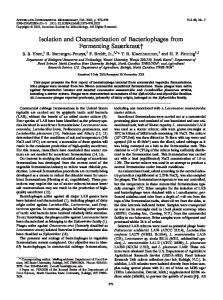
FIG.3 (left). SDS-PAGE:of the Cohn IV-1 enzyme, 53,000 and 33,000 urokinase purified by preparative SDS-PAGE. A, Cohn IV-I activator; B, 53,000 urinary urokinase; C, 33,(xK) urinary urokinase. FIG.4 (right). Immunodiffusion. The immunodiffusion was performed as described under “Methods.” Well 1 (the center well) contained anti-urokinase-Ig purified by DEAE-Sephacel and affinity chromatography on a column of highly purified urinary urokinaseSepharose 4B; wells 2 and 5, 33,000 urinary urokinase; well .3, Cohn fraction IV-1 53,000 enzyme; well 4,53,000 urinary urokinase.
nated peptides resulting from tryptic digestion of the lzSIlabeled proteins. Amidolytic Activity, Plasminogen Actiuation, a n d Effect of Some Macromolecular Inhibitors-The Cohn fraction urokinaseisolated afterpreparativeSDS-PAGEretained full activity for -3 weeks when stored in 0.1% SDS at 4 “C and slowly lost activity during a 6-month period. This concentration of SDS blocks all enzymatic functions, but activity was fully restored if SDS was removed from the protein by sequestration in micelles of nonionic detergents. We found that a final concentration of 0.5% Triton X-100 fully reactivated both urinary and Cohn fraction urokinase, and the two enzymes were accordingly compared under these conditions. The exact molarity of active enzyme in each case was determined by active site titrationusing hydrolysis of MUGB (29), and theamidolytic activity was then established by measuring the rates of hydrolysis of the urokinase substrate S-2444. When normalized fordifferences in molaritymeasured by active site titration the results (Table11) showed that, within the limits of experimental error, Cohn fraction and urinary urokinase were identical in amidolytic activity, both being less active, on a weight basis, than the 33,000 speciesurinary urokinase. The values observed here for urinary urokinase were very close to those reportedby others (11). Plasminogen activation by these enzymes was assayed by a TABLE I1 method in which theproduct plasmin, was measured by Apparent specific activity of urokinase and Cohn IV-1 activator hydrolysis of the chromogenic substrate S-2251. The data Apparent specific activity” (Table 11) again show identical activity for Cohn fractionand S-2444 assay S-2251 &.say 53,000 urinary urokinase. The 33,000 component of urinary PIoug units/mg urokinase was less efficient in plasminogen activation than 89,500 f 2,600 207,000 f 7,000 Urokinase, 33,000 dalton superiority in the 53,000 enzymes, in contrastbothtoits 109.500 f 4,600 119,000 f 5,000 Urokinase, 53,000 dalton amidolysis of the small substrate S-2444, and to the reportof 119,000 f 4,000 Cohn IV-1, 53,000 dalton 122.000 f 3,600 Barlow and White(11). The different assaysused may account “The activities were assayed by hydrolysis of S-2444 or S-2251 for this discrepancy. using urokinase (Leo) as reference standard enzyme. Concentration Resistance tomost macromolecular protease inhibitors is a was measured by MUGB titration (as described under “Methods”) striking characteristic of urinary urokinase and analogous using a 30-min incubation period at room temperature. Each specific activity value reflects the average of three independent determina- plasminogen activators of murine origin (36). We compared the effects of four inhibitors on Cohnfraction and 53,000 tions.
Downloaded from www.jbc.org by guest, on July 12, 2011
by incubation a t 37 “C overnight in 1 ml of NH.8HCO:3(50 mM), and the solutionswere lyophilized and then reanalyzed by SDS-PAGE and zymographywithfibrin-agaroverlays. The 53,000 lytic species had retained its mobility; it was not converted to any other detectable components. In contrast, the 100,000 band had disappeared completely and been replaced by a component with mobility inseparable from 53,000 urokinase,suggesting that the largerspecies, whateverits nature, might be a precursor to53,000 urokinase. The material recovered after two cycles of affinity chromatography was concentrated and subjected to preparative SDS-PAGE. Fractions of 1 ml were collected and an aliquot of each wasanalyzedbyzymographyfor the presence of fibrinolytically active materialmigrating a t 53,000. The active fractions containing 53,000 enzyme were pooled and assayed. A summary of the purification of Cohn fraction IV-1 enzyme is given in Table I: 47 pg of apparently homogeneous urokinase wererecovered from a total of 3 kg wetweight of Cohn fraction IV-1. Because no quantitative assayof urokinase-like activity could be performed on the crude starting material, we do not have an exact estimate of the overall yield or degree of purification. The specific enzymatic activity of Cohn fraction urokinase was first determined by direct hydrolysis of the synthetic substrate S-2444. This procedure gave a value of 28,000 Ploug units/mg of protein (Table I), which was considerably below that previously reported for 53,000 urinary urokinase (12). However, when this value was normalized to the concentration of active enzyme measured by active site titration, the specific activity was identical with that of 53,000 urinary urokinase (Tables I and 11). Thus, a large fraction of Cohnfraction enzymeconsisted of enzymaticallyinactive protein. The inactive protein is almost certainly urokinase that was denatured and inactivated during immunoaffinity chromatography, rather than contaminating unrelated protein: this was demonstrated by subjecting highly purified 53,000 urinary urokinase tochromatographyonthesame immunoaffinity columns used for isolating Cohn fraction enzyme, the specific activity of the urinary enzyme recovered after elutionwas reduced to 30,000 Ploug units/mg of protein, but was unchanged after correction by active site titration. Evidently, a largefraction of adsorbed urokinaseis irreversibly denatured during adsorption and elution from the anti-urokinasecolumn. Theelectrophoretic mobilities of purified Cohn fraction enzyme, 53,000 and 33,000 urinary urokinase are compared in Fig. 3. The apparent molecular weights of Cohn fraction and 53,000 urinary urokinase areidentical. Comparison of Cohn Fraction Urokinase-like Enzyme and Urinary Urokinase-The parameters used for this comparison were catalytic activity with both small substrates and plasminogen, susceptibility to several macromolecular inhibitors, immunological specificity, electrophoretic mobility, amino acid composition, and two-dimensional maps of iodi-
Human Plasma Urokinase
3281
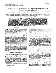
urinary urokinase, using the amidolytic assay with S-2444 as substrate: both enzymesbehavedidentically and were not inhibited by soybean trypsin inhibitor (20 pg/ml), lima bean trypsin inhibitor (30 pg/ml), Trasylol (0.7 pg/ml), and Erythrina inhibitor (30 pg/ml). Immunological Properties-Whenanalyzed by Ouchterlony double immunodiffusion using either rabbit antisera or affinity purified antibodies, Cohn fraction urokinase gave a line of identity with 53,000 urinaryurokinaseandpartial identity with 33,000 urinary urokinase (Fig. 4). When incorporated into the fibrin agar overlay used for
50min
B
120 min
" A B a b A' 6' a' b'
MW
1
zymographic detection of fibrinolysis affinity purified antiurokinaseinhibitedCohnfractionenzyme as well as both components of urinary urokinase (Fig. 5). AminoAcidAnalysis a n d Tryptic Peptide MappingThese two structural parametersprovide further evidence for -Ab +Ab -Ab +Ab the identityof Cohn fraction and urinary urokinase. The data FIG. 5. Inhibition of the fibrinolytic activity of urinary uro- in Table I11 document thesimilarity betweenthe two enzymes kinase and the activator from Cohn fraction IV-1 by aftinity andare generally in accordwith the previously reported purified anti-urokinase-Ig. SIX-PAGE and the zymography were values in the literature (11, 37). For several amino acids our performed as described under "Methods." A, a, A', and a' contain values appear to differ significantly from those of previous equal amounts of immunoaffinity-purified activator from Cohn IV-1. workers. These differences may reflectslightvariations in B, h, B', and h' contain equal amounts of benzamidine-sepharose- conditions of hydrolysis, and they do not obscure the overall purified urokinase. The fibrin-agar for a. h, a', and h' contains 0.01 similarity in amino acid composition between the plasma and mg/ml of affinity-purified anti-urokinase-Ig;that for A, B,A ' , and B' urinary enzymes. does not. A, B,a, and h were incubated for 50 min. A', B',a', and h' To obtain tryptic peptide maps, samples of Cohn fraction were incubated for 120 min. TABLE 111 Amino acid composition of urokinase and Cohn IV-1 activator Amino acid residues per 33,000 urokinase
This papeP
White el al. (11)
Asph 19.0 Thr 34.3' Ser Glu 28.8 Pro 17 46.0 GlY 27 Ala CYS9 22.7 Val 11 Met 18.7 Ile 15 36.7 Leu 24 18.3 Tyr 12 Phe His LYS Arg Try
19.1 25.0 f 0.1' 20.0' 2220.2' 28.3 33.9 & 0.2 16.4 15.2 f 0.1 22.4 29.4 f 1.9 13.9 f 0.5 9.0 15.1 f 0.1 10.9 11.5 f 0.1 4.5 4.3 f 0.3 15.6 12.8 f 0.1 20.6 24.4 f 0.2 12.8 11.8 +- 0.1 9.8 f 0.1 9.0 10f 0.1 17.4 19f 0.3 15.5 f 0.3 N.D.
53,000 urokinase Johnson et al. (37)
24
This
45.2 f 0.3"
White et al. (11)
Johnson et al. (37)
40.4
43
Cohn IV activator
42.4
21.8 33
51.849.4 f 0.148 44.1 27.0 f 0.130 28.7 45.3 45 41.1 f 0.1 10.1 24.0 26.0 14 23 20.2 f 0.4 20 19.4 26.4 f 0.1 24.6 f 1.0 22 23.2 5 7.7 6.4 f 0.5 8 17.2 18 20.4 & 0.2 33.6 f 0.1 33 17.8 19 19.9 f 0.6 9.3 9 14.3 14 15.2 & 1.2 15.7 10.2 15.9 23 19.7 16.8 f 0.1 17.3 27.1 f 0.3 27.4 35 31.0 14.3 15 21.4 2522.5 +- 0.3 22.1 N.D. 4.9 N.D. N.D. 9.6 " These values were obtained from 24-, 48-, and 72-h hydrolysates extrapolated to zero time unless otherwise indicated. Values from performic acid oxidation; relative to aspartic acid. ' Standard deviation. Values from 24-h hydrolysates.
24.7 6.4 37.7
N.D.
Downloaded from www.jbc.org by guest, on July 12, 2011
FIG. 6 . Tryptic peptide maps of the urinary urokinase and the activator from the Cohn fraction IV-1.A, 33,000 urinary urokinase; B,53,000 urokinase; C, 53,000 enzyme from Cohn fraction IV-1. D,diagram of the major peptides in autoradiogramA to C. The shaded spots are missing in 33,000 urinary urokinase.
3282
Human Plasma Urokinase
Downloaded from www.jbc.org by guest, on July 12, 2011
of it is present in an inactive form. However, since we have not yetisolated an inactive form from plasmawe do not know whether it is a proenzyme or exists as a complex with a specific inhibitor. Whatever the precise nature of the inactive circulating form, it does seem likely that it is an intrinsic physiological constituent of plasma, and not simply a transient inhibitor complex targeted for removal and breakdown. Because numerous tissues secrete enzymes indistinguishable from urokinase to generate the localized proteolysis needed for a wide range of physiological processes, it might be considered that the enzyme we have isolated is merely released from a complex LOG C formed with one of the many protease inhibitors that are FIG. 7. Logit BT versus log c (ng/ml) plot of a radioimmunoassay for urokinase in human plasma. All concentrations were present at high concentration in blood and body fluids. This appears unlikely, because all of the known protease inhibitors, assayed in duplicate. The slope of the curve was obtained by linear regression analysis of the mean values of logit BT. Crosses indicate several of whichbind rapidly to urokinase, form covalent the standard values and open circles indicate the values obtained for complexes that are not dissociated under our conditions of pooled plasma samples. enzyme purification. It also seems unlikely that theenzyme is liberated by proteolysis of such complexes, becauseproteolysis and urinary urokinase were concentrated by SDS-PAGE, would be expected to yield a population of enzyme molecules recovered in slices containing the respective bands, iodinated, that was either very heterogeneous in size, or contained modigested with trypsin, and analyzed by two-dimensional elec- lecular species larger than urokinase, or both. In fact, the trophoresis and chromatography as described by Elder et al. enzyme obtained from Cohn fractions or observed in zymog(30). The pattern of iodinated tryptic peptides was then ob- raphy of plasma is appreciably less heterogeneous on electrotained by autoradiography. The autoradiograms in Fig.6 show phoresis than the well characterized urinary urokinase. identical patterns for Cohn fraction and 53,000 urinary uroWe cannot as yet draw any conclusionsconcerning the kinase, and a largely, but notentirely similar iodinated peptide physiological significance of plasma urokinase or prourokinase, if the latter exists. Our immunoassay data indicate that map for 33,000 urinary urokinase. plasma contains at least 10 pg/liter of urokinase-reactive Radioimmunoassay of Urokinase in Human PlasmaSamples of fresh, pooled human plasma were obtained from material, a figure that is consistent with the levels of enzyme a blood bank and assayed for urokinase-like antigens. The extrapolated from the quantities observed in Cohn fraction. results of the radioimmunoassay are given in Fig. 7 and yield Given the catalytic efficiency of urokinase, even this low concentration ( a )could rapidly generate appreciable amounts a mean value of 12 pg of urokinase/liter of plasma. of plasmin, especiallyif activation was localized,and ( b )would DISCUSSION yield a level of fibrinolytic activity significantly greater than Our data provide for the first time a substantial body of that which could begenerated by the much larger amounts of coherent evidence forthe presence of a urokinase-like enzyme circulating prekallikrein, assuming that the latter is capable in human plasma. That being so, our results should be assessed of functioning inblood as a plasminogen activator. These in relation to three questions. 1) Are the enzymes we have considerations suggest that urokinase, or its inactive precurisolated from Cohn fractions, and the activity generated from sor, should tentatively be consideredas a plausible, physiologplasma, essentially identical with urinary urokinase? 2) If so, ical activator of fibrinolysisintrinsic to plasma, although other what is the form in which this urokinase, or a closely related physiologically significant activators may also bepresent. molecule circulates in the plasma? 3) What might be the Acknowledgments-We thank Dm. Peter Blackburn and Stanford physiological relevance, if any, of the presence of urokinase, Moore for help with the amino acid analysis and Kart Grizzutti and or some precursor form in plasma? Marcia Lipski forexcellent technical assistance in the initial phase of The answer to thef i t question can be obtained by consid- this work. ering the aggregate of our findings which show that urinary REFERENCES urokinase (53,000) and the Cohn fraction enzyme are indistin1. Robbins, K. C., and Summaria, L. (1970) Methods Enzymol. 19, guishable by chromatographic, immunological, electropho184-199 retic, catalytic, and structural criteria. Although we have not 2. Landmann, H. (1978) in Fibrinolytic Enzymes and Anti-fibrinoexcluded minor differences in glycosylation orother analogous Zytics (Markward,F., ed) pp. 3-48, Springer Verlag, New York post-translational modifications, we conclude that these en3. Collen, D. (1980) Thromb. Haemostasis 43,77-81 zymes are otherwise essentially identical. Owing to the limit4. Ratnoff, 0. D., and Naff, G . B. (1967) J. Exp. Med. 165,337-358 ing amounts of material that could be purified directly from 5. Habal, F. M., Burrowes, C. E., and Movat, H. Z. (1976) in Kinins, Pharmacodynamics and Biological Role (Sicuteri, F:, Back, plasma we were unable to obtain as extensive a comparison N., and Haberland, G. L.,eds) p. 23, Plenum Press, New York between plasma and urinary urokinase: in this case, apparent 6. Virji,M. A. G., Vassalli, J.-D., Estensen, R. D., and Reich, E. identity was observed onlyin electrophoretic, chromato(1980) Proc. Natl. Acad.Sei. U. S. A . 77,875-879 graphic, and immunological behavior. However, since the 7. Reich, E. (1978) in Biological Markers of Neoplasia (Ruddon, Cohn fractions are derived entirely from plasma, there is no R. W., ed) pp. 491-500, Elsevier, New York 8.Astrup, T. (1978) in Progress in ChemicalFibrinolysis and reason to doubt that the enzymes obtained from the two Thrombolysis (Davidson, J. F., Rowan, R. M., Samama, M. M., sources are both the same. and Desnoyers, P. C., eds) Vol. 111, pp. 1-57, Raven Press, New Although it seems safe to conclude that urokinase, or a York molecule very closely related to it, is present in human plasma, 9. Christman,J. K., Silverstein, S. C., and Acs, G . (1977) in Research we cannot yet identify the form in which it is circulating. Monographs in Cell and Tissue Physiology (Barrett, A. J., ed) Urokinase-like activity can be detected in zymographic gels Vol. 11, pp. 90-149, North Holland, Amsterdam after aging a plasma sample, and the enzyme shows latency 10. Bernik, M. B., White, W. F., Oller, E. P., and Kwaan, H. C. (1974) J.Lab. Clin. Med.84,546-558 during the early stages of fractionation, indicating that most
Human Plasma Urokinase 11. White, W. F., Barlow, G. H., and Mozen, M. M. (1966) Bzochemi s t v 5,2160-2169 12. Rijken, D. C., Wijngaards, G., Zaal-de Jong, M., and Welbergen, J. (1979) Bwchim. Biophys. Acta 580, 140-153 13. Cole, E. R., and Bachmann, F. W. (1977) J. Biol. Chem. 252, 3729-3737 14. Nolan, C., Hall, L. S., Barlow, G. H., and Tribby, I. I. E. (1977) Biochim. Biophys. Acta496, 384-400 15. Binder, B. R., Spragg, J., and Austen, K.F. (1979) J.Biol. Chem. 254, 1998-2003 16. Radcliffe, R., and Heinze, T. (1978)Arch. Biochem. Biophys. 189, 185-194 17. Colman, R. W. (1969) Biochem. Biophys. Res. Commun. 35,273279 18. Kaplan, A. P., and Austen, K. F. (1972) J. Exp. Med. 136, 13781393 19. Venerrod, A. M., and Laake, K. (1976) Thromb. Res. 8,519-522 20. Laake, K., and Venerrod, A. M. (1974) Thromb. Res. 4,285-302 21. Mandle, R. J., Jr., and Kaplan, A. P. (1977) J. Biol. Chem. 252, 6097-6104 22. Goldsmith, G., Saito, H., and Ratnoff, 0.(1977) Thromb. Haemostasis 38, 136 (Abstr.) 23. Astedt, B. (1978) in BiologicaZMarkers of Neoplasia: Basic and Applied Aspects (Ruddon, R. W., ed) pp. 481-488, Elsevier,
North Holland, New York 24. Schleuning, W.-D., and Fritz, H. (1974)Hoppe-Seyler’s Z. Physiol. Chem. 355, 125-130
3283
25. Laemmli, U. K. (1970) Nature (Lond.) 227,680-685 26. Granelli-Piperno, A., and E. Reich, E. (1978) J. Exp. Med. 148, 223-234 27. Unkelesa, J., Dang, K., Kellermann, C. M., and Reich, E. (1974) J. Biol. Chem. 249,4295-4305 28. Jameson, G. W., Roberts, D. V., Adams, R. W., Kyle, W. S. A., and Elmore, D. T. (1973) Biochem. J . 131, 107-117 29. Bohlen, P., Stein, S., Dairman, W., and Udenfriend, S. (1973) Arch. Bwchem. Bwphys.155,213-220 30. Elder, J. H., Pickett, R. A., 11, Hampton, J., and Lerner, R. A. (1977) J. B i d . Chem. 252,6510-6515 31. Moore, S., and Stein, W. H. (1963) Methods Enzymol. 6,819-831 32. Hirs, C. H. W. (1956) J. BWZ. Chem. 219,611-621 33. Ouchterlony, O., and Nilsson, L.A. (1973) in Handbook of Experimental Immunology (Weir, D. W., ed) 2nd Ed, Chap. 19, Blackwell, Oxford 34. Krohn, K., Sherman, L., and Welch, M.(1972) Biochim. Biophys. Acta 285,404-413 35. Rodbard, D., Rayford, P. L., Cooper, J . A., and Roos, G . T. (1968) J. Clin. Endocrinol. Metab. 28, 1412 36. Dano, K., and Reich, E. (1975) in Proteases and Biological Control (Reich, E., Rifkin, D. B., and Shaw, E., eds) pp. 357366, Cold Spring Harbor, New York 37. Johnson, A. J., Soberano, M., Ong, E. B., Levy, M., and Schoellman, G. (1977) in Thrombosis and Urokinase (Paoletti, R., and Sherry, S., eds) pp. 59-67, Academic Press, New York Downloaded from www.jbc.org by guest, on July 12, 2011