GS3 rotor for 10 min at 4°C. The pellet was resuspended in denaturing solution [4 M guanidinium thiocyanate, 25 mM sodium citrate, pH 6.5,0.5% (masshol.) ...
Eur. J. Biochem. 219,441-448 (1994) 0 FEBS 1994
Isolation and complete sequence of CBR, a gene encoding a putative cytochrome b reductase in Saccharomyces cerevisiae Michael CSUKAI, Michael MURRAY and Elisha ORR Department of Genetics, University of Leicester, Leicester, England (Received September 7, 1993) - EJB 93 1359/2
We have isolated and characterised a novel yeast gene, CBR (cytochrome b reductase), encoding a 35-kDa yeast novobiocin-binding protein. The predicted protein sequence of CBR displays considerable similarity to both plant nitrate reductases and mammalian cytochrome b, reductases indicating that it is a putative member of the flavoprotein pyridine-nucleotide-cytochrome-reductase family. Disruption of CBR is not lethal under various growth conditions, suggesting the presence of some functional overlap with other reductases, possibly with the cytochrome P-450 reductase.
Coumarin antibiotics such as novobiocin are thought to affect DNA metabolism in bacteria [ l ] by competing with ATP for the nucleotide-binding site in the B subunit of DNA gyrase. This enzyme is a type I1 topoisomerase which introduces negative supercoils into relaxed, covalently closed, double-stranded DNA molecules [2, 31. Novobiocin also inhibits some eukaryotic type I1 topoisomerases in vitro, although at 1000 times the concentration required to inhibit bacterial gyrases [4]. Similar to its activity in bacteria, the drug inhibits a number of cellular processes in higher eukaryotes, such as DNA synthesis [5-91, transcription [lo, 111, differentiation [12] and alters chromatin structure [13, 141. This activity and the high degree of sequence similarity between gyrases and eukaryotic type I1 topoisomerases [15] have led to the presumption that the eukaryotic enzyme is the target of the drug. Nevertheless, a number of studies have indicated that topoisomerase I1 is not the target of novobiocin in vivo or in vitro [13, 14, 161. These observations are consistent with the response of the yeast Saccharomyces cerevisiae to the antibiotic. In drugsensitive strains of S. cerevisiae, novobiocin arrests cells in the G1-phase of the cell cycle [17]. This phenotype differs from the terminal phenotype of yeast top2 (ts) mutants. Furthermore, the screening of hundreds of novobiocin-resistant mutants failed to identify any yeast mutant defective in type I1 topoisomerase 117, 181. Finally, unlike bacterial gyrase, the yeast topoisomerase I1 does not specifically bind novobiocin in vitro [19]. Recently, interest in the use of novobiocin in cancer chemotherapy has been revived by the observations that novobiocin inhibits, synergistically with a number of drugs, the proliferation of cancer cells [20-221 and reduces cell killing mediated by cytotoxic T-cells 1231. In an attempt to identify the biochemical target(s) of novobiocin in yeast, we have Correspondence to E. Orr, Department of Genetics, University of Leicester, Leicester, England LE1 7RH Abbreviations. YPD, yeast peptone dextrin; PAP, peroxidase anti-peroxidase IgG. Note. The novel nucleotide sequence data published here have been submitted to the EMBL sequence data bank(s) and are available under accession number(s) 228365.
isolated eight proteins from total cell extracts which bind novobiocin-Sepharose. Of these, three have previously been characterised: the yeast myosin heavy chain 124-271; Sup45, a protein conferring novobiocin-resistance in vivo [18]; the P-subunit of the mitochondria1 F,-ATP synthetase [19]. The remaining five proteins include a novel 200-kDa structural protein (our unpublished results) and four novel proteins, two of molecular masses 20 kDa, 29 kDa and two of 35 kDa. In this study we describe the purification and characterisation of the 35-kDa protein which has been identified as a novel yeast cytochrome reductase. The predicted amino acid sequence of the yeast gene shows 34-37% identity to the catalytic domain of mammalian NADH-cytochrome b, reductases, as well as 30-33% identity to the C-terminal, FAD-binding domain of Arabidopsis and tomato nitrate reductases. The mammalian cytochrome b, reductase is a component of the endoplasmic reticulum electron-transport chain, involved in the desaturation and elongation of fatty acids [28-301, cholesterol biosynthesis [31] and in the detoxification of xenobiotics [32, 331. The yeast gene includes 13 of the 14 amino acids characteristic of a family of flavoenzymes, the flavoprotein pyridine-nucleotide-cytochrome reductases, which includes the cytochrome b, and nitrate reductases [34]. The fourteenth amino acid in the yeast sequence, isoleucine, is a conservative substitution for leucine, found at the corresponding position in other cytochrome reductases belonging to the same family.
MATERIALS AND METHODS Strains Saccharomyces cerevisiae, 842, 8 4 2 ~ '[25], 483 (MATa, SUQS, ade2-1, his5-2, lysl-1, canl-100, ura3-1, PNMlly-) and 8HA (MATa, leu2, adel, ura3, trpl) strains were routinely grown on YPD (l%, masslvol., yeast extract, 2%, mass/vol., bactopeptone and 2%, masshol., glucose). Escherichia coli, RR1 2 ~ [35] 1 were ~ 'grown ~ ~ on LuriaBertani medium (1% Difco bacto tryptone, 0.5%, masslvol., Difco bacto yeast extract and 0.5%, masslvol., NaCl).
442
Molecular biological techniques Standard molecular biological techniques were as described by Sambrook et al., [36], unless otherwise stated. Preparation of novobiocin-Sepharose Novobiocin was coupled to Sepharose following the procedure described by Staudenbauer and Orr [37]; 10 g epoxyactivated Sepharose CL 6B (Pharmacia) was swollen in 1.O 1 distilled water for 60 min at room temperature and washed on a scintered glass filter with 1.0 1 0.3 M Na2C0,, pH 9.5 (buffer A). The gel was mixed with a solution of 1 g novobiocin (Sigma) in 30 ml buffer A and gently shaken for 16 h at 37°C. Excess epoxy groups were blocked by the addition of ethanolamine (final concentration 1 M) and shaking was continued for a further 4 h at 37 "C. The product was washed sequentially in 1 1 0.5 M NaCl in buffer A, distilled water, 0.5 M NaCl in 0.1 M Sodium acetate, pH 4.0 and distilled water. Care was taken to protect the novobiocin from strong light at all times. Fractionation of yeast extract Yeast cells were grown in YPD medium to rniddlellatelogarithmic (fermentative) phase, harvested by centrifugation and the pellet was frozen at -20°C. Cells were broken using a Hughes press, cooled to -20°C. The resulting cell lysate was made to 5 mM MgCI, (final concentration) and incubated with DNAse I on ice for 15 min with protease inhibitors ; soya bean trypsin inhibitor (10 pg/ml), phenylmethylsulphonyl fluoride (0.5 mM) and benzamidine (1 mM). An equal volume of 2 M KCI was added and the lysate was centrifuged at 185000Xg for 60 min at 4°C. The clear fraction of the supernatant was loaded onto a novobiocin-Sepharose affinity column. Proteins were eluted in a series of steps in the presence of, respectively, 1 M KC1, 2 M KC1, and finally 5 M urea in buffer B (25 mM Hepes, pH 8.0, 1 mM dithiothreitol, 1 mM EDTA, lo%, by vol., ethylene glycol and 100 mM KC1) at 4°C. Proteins were precipitated overnight at 4°C in the presence of 5% (masshol.) trichloroacetic acid (final concentration) and pelleted by centrifugation in a bench-top centrifuge (13 000 X g ) at room temperature for 10 min. The proteins were resuspended in protein sample buffer [0.5 M TriskICI, pH 6.8, 25% (by vol.) glycerol, 6% (masshol.) SDS, 0.14 M 2-mercaptoethanol, 0.025% (mass/ vol.) bromophenol blue]. Samples were neutralised by the addition of a minimum volume of saturated Tris solution. Recovery of antigen and the raising of antiserum Antibodies were raised in rabbits after several injections of 30-50 pg protein. Proteins in the 5 M urea fraction eluted from the novobiocin column were separated by SDSPAGE. Slices containing the 35-kDa proteins were cut from stained gels, destained and soaked successively in ethanol and sterile distilled water for several hours to remove residual acid. The gel slices were mixed with an equal volume of sterile distilled water and liquidised by sonication. The solution was mixed with an equal volume of Freund's complete Adjuvant further sonicated and passed several times through an 18G needle before injection. After the primary injection, the antigen was suspended in Freund's incomplete Adjuvant for subsequent injections. The anti-(35-kDa) antibodies were affinity purified by incubating the rabbit serum with the 35-kDa proteins blotted
onto a nitrocellulose strip. The strip was washed four times in NaClRris (150 mM NaC1, 10 mM Tris/HCl, pH 7.4). Antibodies were eluted with cooled 0.2 M glycineLHC1, pH 2.2, at 4 "C for 5 min, immediately neutralised with 2 M Tris base and mixed with bovine serum albumin 1% (final concentration) and dialysed against NaCVTris containing 1OmM sodium azide.
Western blots Proteins were electroblotted from SDS/acrylamide gels onto nitrocellulose membranes (Schleicher & Schuell) at 300 mA for 20-30 min [38]. Proteins were visualised with Ponceau-S (BDH) which was subsequently removed by washing in NaCIRris. The membrane was incubated overnight at 4°C in NaCl/Tris containing 3% (mass/vol.) bovine serum albumin, washed twice at room temperature in NaCl/ Tris and incubated with primary antibodies, diluted in NaCl/ Tris containing 1% (mass/vol.) bovine serum albumin, for 2 h at 37°C. Following four washes in NaCl/Tris at room temperature, the membrane was incubated with secondary IgG (goat anti-rabbit) in NaClRris for a further 2 h at 37"C, washed four times in NaCUTiis and incubated with the tertiary reagent, rabbit peroxidase anti-peroxidase IgG (PAP) diluted 1: 1000 in NaCVTris. The membrane-bound complex was visualised at room temperature after incubation in the developer, 30 mg of 4-chloro-1-naphthol dissolved in 10 ml of methanol mixed with 30 ml NaCI/Tris containing 0.1% (by vol.) H,O,.
Construction and screening of pEX expression library S. cerevisiae genomic DNA from strain 8 4 2 ~ 'was partially digested with Sau3AI and ligated into the expression vectors pEXl, pEX2 and pEX3 [39], digested with BamHI and dephosphorylated. Recombinant plasmids were transformed by electroporation into E. coli RR1 Transformants were selected on ampicillin-containing medium at 30°C and lifted onto nitrocellulose filters (Schleicher & Schull) which were placed, colonies facing upward, onto fresh ampicillin-containing plates for a further 4 h at 30°C. The synthesis of recombinant proteins was induced at 42°C for 2 h. Filters were transferred to 3MM paper (Whatman) saturated with 0.1% (mass/vol.) SDS for 15 min and suspended in chloroform tanks for 20 min. The filters were incubated at 4°C overnight in blocking solution [NaClRris containing 2% (masshol.) Marvel MilkTM,3% (madvol.) bovine serum albumin]. Following four washes at room temperature in NaCl/Tris, the filters were incubated with anti-(35kDa) antibodies, diluted in NaCl/Tris, for 3 h at room temperature. The filters were incubated with secondary anti-rabbit antibodies, diluted in NaCl/Tris, for 3 h at room temperature then with rabbit PAP as described above. Positive clones were amplified and purified by further immunological screening.
Construction and screening of Agtll genomic libraries Genomic libraries were constructed using standard techniques [36]. Genomic DNA was isolated from 3. cerevisiae 8 4 2 ~ 'and partially digested with EcoRI. The digestion products were ligated into the EcoRI site of the Agtll vector [40] and packaged. The libraries were screened with a ,*P-labelled DNA probe [41].
443
Isolation of total yeast RNA A 500-ml culture of yeast was grown in YPD at 30°C to an A,, of 1.0. The cells were harvested at 7000 rpm in a GS3 rotor for 10 min at 4°C. The pellet was resuspended in denaturing solution [4 M guanidinium thiocyanate, 25 mM sodium citrate, pH 6.5,0.5% (masshol.) sodium sarcosyl and 100 mM 2-mercaptoethanol] and frozen at -20°C. The cells were broken in a Hughes press, cooled to -20°C. The cell lysate was placed on ice, mixed with 0.1 X vol. 3 M sodium acetate, pH 4.0, followed by an equal volume of water-saturated phenol and 0.1 Xvol. chloroforndisoamyl alcohol (24: 1). The mixture was placed on ice for 10 min, incubated at 60°C for a further 10 min and centrifuged at 9000 rpm in a HB4 rotor. The aqueous phase was removed and extracted with an equal volume of phenol as before. The RNA was pelleted, after incubation with an equal volume of propan-201 for an hour on ice, at 10000rpm in a Sorval SS34 rotor for 10 min. The pellet was washed in 80% (by vol.) ethanol, dried and resuspended in diethyl-pyrocarbonate-treated water.
Northern blot analysis RNA was denatured, prior to electrophoresis, by incubation at 55°C in 1-3 vol. of GFM buffer [1.1 M deionised glyoxal, 0.6X Mops buffer (200mM Mops, 50mM NaAc, 1 mM EDTA, pH 7.0), 78% (by vol.) deionised formamide] for 15 min. Total RNA, dissolved in 0.1X vol. of sample buffer [40% (by vol.) deionised formamide, 50% (by vol.) glycerol, 1X Mops buffer, 0.05 % (mass/vol.) xylene cyan01 and 0.05% (mass/vol.) bromophenol blue], was electrophoresed through a 1.5% agarose gel for 2.5-3 h in Mops buffer. If RNA was to be examined directly, 0.5 pgiml ethidium bromide was added to the GFM buffer prior to the incubation at 55 "C. Following electrophoresis, RNA was transferred onto Hybond-IF" membrane, fixed to the filter by exposure to ultraviolet light and the filter baked for 1 h at 80°C. Hybridisation was carried out in 6X NaCVCit, (NaCV Cit, 150mM NaC1, 15 mh4 sodium citrate, pH7.4), 5X Denhardt's solution, 50% (by vol.) deionised formamide, 0.5% (masshol.) SDS and 100 pg/ml sheared single-stranded herring sperm DNA overnight at 42°C. The filter was washed twice in 2X NaCVCit, 1% (masshol.) SDS at 42°C; twice in 2X NaCl/Cit, 1% (masshol.) SDS at 65°C; twice in 0.5X NaCYCit, 0.1% (masshol.) SDS at 65°C and autoradiographed using Fuji RX film.
L a n e M
1
2
3
4-ZODkDa
-52kDa
4-35
Ma
+20kDa
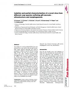
Fig. 1. SDSPAGE analysis of proteins eluted from a novobiocinSepharose column. Yeast proteins prepared from the Amyol strain, Znld, were applied to the novobiocin-Sepharose column and eluted in buffer containing KC1 or urea. Proteins were separated by electrophoresis on 15% SDUpolyacrylamide gels and stained with Coomassie blue dye. Lane l, crude extract; lane 2, 2 M KCl fraction; lane 3, 5 M urea fraction containing proteins of 20 kDa, 35 kDa, 52 kDa (the /l subunit of the mitochondrial F,-ATP synthetase) and 200 kDa.
Gene disruption Disruption of the wild-type allele encoding the p35 protein was achieved by subcloning the LEU2 cassette from the plasmid YDp-L [46] into a unique BamHI site in the CBR gene as indicated in Fig. 5a. The diploid strain 842 was transformed with linear DNA by electroporation [47]. The replacement of one wild-type allele with the cbr: :LEU2 allele was confirmed by tetrad dissection and Southern blot analysis.
RESULTS Purification of high-affinity novobiocin-binding proteins A number of major novobiocin-binding proteins have been purified from crude yeast extract by affinity chromatogChromosome assignment raphy on a novobiocin-Sepharose column. Tightly bound Yeast chromosomes, inmobilised onto Hybond-IFM proteins were eluted from the column in either 2 M KC1, or (Amersham, a kind gift from M. Pocklington) were probed 5 M urea, washed and analysed by SDS/PAGE. The 2 M salt with a 4-kb SaZI-Hind111 fragment using essentially the buffer was previously shown to elute two major proteins: the 200-kDa yeast myosin heavy chain [24-271 and the 52-kDa same conditions applied for Southern blots [42]. Sup45, the product of a gene conferring novobiocin resistance to yeast [18]. In the interest of simplifying the purificaDNA sequencing and generation tion procedure, the presence of the 200-kDa myosin heavy of unidirectional-nested-deletionclones chain was avoided by using Znld, a Amy01 mutant strain, Double stranded DNA sequencing [43] was performed lacking the myosin heavy chain [25]. Due to their greater by the dideoxynucleotide chain-termination method [44] affinity for novobiocin, elution of the other novobiocin-bindusing T7 polymerase (Pharmacia) and universal M13 prim- ing proteins [the 200-kDa (p200), the 50-kDa (the p subunit ers. Nested deletions of the insert DNA were produced with of the mitochondria1 F,-ATP synthetase) and the 35-kDa proa commercial kit (Pharmacia). All DNA sequence data, nu- teins] from the column required denaturation with urea cleotide and peptide data manipulations were carried out on (Fig. 1). a VAX cluster using the Genetics Computer Group sequenceThe p35 proteins were isolated from acrylamide gels to analysis software package [45]. raise antibodies. The serum recognised a 35-kDa protein, al-
444 a
h
1
2
b 3 4
isolated. The restriction map of this insert, including the region which hybridised to the probe, is shown in Fig. 3. Northern blot analysis of total yeast RNA shows a single transcript of 1.2-kb, consistent with a gene encoding a 35kDa protein. The gene has been assigned, through chromosome hybridisation, to chromosome IX.
Sequence analysis of the yeast gene Both strands of the pCBR fragment, which hybridised to the original positive pEX clone, were sequenced from convenient restriction endonuclease sites or from nested deletions (Fig. 3). A 966-bp open reading frame, identified on pCBR, was translated into a polypeptide of 322 amino acids, with a calculated molecular mass of 36 196 Da. The predicted amino acid sequence of the open reading frame, shown in Fig. 4, displays significant similarity (3437% identity, 61 -63% similarity) to known mammalian cytochrome b, reductases. Furthermore, the yeast protein is similar in size to mammalian cytochrome b, reductases and contains a highly hydrophobic N-terminal region similar to the small hydrophobic N-terminal membrane-binding domain characteristic of cytochrome b, reductases [48]. In addition to its similarity to mammalian cytochrome Fig. 2. Immunoblot of pEX clones. Anti-p35 antibodies (serum and reductases, the yeast gene product displays significant simiaffinity-purified IgG) were incubated with: (a), a blot of yeast proteins eluted by 5 M urea from novobiocin-Sepharose, (b), a blot of larity (30-33% identity, 53-58% similarity) to the FADan E. coli extract containing p galactosidase-fusion proteins which binding domain of nitrate reductases (Fig. 4). The similarity are expressed from the positive pEX clones, identified in the expres- of the yeast gene product to both cytochrome b, reductases sion library. Lanes 1 and 3, anti-p35 serum; lanes 2 and 4, affinity- and the FAD domain of nitrate reductase is not surprising as purified anti-p35 antibodies. Note that both a 35-kDa and 29-kDa the similarity between these two enzymes has previously protein are recognised (in lane 1)by antibodies in the serum whereas been noted [49-511. only the 35-kDa protein specifically cross-reacts with affinity-puriComparison of the predicted amino acid sequence of fied anti-p35 antibodies. Cbrp with alignments produced by Hyde et al., [34] indicates that the yeast gene contains 13 of the 14 amino acids characteristic of a family of flavoenzymes named flavoprotein pyrithough it additionally cross-reacted with a 29-kDa protein dine-nucleotide-cytochrome reductases (Fig. 4). The fourpresent in the 5 M urea fraction. The observation that affini- teenth amino acid, leucine, (residue 745 of tomato nitrate ty-purified anti-p35 antibodies only recognise a 35-kDa pro- reductase, 130 in the human cytochrome b, reductase, corretein, strongly indicates that the two proteins are not related, sponding to residue 159 in the yeast protein) is replaced in i.e. the 29-kDa protein is not a degradation product of p35 the yeast protein by an isoleucine residue (Fig. 4). Since the (Fig. 2). role of this leucine residue is unclear it is impossible to assess the importance, if any, of this substitution. Isolation of the gene encoding p35 Approximately lo5colonies of the yeast genomic expres- CBR disruption The sequence analysis of CBR identified a unique BamHI sion library constructed in pEX vectors were screened with anti-p35 antibodies. One positive clone, containing a 120-bp site in the gene. To demonstrate that the CBR gene encodes Sau3AI insert, produced a lacZ-fusion peptide which was the p35 protein, we cloned the yeast LEU2 gene as a selectarecognised by both the serum and affinity-purified anti-p35 ble marker into the BamHI site (Fig. 5a). A linearised fragantibodies (Fig. 2). The 120-bp Suu3AI fragment was used ment containing the LEU2-disrupted cbr was used to transas a probe to screen a yeast genomic library constructed in form the diploid strain 842. Replacement of a wild-type alAgtll. A 4-kb EcoRI positive insert, designated pCBR, was lele with the disrupted one was verified by Southern blot
Scale:-=
200bp
Fig.3. Restriction map and sequencing strategy of pCBR. B, BarnHI; Bg, BglII; E, EcoRI; K, KpnI; H, HindIII; S, SulI; X, XhoI. The bold bar, marked P, shows the regions of pCBR which hybridises to the 120-bp Sau3AI probe. The small arrows indicate the region and direction of sequencing. The bar indicates 200 bp. ORF, open reading frame.
50
1
Nia1:Sonr Nia:Cucma Nia2:Toba Nia1:Arat Nia:Lvces YeastcSb CB5R :Hum CB5R:Rat CBSR:Bov Consensus
inagtdctee FBaiHsDKaK inagtdctee FdaiHsnKaX inagtdctee FdaiHsDKaK inagtdctee FeaiHsDKaK inaatdctee FdaiHsDKaK -mykysy YirrKnEReK
rLLedfrige ListGyTsds Bspgnsvhgg sVysglagLA KmLedyrige LitLGyAsds BsnSpnnsth gas..nfshl KLLedfrige LitL2yTsds PgnSVhGsss fss . . . . fLA XLLedyrige LiLtGy..ds Spnvsvhgas r.fg...pLLA KLLedfrige LlttGvTsds SwSsvhass sIs...sfLA mkvciqya LqqeGSikq S-kmAidAGKL W v i V i v W P lgHm VLfpVwfLys gaqlstlsRV VLSPVwfVyS gaqlstlgHV VLSPLwfLys tdctee fdaihsdkaK klLedfrige Littgytsds s.nss.1g.svls...flla
+
100
pIteaVplrn valn. ..pRv ki.PckLIBK VSLSBDvrrF RFOLPseDQV apiRea.Pvs rrvaLa.pnE ki.PckLISX TsISBDvrVF RFALPGGQDq pIkelV.PAq rsvaLi.pRE ki.PckLIDK QsISBDvrkF RFALPseDQV pIkel..TPq kniaLvnpRE ki.PvrLIEK TsISBDvrkF RFALPseDQq DIkelVQTPt rsvaLi.p,RE ki.PckLVDK qsISHDvrkF KFALPseDQV LLfKf I iGPk TkpvLDPkRN dFQSfpLVBK TILTHNTsKY KFOLPhADW CB5R:Hum LImRlfqrSt PairLEspdi kY.PlrLIDR eILSHDTrrF RFALPsPQhI CB5R:Rat LfmXl€qrSs PaitLEnpdi kY.PlrLIDK e:ISHDTrIF RFALPsPQhI C35il:Bov LImKlfqrSr TaitLEnpdi kY.PlrLIDK eVISHDTrrF RFALPsPEhI Consensus .i..l.qr.t .aiale.pre ki.pc Lidi .s:sBdvrrf rFaLPsedqv
Nial :Sonr Nia :Cucma Nia:SToba Nia:SArat Nia :Lyces Yeastc5b
.. .
Nial :Sonr Nia:Cucma Nia2 :Toba Xial :Arat Nia :Lyces Yeastc5b
__
150
fLcNWLMKl cmRAYTPSB. tiDwOyfDL WKvYfkdvh fIcATVWK1 mRAYTPT8. siDernQfFBL VVKvYfkgvh FLcAVIDdKl cmRAYTPT8. LiDevOyFBL WiYfkgih fVcAllIlJdK1 c1RAYTPTB. a:DAvGhi=L WKvYZkdvh fLcATVWX1 cmRAYTPTS. tvDeviJfFEL WKiYfkgvh vIkANINOKd ItRSYTPTSl dgDTKQn?EL LVKsY. . . yLsARIDOnl VvRPYTPiS. sdDdKQfvDL VIKvYfkdth CBSR :H m YLsTRIWnl VIRPYTPVS. sdDdl(QlV3L WKvYfkdLh CBSR :Rat yLsARIWn1 ViRPYTPvB. sdDdKQfvDL VIKvYfkdth CBSR :Bov consensus flca.tdgk1 cmRaYTPC8. siD..GffdL wKvYfkdvh 0
.
.LQLPVOkHI aLQLPVQka1 .LQLPVOkaI .LOLPVGkHV .LOLPVOkHI .LQLPIQQHI .LOLWQ(pII .LOLPIQOHI .LOLPVWI .LOLPvOkHi
+ prfpnGOvM8 qhLdsLsLa pkfpnGOiMP qyLdsMeVOs pkfpnGOqMB qyMsMpLOs prfpr.GOXS qhLdsLpIOs pkfpnGOqMB qhLdsLpIOa . . . . PTOnVS kmIgELkIOD pkfpACQkMS qyLesMqIQD pkfpAGOkM8 qYLeNHnzQD pkfpAGQkMP qyLeSMgIQD pkfpngOkm8 qyldsmpio.
* + +
200
vhgkpkfa.. . . . . kKLAHI sQQMITPIY vhgkprfa.. . . . . rRLAML AOQMITPIY vhgkqkfa.. . . . .kKLAMI AOQMITPVY vsgkpkfa.. . . . .kKLAUL AOQMITPIY vhgkqkfa. . . . .kKDANI AOQMITPVY . . . . . . . . . . . . . .s H U M AOQMIAPYP irpdkksnpi irtvksVOYT AQQMITPML iradkksnpv vrtvksVClYI AOQMITPML irpdsksdpv iktvksV(M AOQNIITPHL vhgkpkfa.. . . . .kklaYi aOQMItPmy
Nial :Sonr Nia :Cucma Nia2 :Toba Nial :Arat Nia:Lyces Yeastc5b CBSR :Hum CB5R:Rat CB5R : Bov Consensus
iVDVKOPlOh ieylgkqnft TLDVKOPlOh ieltgrqnfm fLDVXOPlOh ieYQgkyxf1 mIDIKQPlOh :eYkgkqn€l fLDVKGPlOh ieYQgkgrf1 SIQIKGPrOn yhYErncR.. TIEfROPsOl 1vYQgkgKfa TIEfRQPnQl LvYQgkgKfa TIEfROPnQl IvYQgkgKIa cidvkQPlOh ie'lqgkgnf.
Nia1:Sonr Nia:Cucma Nia2 :Toba Nial :Arat Nia:Lyces Yeastc5b CB5R:Hum CB5R:Rat CB5R:Bov Consensus
eDkTeMhWY eDeTeMVNY eDdTeM$VVY eDeTeMyWY eDdTeMyVW hDtTkVsLVF dDhTvchLLF nDhTvcyLLF zIDhTvchLLF eDhTemyvvy
Nial :Sonr Nia :Cucma Nia2 :Toba Nia1:Arat Nia:Lyces YeastcSb CB5R:Hum CB5R:Rat CB5R:Bov Consensus
+* 0 300 322 LRDHVPAVGd dv.laLcCOP PPMIqfavqP nLD......KmgfdIkEQLl IF* LRmIPAAAe dt.laLaCOP PAHIqfavqP nLE.... . .K mnydtknsLl VF* LREIlIPePSh tt.laLaCOP PPWqfavnP nLE......K mgydIkDSL1 VF* LRZHAIPeGle gEslaLaCQP PMIqfalqP nLE.. . . . .X mgynVkEDL1 IF* LREIlIPePSh tt.laLaCOP PPMIqfainP nLE......K mgydIkEELl VF* I K m P M T m DNvqILICOP PAHVasvrrS tVDlgfrrsK pLSKMeDQVP VF* IRDHLPPPee Ep,lVLMCOP PPMIqyaclP nLD.......hVGHptErcF VF* IRDHLPPPGe Et.1ILMCOP PPldIufaclP nLE...... . rVGHDkErcP tF* VRDHLPPPee Et .lVLMCOP PPMIkaclP nLD. . . . . . . rVGHbkErcF aF* 1reH.P.p.e .t.laLaCOP PpMiqfa..p nle......kmgyd.kerl1 vF*
+ ANrtBBDILL ANrtBDDILL ANrtlDDILL ANrtEDDILV ANrtEDDILL GlJvhBEDILL AIPqtEkDILL ANqsBkDILL ANqtEkDILL aNrtBdDIL1
QVMqAIlkDP QWqAIlkDP QVXqAIlkDP QIIqSIlsDP QVXqSIlkDP QImUIamDP QVIRAImkDP PVIRAVlkDP QVIRAImkDP Qv.qailkDP
250 ReELDkwade frdrvKvwYV V.ekaeEgNK ydtOFISek1 RdE1BLDtwa.k KnurlKVwYV VQeSiRBuWE YSVOFITeNI KeBLDSwaek iPirvKVwW VQdSiK% GSIOFITeaI ReBLBGwask HkerlKIwYV V.eiaKlgWS yStQFITeaV KdELDAwaeq VPnrvKVwYV VQeSitQgUK yStQFVTes1 KkBLKALvam KPSqFKIvYY LDsPdRBdWT gOVOYITkW RpBLBeLrnk HSArFKLwYT LD.rapBaWJ yOqOFVneEM RpBLBeLrne HSSrFKLwYT VD.kapDaWD ySqQFVneEM XpELEeLrde tiShrFLwYT VD.kapBaWD ySqQNne'M r.BLeewa.kh..rfKvwYv vde.apem. ys.Ofiteei
Fig. 4. Line-up comparison of the predicted amino acid sequence of CBR with mammalian NADH-cytochrome b, reductases and the FAD-binding-domainof plant NADH-nitrate reductases. The predicted amino acid sequence of the yeast CBR gene is aligned with the following cytochrome b, and nitrate reductases: human cytochrome b, reductase (CB5R:Hum) [60] ; rat cytochrome b, reductase (CB5R:Rat) [61] ; bovine cytochrome b, reductase (CB5R:Bov) [62] ; the spinach nitrate reductase FAD-binding-domain (Nial :Sonr) [63] ; the pumpkin nitrate reductase FAD-binding-domain (Nia:Cucma) [64]; the tobacco nitrate reductase FAD-binding-domain (Nia2 :Tobac) [49]: the Arubidopsis nitrate reductase FAD-binding-domain (Nial :Arat) [65] ; and the tomato nitrate reductase FAD-binding-domain (Nia:Lyces) [51]. In this alignment, only amino acid residues which show similarity to the yeast CBR protein are shown: conserved amino acids are in bold, conservative changes in upper case. The 14 conserved amino acids described by Hyde et al., [34] are marked with asterisks (*).
analysis of total yeast DNA (Fig. Sb). The transformed diploid strain, containing the cbr: :LEU2 allele, was sporulated and the 2 :2 segregation of Leu2' : L e u 2 cells indicated that integration of the LEU2 gene had occurred at the single CBR locus. All four haploids were viable, demonstrating that the
CBR protein is not essential for growth under the conditions used in this work. Crude protein extracts, prepared from disruptant and wild-type strains, were analysed by Western blots using the anti-(35-kDa) antibodies. The absence of the 35-kDa protein
446 from the disruptant crude extract (Fig. 5c) strongly suggests that the CBR gene encodes the 35-kDa protein.
Comparison of CBR with other novobiocin-binding proteins
Lane
1
2
B
3
Intriguingly, all the novobiocin-binding proteins isolated so far, contain nucleotide-binding sites. It has already been proposed that these sites may form the binding domains for novobiocin [3, 37, 521. The myosin heavy chain, the p subunit of the mitochondria1 F,-ATP synthetase and DNA gyrase all bind ATP, whereas the cytochrome reductase contains FAD-binding and NADH-binding sites. Nevertheless, these proteins display no common sequence domain which could be considered as the antibiotic-binding site.
DISCUSSION
W
D
D
W
C
Fig. 5. Disruption of the CBR gene. (A) Strategy for disrupting the CBR gene. The position of the yeast LEU2 insertion into the BamHI site of CBR is indicated. ORF, open reading frame. (B) Southern blot analysis. Genomic DNA, prepared from haploid and diploid strains transformed with the LEU2-disrupted CBR gene, digested with the restriction endonuclease HindIII, separated by 1% agarose gel electrophoresis and blotted onto a nylon filter. The filter was hybridised to a HindIII fragment containing the entire CBR gene. Lane 1, haploid wild-type yeast strain (842H): lane 2, Leu2+ diploid strain; lane 3, Leu2' haploid strain. (C) Western blot analysis of the CBR-disruptant strain. Total yeast protein extracts were electrophoresed on a 10% SDS/polyacrylamide gel and blotted onto a nitrocellulose filter. The blotted proteins were incubated with anti-p35 antibodies, followed by goat anti-rabbit IgG and rabbit PAP, and developed. Lanes marked W contain total proteins of the wild-type strain: lanes marked D contain total protein extracts from yeast cbr mutants. The anti-p35 antibodies detect both a 35-kDa and a 29-kDa protein in the wild-type strain, whereas only the 29-kDa protein was detected in the mutant extract.
Our work, described here, demonstrates that one of the yeast novobiocin-binding proteins is a novel yeast cytochrome b reductase (CBR). This conclusion is based on the sequence similarity of the yeast gene to previously cloned mammalian cytochrome b, reductases. As a component of the microsomal electron-transport system, NADH-cytochrome b, reductase is involved in the desaturation and elongation of fatty acids [28-301, cholesterol biosynthesis [31, 531 and drug metabolism [32, 331. The enzyme is an amphipathic membrane-bound flavoprotein containing a large hydrophilic catalytic domain and a small hydrophobic membrane-binding segment. Similarly, the predicted amino acid sequence of the yeast protein shows that, in addition to the large hydrophilic domain, it contains a small hydrophobic region, suggesting that the yeast protein is also membrane associated. Sequence analyses of known reductases indicate that the yeast gene belongs to the flavoenzyme gene family described by Karplus et al., [54] and Hyde et al., [34], termed flavoprotein pyridine-nucleotide-cytochrome reductases. It is unlikely that the CBR gene product is the main target of novobiocin in vivo since haploid yeast mutants, in which the wild-type CBR allele is disrupted, are viable and are not defective in either DNA replication or transcription (data not shown). We have identified the second 35-kDa novobiocinbinding protein as an enzyme essential for haem biosynthesis (data not shown). The lack of an obvious phenotype of d cbr mutants is not particularly surprising as a number of studies suggest that one cytochrome reductase activity can be replaced by another. For example, the mammalian cytochrome b, reductase can be substituted by cytochrome P-450 reductase in vitro [28, 29, 311 and the disruption of the yeast gene encoding the P-450 reductase does not entirely abolish the activity of cytochrome P-450 [55]. Furthermore, cell viability is not affected by the deletion of the P-450 reductase gene, although it leads to a marked reduction in growth rate [55]. Double disruption of the yeast CBR and the P-450 reductase gene leads to the loss of viability, suggesting that indeed one product can compensate for the loss of the other (our unpublished results). In early work, the presumed target of novobiocin was topoisomerase I1 [56]. This presumption was based on the sequence similarity of this enzyme with the bacterial DNA gyrase, as well as on the observation that high concentrations of the drug inhibit eukaryotic topoisomerase I1 [4]. Our previous results [17] and those of others [57-591 show that,
447
although the main target of novobiocin is not topoisomerase 11, the true target must affect DNA replication and transcription. We have previously reported that the P-subunit of the mitochondrial F, ATP synthetase is a major novobiocin-binding protein [19]. Interference with this ATP-synthesising enzyme should be sufficient to arrest growth in higher eukaryotes. However, yeast can grow on fermentative media without energetically functioning mitochondria. Yet, even under these conditions, novobiocin still inhibits cell growth, indicating that its poisoning effects in yeast are exerted via other components of the cell. Finally, it is interesting to note that the only common feature of all the novobiocin-binding proteins characterised to date is the presence of a nucleotide-binding site. No shared sequence motif which might account for the novobiocinbinding activity can be identified in bacterial gyrases and the yeast proteins. Presumably, the tertiary structures of these proteins must play a part in binding the drug. The continued characterisation of the other novobiocin-binding proteins should assist in clarifying this enigma. This work was supported by the Wellcome Trust (grants no. 030128 and 16745A.18). MC acknowledges the receipt of a studentship from the Science and Engineering Research Council. It is a pleasure to thank Mick Pocklington and Sean Donnelly for their help and advice throughout the work.
13. 14. 15.
16. 17. 18. 19. 20.
21. 22.
REFERENCES 1. Orr, E., Lother, H., Lurz, R. & Wahle, E. (1984) Escherichia coli DNA gyrase, in Proteins involved in DNA replication (Hubscher, U. & Spidari, S., eds) vol. 179, pp. 395-407. Plenum Publishing Corporation, New York and London. 2. Gellert, M., Mizuuchi, K., O'Dea, M. H. & Nash, H. A. (1976) DNA gyrase: an enzyme that introduces superhelical turns into DNA, Proc. Natl Acad. Sci. USA 73, 3872-3876. 3. Mizuuchi, K., O'Dea, M. H. & Gellert, M. (1978) DNA gyrase: subunit structure and ATPase activity of the purified enzyme, Proc. Nut1 Acad. Sci. USA 75, 5960-5963. 4. Liu, F. L., Liu, C.-C. & Alberts, M. (1980) Type I1 DNA topoisomerase: enzymes that can unknot a topologically knotted DNA molecule via a reversible double-strand break, Cell 19, 697-707. 5. Collins, A. & Johnson, R. (1979) Novobiocin: an inhibitor of the repair of UV-induced but not X-ray-induced damage in mammalian cells, Nucleic Acids Res. 7, 1311-1320. 6. Lavin, M. F. (1981) Effect of novobiocin on DNA synthesis and structure in human lymphoblastoid cells, Biochem. Biophys. Res. Commun. 100, 328-335. 7. Collins, A. R. S., Squires, S. & Johnson, R. T. (1982) Inhibitors of repair DNA synthesis, Nucleic Acids Res. 10, 1203-1212. 8. Mattern, M. R., Paone, R. F. & Day, R. S. (1982) Eucaryotic DNA repair is blocked at different steps by inhibition of DNA topoisomerase and DNA polymerases a and b, Biochim. Biophys. Acta 677, 6-13. 9. Clarkson, J. M. & Mitchell, D. L. (1983) The effect of various inhibitors of DNA synthesis on the repair of DNA photoproducts, Biochim. Biophys. Acta 740, 355-361. 10. Mattern, M. R. & Scudiero, D. A. (1981) Characterisation of the inhibition of replicative and repair-type DNA synthesis by novobiocin and nalidixic acid, Biochim. Biophys. Acta 653, 248-258. 11. Aller, P. & Baserga, A. (1986) Selective increase of c-myc mRNA levels by methylglyoxyal-bis (guanylhydrazone) and novobiocin in serum-stimulated fibroblasts, J. Cell Physiol. 128, 362-366. 12. Flickinger, R. A. & Richman, R. (1984) The effect of induction of hemoglobin synthesis in cultured Friend cells on the
23.
24. 25. 26. 27. 28. 29.
30.
31.
32.
33.
number of initiation sites for replication and transcription, Cell Direr. 14, 59-71. Han, S., Udvardy, A. & Schedl, P. (1985) Novobiocin blocks the Drosophila heat-shock response, J. Mol. Biol. 183, 1329. Kmiec, E. B., Ryoji, M. & Worcel, A. (1986) Gyration is required for 5s RNA transcription from a chromatin template, Proc. Natl Acad. Sci. USA 83, 1305-1309. Lynn, R., Giaevez, G., Swanberg, S. L. & Wang, J. C. (1986) Tandem regions of yeast topoisomerase I1 share similarity with different subunits of bacterial gyrase, Science 233, 647649. Collins, A. (1990) Topoisomerase 11 can relax: Novobiocin is a mitochondrial poison after all, BioEssays 12, 493-494. Pocklington, M. J., Jenkins, J. R. & On; E. (1990) The effect of novobiocin on yeast topoisomerase type 11, Mol. & Gen. Genet. 220, 256-260. Pocklington, M. J., Johnston, L., Jenkins, J. R. & Om, E. (1990) The omnipotent suppressor sup45 affects nucleic acid metabolism and mitochondrial structure, Yeast 6, 441 -450. Jenkins, R. J., Pocklington, M. J. & Orr, E. (1990) The F, ATP synthase 8-subunit : a major yeast novobiocin binding protein, J. Cell Sci. 96, 675-682. Eder, J. P., Teicher, B. A,, Holden, S. A., Cathcart, K. N. S., Schnipper, L. E. & Frei 111, E. (1989) Effect of novobiocin on antitumor activity and tumor cell and bone-marrow survivals of three alkalating agents, Cancer Res. 49, 595-598. Eder, J. P., Wheeler, C. A., Teicher, B. A. & Schnipper, L. E. (1991) A phase I clinical trial of novobiocin, a modulator of alkylating agent cytotoxicity, Cancer Res. 51, 510-513. Nordenberg, J., Albukrek, D., Hadar, T., Fux, A., Wasserman, L., Novogrodsky, A. & Sidi, Y. (1992) Novobiocin-induced anti-proliferative and differentiating effects in melanoma B16, BE J. Cancer 65,183-188. Wood, P. J. & Stansfield, A. G. (1992) Inhibition of T-cell mediated cytotoxicity by novobiocin suggests multiple pathways for both CD4' and CD8' cytotoxic T cells, Immunology 76, 460-465. Watts, F. Z . , Miller, D. M. & Orr, E. (1985) Identification of myosin heavy chain in Saccharomyces cerevisiae, Nature 316, 83-85. Watts, F. Z., Shiels, G. & Orr, E. (1987) The yeast MY01 gene encoding a myosin-like protein repaired for cell division, EMBO J. 6, 3499-3505. Sweeney, F. P., Watts, F. Z., Pocklington, M. J. & Orr, E. (1990) The MY01 gene from Saccharomyces cerevisiae: its complete nucleotide sequence, Nucleic Acids Res. 18, 7147. Sweeney, F. P., Pocklington, M. J. & Orr, E. (1991) The yeast type11 myosin heavy chain: analysis of its predicted polypeptide sequence, J. Muscle Res. & Cell Motil. 12, 61 -68. Oshino, N., Imai. Y. & Sato, R. (1971) A function of cytochrome b, in fatty acid desaturation by rat liver microsomes, J. Biochem. (Tokyo) 69, 155-167. Keyes, S. R. & Cinti, D. L. (1980) Biochemical properties of cytochrome &-dependent microsomal fatty acid elongation and identification of products, J. Biol. Chem. 255, 1135711364. Dailey, H. A. & Strittmatter, P. (1980) Characterization of the interaction of amphipathic cytochrome b5 with stearyl coenzyme A desaturase and NADPH: cytochrome P-450 reductase, J. Biol. Chem. 255, 5184-5189. Reddy, V. V. R., Kupfer, D. & Caspi, E. (1977) Mechanism of C-5 double bond introduction in the biosynthesis of cholestrol by rat liver Microsomes. Evidence for the participation of microsomal cytochrome b,, J. Biol. Chem. 252, 2797-2801. Hildebrandt, A. & Estabrook, R. W. (1971) Evidence for the participation of cytochrome b5 in hepatic microsomal mixedfunction oxidation reactions, Arch. Biochem. Biophys. 143, 66-79. Aoyama, T., Nagata, K., Yamazoe, Y., Kato, R., Matsunaga, E., Gelboin, H. V. & Gonzalez, F. J. (1990) Cytochrome b, potentiation of cytochrome P-450 catalytic activity demon-
448 strated by a Vaccinia virus-mediated in situ reconstitution system, Proc. Natl Acad. Sci. USA 87,5425-5429. 34. Hyde, G. E., Crawford, N. M. & Campbell, W. H. (1991) The sequence of squash NADH-nitrate reductase and its relationship to the sequences of other flavoprotein oxidoreductases, J. Biol. Chem. 266, 23542-23547. 35. Bolivar, F., Rodrigez, R. L., Green, P. J., Betlatch, M. C., Heyneker, H. L. & Boyer, H. W. (1977) Construction and characterisation of new cloning vehicles. A multipurpose cloning system, Gene (Amst.) 2, 95-110. 36. Sambrook, J., Fritch, E. F. & Maniatis, T. (1989) Molecular cloning: a laboratory manual, 2nd edn, Cold Spring Harbour Laboratory Press, Cold Sping Harbour, NY. 37. Staudenbauer, W. L. & Orr, E. (1981) DNA Gyrase: affinity chromatography on novobiocin-Sepharose and catalytic properties, Nucleic Acids Res. 9, 3589-3603. 38. Towbin, H., Staehelin, T. & Gordon, J. (1979) Electrophoretic transfer of proteins from polyacrylamide gels to nitrocellulose sheets: procedure and some applications, Proc. Natl Acad. Sci. USA 76, 4350-4354. 39. Stanley, K. K. & Luzio, J. P. (1984) Construction of a new family of high efficiency bacterial expression vectors : identification of cDNA clones coding for human liver, Proteins 3, 1429-1434. 40. Young, R. A. & Davis, R. W. (1983a) Efficient isolation of genes by using antibody probes, Proc. Natl Acad. Sci. USA 80, 1194-1198. 41. Feinberg, A. P. & Vogelstein, B. (1984) A technique for radiolabelling DNA restriction endonuclease fragments to a high specific activity (Addendum), Anal. Biochem. 137, 266-267. 42. Church, G. M. & Gilbert, W. (1984) Genomic sequencing, Proc. NatI Acad. Sci. USA 81, 1992-1995. 43. Chen, E. Y. & Seeburg, P. H..(1985) Supercoil sequencing: a fast and simple method for sequencing plasmid DNA, DNA (NY) 4,165-170. 44. Sanger, F., Nicklen, S. & Coulson, A. R. (1977) DNA sequencing with chain-terminating inhibitors, Proc. Natl Acad. Sci. USA 74, 5463-5467. 45. Devereux, J., Haeberli, P. & Smithies, 0. (1984) A comprehensive set of sequence analysis programs for the VAX, Nucleic Acids Res. 12, 387-395. 46. Berben, G., Dumont, J., Gilliquet, V., Bolle, P.-A. & Hilger, F. (1991) The YDp plasmids: a uniform set of vectors bearing versatile gene disruption cassettes for Sacchammyces cerevisiae, Yeast 7, 475-477. 47. Becker, D. M. & Guarente, L. (1991) High-efficiency transformation of yeast by electroporation, Methods Enzymol. 194, 182- 187. 48. Spatz, L. & Strittmatter, P. (1973) A form of reduced nicotinamide adenine dinucleotide-cytochrome b, reductase-containing both the catalytic site and an additional hydrophobic membrane-binding segment, J. Biol. Chem. 248, 793 -799. 49. Calza, R., Huttner, E., Vincentz, M., Rouze, P., Galangau, F., Vaucheret, H., Cherel, E., Meyer, C., Kronenberger, J. & Caboche, M. (1987) Cloning of DNA fragments complementary to tobacco nitrate reductase mRNA and encoding epitopes common to the nitrate reductases from higher plants, Mol. & Gen. Genet. 209, 552-562. 50. Crawford, N. M., Smith, M., Bellissimo, D. & Davis, R. W. (1988) Sequence and nitrate regulation of the Arabidopsis
thaliana mRNA encoding nitrate reductase, a metalloflavoprotein with three functional domains, Proc. Natl Acad. Sci. USA 85, 5006-5010. 51. Daniel-Vedele, F., Dorbe, M. F., Caboche, M. & Rouze, P. (1989) Cloning and analysis of the tomato nitrate reductaseencoding gene: protein domain structure and amino acid homologies in higher plants, Gene (Amst.) 85, 371-380. 52. Sugino, A,, Higgins, N. P., Brown, P. O., Peebles, C. C. & Cozzarelli, N. R. (1978) Energy coupling in DNA gyrase and the mechanism of action of novobiocin, Proc. Natl Acad. Sci. USA 75,4838-4842. 53. Fukushima, H., Grinstead, G. F. & Gaylor, J. L. (1981) Total enzymic synthesis of cholesterol from lanosterol, J. Biol. Chem. 256,4822-4826. 54. Karplus, P. A,, Daniels, M. J. & Herriott, J. R. (1991) Atomic structure of ferredoxin-NADP' reductase: prototype for a structurally novel flavoenzyme family, Science 251, 60- 66. 55. Sutter, T. R. & Loper, J. C. (1989) Disruption of the Saccharomyces cerevisiae gene for NADPH-cytochrome P450 reductase causes increased sensitivity to ketoconazole, Biochem. Biophys. Res. Commun. 160, 1257- 1266. 56. Mattern, M. R. & Painter, R. B. (1979) Dependence of mammalian DNA replication on DNA supercoiling. 11. Effects of novobiocin on DNA synthesis in Chinese hamster ovary cells, Biochim. Biophys. Acta 563, 306-312. 57. Gottesfeld, J. M. (1986) Novobiocin inhibits RNA polymerase 111 transcription in vivo by a mechanism distinct from DNA topoisomerase 11, Nucleic Acids Res. 14, 2075 -2088. 58. Ishida, R., Nishizawa, M., Fukami, K., Maekawa, K., Takahashi, T. & Nishimoto, T. (1987) Isolation and characterization of novobiocin-resistant BHK cells, Somatic Cell Mol. Genet. 13, 11-20. 59. Webb, M. L., Maguire, K. A. & Jacob, S. P. (1987) Novobiocin inhibits initiation of RNA polymerase I1 directed transcription of the mouse metallothionein-I gene independent of its effect on DNA topoisomerase 11, Nucleic Acids Res. 15, 85478560. 60. Yubisui, T., Miyata, T., Iwanaga, S., Tamura, M. & Takeshita, M. (1986) Complete amino acid sequence of NADH-cytochrome b, reductase purified from human erythrocytes, J. Biochem. (Tokyo) 99,407-422. 61. Zenno, S., Hattori, M., Misumi, Y., Yubisui, T. & Sakaki, Y. (1990) Molecular cloning of a cDNA encoding rat NADHcytochrome b5 reductase and the corresponding gene, J. Biochem. (Tokyo) 107, 810-816. 62. Ozols, J., Korza, G., Heinemann, F. S., Hediger, M. A. & Strittmatter, P. (1985) Complete amino acid sequence of steer liver microsomal NADH-cytochrome b, reductase, J. Biol. Chem. 260, 11953-11961. 63. Prosser, I. M. & Lazarus, C. M. (1990) Nucleotide sequence of a spinach nitrate reductase cDNA, Plant Mol. Biol. 15, 187190. 64. Crawford, N. M., Campbell, W. H. & Davis, R. W. (1986) Nitrate reductase from squash: cDNA cloning and nitrate regulation, Proc. Natl Acad. Sci. USA 83, 8073-8076. 65. Cheng, C., Dewdney, J., Nam, H., den Boer, B. G. W. & Goodman, H. M. (1988) A new locus (NIA1) in Arabidopsis thaliana encoding nitrate reductase, EMBO J. 11, 3309-3314.