Feb 19, 1988 - This fimbrial adhesin was classified as F17 (P. F. Lintermans, P. Pohl, A. ...... F17 GVKA I KLGQAAAAQSVI)TV-- A I NANVtLRYNAQYY T- GVA- ...
Vol. 56, No. 6
INFECTION AND IMMUNITY, June 1988, p. 1475-1484
0019-9567/88/061475-10$02.00/0 Copyright © 1988, American Society for Microbiology
Isolation and Nucleotide Sequence of the F17-A Gene Encoding the Structural Protein of the F17 Fimbriae in Bovine Enterotoxigenic Escherichia coli P. LINTERMANS,l* P. POHL,' F. DEBOECK,2 A. BERTELS,l C. SCHLICKER,l J. VANDEKERCKHOVE,3 J. VAN DAMME,3 M. VAN MONTAGU,23 AND H. DE GREVE2 Laboratory of Bacteriology, The National Institute for Veterinary Research, Groeselenberg 99, B-1 180 Brussels'; Laboratory of Genetic Virology, The Free University of Brussels, B-1640 St.-Genesius-Rode2; and Laboratory of Genetics, The State University of Ghent, B-9000 Ghent,3 Belgium Received 28 October 1987/Accepted 19 February 1988
The genetic determinant for production of the fimbrial F17 adhesive antigen was isolated from a bovine enterotoxigenic Escherichia coli strain. The F17-A gene, coding for the structural component of the F17 fimbrial adhesin, was cloned and sequenced. An open reading frame of 540 base pairs encoding a polypeptide of 180 amino acids, of which the NH2-terminal 21 residues are characteristic of a signal sequence, has been characterized. The mature protein lacks histidine, methionine, and tryptophan. A possible promoter and ribosome binding site as well as a possible site for termination of transcription are proposed. An important homology of the F17-A protein with fimA and papA fimbrial proteins was found. The N-terminal sequence of the mature F17-A pilin is extremely similar to the N-terminal sequence of the G fimbriae identified on human pyelonephritogenic E. coli strains.
Pathogenic bacteria often need an intimate association with the mucosa they colonize or invade. Such an association is established by the production of adhesins, which makes the expulsion of the bacteria by peristaltic or ciliary movement or other mechanisms inefficient (4, 7). Enterotoxigenic Escherichia coli strains induce diarrhea by colonizing the small intestine of several mammals and by secreting one or several enterotoxins which provoke the net influx of sodium ions and water into the gut lumen (7, 34). Other factors may influence the onset of diarrhea. The colonization of enterotoxigenic E. coli occurs by means of various host-specific fimbrial adhesins (7, 36). The number of newly discovered fimbrial adhesins is increasing rapidly. Bovine neonatal colibacillosis is caused by enterotoxigenic E. coli strains producing the K99 and F41 adhesins (7, 10, 30). Recently, another fimbrial adhesin, originally called Att25 (Belgium) or F(Y) (France), was described in bovine strains associated with neonatal calf diarrhea (10,31). This fimbrial adhesin was classified as F17 (P. F. Lintermans, P. Pohl, A. Bertels, G. Charlier, J. Vandekerckhove, J. Van Damme, J. Schoup, C. Schlicker, T. Korhonen, H. De Greve, and M. Van Montagu, Am. J. Vet. Res., in press). The presence of F17 fimbriae could be linked to pathogenicity (30). Since these fimbriae are a potent immunologically active polypeptide, F17-positive E. coli are already widely used in bovine diarrhea vaccines. In this paper we describe the isolation and molecular cloning of the genes required for F17 production. The F17 pilin gene was localized and sequenced, and the amino acid sequence was compared with the K88ab (small subunit), K99, PapA, FimA, and G pilin sequences (1, 20, 25, 26, 29, 32, 33). These studies revealed a strong amino acid homology between the F17 fimbriae and the PapA, G, and FimA pilins. A potential antigenic determinant on the F17 pilin is predicted on the basis of the hydrophilicity plot and on
MATERIALS AND METHODS Bacterial strains and plasmids. E. coli 25KH09st is a nontoxigenic derivative of a bovine enterotoxigenic strain and was serotyped as O101:K+:H- (Lintermans et al., in press). This strain was used as source of total DNA. E. coli strains JM101 (11) and K514 (5) were used as a recipient for transformation. E. coli HB101 (3) was used in studies evaluating the binding properties of the genetic constructions. The plasmid pUC8 (43) was used as the cloning vector. Culture conditions. All strains were grown in LB medium (2; Lintermans et al., in press) at 37°C. Transformants were selected on solid LB medium containing carbenicillin (100 ,ug/ml), 5-bromo-4-chloro-3-indolyl-,3-D-galactopyranoside (X-Gal) (30 ,ul of a 2% solution in dimethylformamide), and isopropyl-,3-D-thiogalactopyranoside (IPTG) (50 pul of a 100 mM solution in water). Enzymes. Restriction enzymes, T4 polynucleotide kinase, calf intestinal phosphatase, and T4 ligase were purchased from Boehringer (Mannheim, Federal Republic of Germany), and incubation conditions were as specified by the supplier. Trypsin was a product of Sigma Chemical Co. (St. Louis, Mo.). DNA electrophoresis. All DNA separation procedures were carried out by a horizontal electrophoresis system (Bio-Rad Laboratories, Richmond, Calif.). Construction of a genomic library. Total bacterial DNA was prepared from strain 25KH09st by the method of Dhaese et al. (6). The DNA was partially digested with the restriction enzyme Sau3A (1 U of Sau3A per 50 ,ug of DNA) and fractionated on a sucrose gradient (22). The samples containing DNA fragments of 15 to 20 kilobases were determined by horizontal agarose electrophoresis. After precipitation with ethanol, this DNA was used in ligation experiments. The cloning vector pUC8 was digested with BamHI and treated with calf intestinal phosphatase as described by Maniatis et al. (22). The restricted and fractionated DNA of strain 25KH09st was ligated to the pUC8 vector. Transfor-
secondary structure predictions. *
Corresponding author. 1475
1476
INFECT. IMMUN.
LINTERMANS ET AL.
y
lOOnm u
M-1l11'11-
If:c .:
,s
.;
_
*
4 .,
.:,,^ Y *
41
4., . *. -.
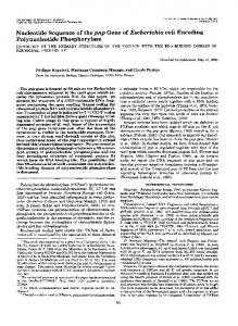
FIG. 1. Electron micrographs after colloidal gold immunolabeling of (A) E. coli K514(pPLHD1), (B) E. coli K514(pPLHD2), (C) E. coli HB101, and (D) E. coli 25KH09st. Bar, 0.1 p.m. mants were selected on LB supplemented with carbenicillin,
IPTG, and X-Gal. White colonies were screened by the colony hybridization procedure. Colony hybridization. Colonies were blotted on nitrocellulose. The nitrocellulose sheets were first rinsed for 15 min with phosphate-buffered saline (PBS) and then incubated with 1% ovalbumin in PBS for at least 1 h (13). The F17 antiserum was applied for 90 min at a 1:500 dilution (Lintermans et al., in press). Sheets were washed with a 0.2% solution of Triton X-100 in PBS, incubated with anti-rabbit peroxidase conjugate (Sigma) at a 1:1,000 dilution, and washed again with the same buffer. The antigen was visualized by soaking the sheet in a solution of 0.75 mg of 4-chloro-1-naphthol (Sigma) per ml-0.02% H202-25% methanol in PBS. In vitro adhesion assay. Intestinal villi were prepared as described by Girardeau (9), with some minor modifications. Briefly, calf intestinal villi (duodenum, jejunum, or ileum)
were mixed with 109 E. coli cells in PBS. This mixture was allowed to incubate at room temperature for 15 min with gentle shaking. Adhesion was followed under a phasecontrast microscope (x600). Inhibition of adhesion was obtained by mixing equal volumes of 100 mM N-acetyl glucosamine and bacteria before adding the villi (10). As a control sugar, (x-methyl-D-mannoside was used at a final concentration of 100 mM. Electron microscopy. (i) Negative staining. Transformants reacting with the F17 antiserum in the colony hybridization assay were stained by the negative staining drop method (Lintermans et al., in press). Observations, were made with a transmission electron microscope (Philips). (ii) Immune electron microscopy. A drop of a washed bacterial suspension was placed on carbon-coated grids for 3 min. Excess liquid was removed, and the grids were placed face down on a drop of a 1/100 dilution of F17 serum for 15 min. Grids were washed three times and placed on a drop of
C~ ~ ~ .
VOL. 56, 1988
.
*9-
,;,
:
t-
*4
..
GENE FOR F17 FIMBRIAL PROTEIN FROM BOVINE ETEC
1477
100 nm
.l,
... h
.;t'
.z
it'..
}W;, :,
..:
!=,
:. 4k 3S;.*;, jX
r
's
,:,
Io0 nm
D
06.,
FIG. 1-Continued
5-nm-gold-labeled SpA (Janssen Biotech-Belgium) for 15 min. After a final washing step, the grids were negatively stained with 1% ammonium molybdate. The incubation and washing solutions contained 2.4 g of Tris and 9 g NaCl per liter at pH 8.2. They were supplemented with 1% bovine serum albumin fraction V and Tween 20 (Sigma) (Lintermans et al., in press). Grids incubated without antiserum were used as negative controls. Immunoblotting. Western blotting (immunoblotting) was performed by the technique of Towbin et al. (39). Purified fimbriae of both the wild-type E. coli 25KH09st and E. coli K514(pPLHD2) (Lintermans et al., in press) were run in duplicate on sodium dodecyl sulfate-polyacrylamide gel electrophoresis (21). The F17 antiserum was applied for the identification of the F17 fimbrial fraction. Partial protein sequence analysis. The F17 fimbrial protein was purified as described previously (Lintermans et al., in press). The protein was performic acid oxidized as described
by Hirs (16). The oxidation was terminated by extensive dialysis against distilled water, and the remaining protein solution was made 0.5% in NH4HCO0 by adding 1/10 volume of a 5% solution and digested with trypsin. The digestion was carried out for 2 h at 37°C, in an enzyme-substrate ratio of 1:50 (by weight). It was terminated by lyophilization, and the peptide mixture was separated by two-dimensional peptide mapping as described previously (42) and detected by a dilute fluorescence staining (41). Peptides were eluted from the paper, hydrolyzed with 6 N HCI, and analyzed with a Biotronik amino acid analyzer equipped with a fluorescence detector measuring the o-phthaldialdehyde amino acid reaction products (2). Peptides containing a high number of amino acids were selected for amino acid sequencing. These analyses were carried out with a gas-phase sequenator equipped with an on-line phenylthiohydantoin-amino acid analyzer (Applied Biosystems Inc.) (15). Synthesis of oligonucleotide probe. A mixed synthetic DNA
A
-t
t
/..r.
.#.0
ti+0X.X 0i
S*L'2w
FIG. 2. (A) E. coli HB101(pPLHD2) adhering to isolated intestinal calf villi. (B) Incubation of E. coli HB101(pPLHD2) with N-acetylglucosamine blocked binding to the calf villi. (C) Binding of E. coli 25KH09st. Magnification, x580. 1478
VOL. 56, 1988
GENE FOR F17 FIMBRIAL PROTEIN FROM BOVINE ETEC
oligonucleotide of 12 nucleotides was synthesized using a 380A DNA synthesizer (Applied Biosystems). The mixed synthetic 12-nucleotide-long DNA fragment was separated from the shorter oligonucleotides by using a high-pressure liquid chromatography column (Varian 5000) of Particil SAX-10,u (0.46 by 25 cm). The oligonucleotides were desalted by running through a Sephadex G-25M column (Pharmacia PD-10). The buffer used was 10 mM triethylammonium bicarbonate (pH 7.0). The crude compound was suspended in a small amount of buffer and loaded on the Sephadex. Fractions of 500 ,ul were collected, and the optical density at 260 nm was measured. The desired fractions were lyophilized and rinsed with ethanol to obtain a salt-free pellet. The desalting step was repeated twice. Labeling of the oligonucleotide probe. The synthetic 12base-long oligonucleotide mixture was labeled with [y32P]ATP using T4 polynucleotide kinase (22) and was used as a probe in hybridization experiments. Southern blot hybridizations. pPLHD2 plasmid DNA was digested with BstEII, BstEII-EcoRI-HindIII, and EcoRIHindIII-BamHI restriction enzymes. After gel electrophoresis, the DNA fragments were transferred to nitrocellulose filters by Southern blot (35). The prehybridization, hybridization, and washing steps were carried out at 27°C. A temperature of 32°C should be applied, according to Wallace et al. (44). Since only two of the four wobble bases for threonine are present in the oligonucleotide probe, a temperature reduction by 5°C to 27°C was used (37). Nucleotide sequence determination. The DNA samples were prepared by the method of Swinghamer (38) and were end labeled with T4 polynucleotide kinase. The DNA sequences were determined by using the chemical method of Maxam and Gilbert (24). Hydrophilicity pattern and prediction of antigenic determinants. The prediction of possible antigenic determinants on the F17 subunit polypeptide was performed on the basis of the nucleotide sequence, using the method of Hopp and Woods (18). Prediction of secondary structures. Secondary structure prediction was carried out using a computer simulation based on the method of Garnier et al. (8). RESULTS Isolation of an F17-positive transformant. A genomic library was constructed with vector pUC8 and total bacterial DNA from strain 25KH09st. Carbenicillin-resistant colonies growing white on LB medium containing IPTG and X-Gal were screened with the colony hybridization assay for the presence of F17 fimbriae. One clone out of the 2,000 tested reacted with the F17 antiserum. DNA analysis of this clone revealed the presence of a 20-kilobase plasmid which was
designated pPLHD1. Preliminary characterization of pPLHD1. The fimbriae found at the surface of strain K514(pPLHD1) could be identified by immunoelectron microscopy as F17 fimbriae (Fig. 1A). These F17 fimbriae were still functional, since strain HB101(pPLHD1) could adhere to intestinal calf villi (results not shown). This adhesion could be inhibited with N-acetylglucosamine but not by ao-methyl-D-mannoside, identifying the adhesive factor as the F17 antigen. Subcloning of the region encoding the F17 fimbriae. Internal deletions were made in the pPLHD1 plasmid with EcoRI, BamHI, and HindlIl restriction enzymes. Cells bearing the BamHI and HindlIl deletions no longer produced F17 fimbriae. The EcoRI deletion resulted in an 8.5-kilobase DNA
1479
fragment which still reacted with the F17 antiserum in the colony hybridization assay. This construction was designated pPLHD2. E. coli HB101(pPLHD2) adhered to intestinal calf villi (Fig. 2A), and binding was blocked by Nacetylglucosamine (Fig. 2B). Electron microscopy showed that the surface of the bacteria was fimbriate. The structures could be identified by immunogold labeling experiments as F17 fimbriae (Fig. 1B). A fimbrial preparation of E. coli K514(pPLHD2) migrated on sodium dodecyl sulfate-polyacrylamide gel electrophoresis at the same position as F17 fimbriae isolated from the wild-type strain E. coli 25KH09st. In Western blots both bands reacted with the F17 antibodies (Fig. 3). We can conclude that the 8.5-kilobase insert DNA fragment present in pPLDH2 encodes intact and functional F17 fimbriae. Localization of the F17-A gene in pPLHD2. The N-terminal sequence analysis of intact F17 pilin was negative, from which we concluded that the N terminus was blocked. We then used the amino acid sequences of two selected tryptic peptides to serve as templates for the synthesis of appropriate DNA probes. Their sequences were Val-Val-Asp-GlnThr-Cys-Ser-Val-Thr-Thr-Glu-Ser-Lys and Leu-Pro-ThrVal-Ser-Ala-Asn-Ser-Leu-Ala-Ser-Ser-Gly-Lys. The AspGln-Thr-Cys amino acid tetrapeptide derived from the first peptide could be encoded by 32 different DNA sequences. The mixture of 16 DNA oligonucleotides, GAT CAA ACA TGC, was chosen on the basis of the codon usage of strongly expressed genes in E. coli (17). This mixture was used to localize the F17-A gene in the plasmid pPLHD2. Since only two Thr codons were incorporated in our probe, the hybridization was carried out at a Tm of 27°C, which was 5°C below the hybridization temperature of the oligonucleotide GATCAAACATGT, which has an A or T in the third position of each codon and thus the lowest T,, of the oligonucleotide mixture (37). The mixed oligonucleotide probe hybridized with the 4,860-base-pair BstEII fragment, the 1,600-base-pair Hind III-BstEII fragment, and the 1,200-base-pair HindIII-BamHI fragment (Fig. 4). Hence, the F17-A gene is located between the HindlIl and BamHI sites of pPLHD2. Nucleotide sequence determination of the F17-A structural gene. The nucleotide sequencing strategy used is shown in
2
3
4
5
FIG. 3. Immunoblot analysis with F17 antiserum of F17 fimbriae isolated from E. coli 25KH09st (lane 1) and E. coli K514(pPLHD2) (lane 2). The same fimbrial preparations, separated on an identical Coomassie blue-stained sodium dodecyl sulfate-polyacrylamide gel, appear in lanes 3 and 4, respectively. Lane 5, Molecular size markers (94, 67, 43, 30, 20.1, 14.4 kilodaltons).
1480
INFECT. IMMUN.
LINTERMANS ET AL. 1
3
2
4
6
be identified. At positions 338 to 400, we located a putative signal sequence. This sequence, like other signal peptides, included a positively charged lysine located near the NH2 terminus, followed by a stretch of hydrophobic amino acids, and ended at an alanine 6 amino acids distal to the last alanine of the hydrophobic helix (Fig. 6 and 7) (12, 19). The remaining amino acid sequence predicts a mature protein of 16,391 daltons. Three stop codons terminated the ORF (at positions 878, 890, and 905). A possible strong transcription terminator was located at positions 913 to 933 (CA iGG.CATTTGCC
7
-
4860
-1600 1200
-
CCGTG). The F17 pilin is transcribed from its natural promoter in the constructed recombinant plasmid pPLDH2, since restriction analysis and sequencing of the vector borders indicated that the F17 pilin gene is not under the control of the lacZ promoter. Deletion of the F17-A gene. pPLHD50 carries an internal BclI-AsuII deletion of pPLHD2. E. coli(pPLHD50) became negative for fimbrial production (electron microscopy, membrane blot) and no longer adhered to calf villi (in vitro adhesion assay). Prediction of secondary structure and hydrophilicity pattern. Figure 7 illustrates the hydrophilicity profile of F17-A as predicted from the nucleotide sequence according to Hopp and Woods (18). The hydrophilicity pattern is characterized by a strong hydrophobic zone at the N- and Cterminal regions of the encoded protein. The N-terminal hydrophobic zone represents the signal peptide. The stretch extending from position 173 to the C-terminal end of F17-A is also highly hydrophobic. Internal hydrophobic regions were localized at amino acids 49 to 57, 63 to 77, 80 to 93, 100 to 122, and 150 to 162. A possible antigenic determinant site was identified at positions 38 to 46 of the F17-A polypeptide. Figure 8 shows the predicted secondary structure of F17-A, calculated according to Garnier et al. (8). At positions 40 to 42 a region with p-turn configuration was found. The amino acid residues in this region are probably oriented towards the outside of the pilin protein. Comparison of the amino acid sequences of PapA, G, type 1, K99, K88ab (small subunit), and F17 subunit protein. A comparison between the amino acid sequences of K88ab (small subunit), K99, PapA, FimA, G, and F17 is given in Fig. 9. There is an important similarity between PapA, FimA, and F17 pilin: 50 and 41 amino acids could be aligned
FIG. 4. Southern blot hybridization of pPLHD2 with the synthetic oligonucleotide GAT CAA ACA TGT. Lanes: 1, lambda PstI digest; 2 and 5, pPLHD2 BstEII digest; 3 and 6, pPLHD2 EcoRIHindIII-BamHI digest; 4 and 7, pPLHD2 BstEII-HindIII-EcoRI digest. Lanes 1 through 4, Agarose gel electrophoresis of restriction fragments; lanes 5 through 7, autoradiograph of blot after Southern hybridization.
Fig. 5. Sequence analysis of the region which hybridized with the labeled oligonucleotide revealed the presence of an open reading frame (ORF) encoding 180 amino acids, starting at the ATG codon after the HindIll site. The nucleotide sequence of the F17-A structural gene and the primary structure of the corresponding protein are given in Fig. 6. The amino acid sequences of two tryptic peptides obtained with the protein sequence analysis were found in this ORF at positions 431 and 469 (Val-Val-Asp-Gln-Thr-Cys-Ser-ValThr-Thr-Glu-Ser-Lys) and at positions 486 to 526 (Leu-ProThr-Val-Ser-Ala-Asn-Ser-Leu-Ala-Ser-Ser-Gly-Lys), respectively. As both were preceded by lysine, they could be identified as true tryptic peptides. The codon ATG (position 338) was preceded by a potential ribosomal binding site (-GAGGA-) (position 329). A possible promoter sequence (TATATT) was localized at position 307. No -35 site could
I~~~~~~~~~~~4.
Hin dill
Clal
BumHI
ScaJ AsuIl kl\71\1\1\1\1\1\\"SIINII.\..N. BcII
PUC8
1
4
3
2
5
6
8 8.5 kb
7
I
I
I
F 17-A -j
ul
! !Li, t
j
I
II
a]
X LL
!LiJm
FIG. 5. Sequencing strategy of the F17-A structural gene located on plasmid pPLHD2. The direction of sequenced DNA fragments is shown by arrows above the restriction map of the sequenced region. The hatched box indicates the F17 pilin structural gene; the white box represents the signal sequence.
VOL. 56, 1988
GENE FOR F17 FIMBRIAL PROTEIN FROM BOVINE ETEC
1481
. . . . . Hindlll PstIl Sall . AAGCTTGGCTGCAGGTCGACGGATCAAAATAATAGTTACAAATAGAAATTAATTTAATTTATTGAATATGAAAAGGTTTTATTTTGCAGAT(;TGTTATTT
I
Clal
.
.
.
.
l(l
CATTTGGCGACGTTGATTCATATATTTTATCGATCC.TTACAGCCGATCG.TTAATTTGTTGATTGATAG,CTTTTACCTAT'CAAATTTTGAATATTGA1'CGT
20il
TlTT'AAC'UTTTTGAAA(.TTTTrTTAACTATGGATGATCTTTTATATAGGCAATTAAATTTATTCTTAATATATTAAGTTAAATTCTTAATT(.CTGCTGTTT -21 Met Gln Lys Ile Gln Phe Ile Leu Gly Ile Leu Ala Ala Ala Ser TTCT ATG CAG AAA ATT CAA TTT ATC CTT GGA ATA CTG GCG GCT GCA TCA
301
TCATGATATATTCTTATTGGTTTTCACT 'GAGGA
383
+1 10. BclI . Ser Ser Ser Thr Leu Ala Tyr Asp Gly Lys Ile Thr Phe Asn Gly Lys Val Val Asp Gln Thr Cys Ser Val Thr TCT TCG TCT ACG CTT GCT TAT GAC GGT AAA ATT ACT TTT AAT GGA AAA GTT - - GTT GAT CAA ACT TGT TCT GTT ACA
458
20 40. 30. Thr Glu Ser Lys Asn Leu Thr Val Lys Leu Pro Thr Val Ser Ala Asn Ser Leu Ala Ser Ser Gly Lys Val Val ACA GAA AGC AAG AAT TTG ACA GTT AAG TTA CCA ACT GTC TCT GCT AAT TCA TTA GCT TCA AGC GGA AAA GTG GTG
533
60. 50. Gly Leu Thr Pro Phe Thr Ile Leu Leu Glu Gly Cys Asn Thr Pro Ala Val Thr Gly Ala Gln Asn Val Asn Ala GGA CTT ACT CCT TTC ACA ATT TTG CTG GAA GGG TGC AAT ACG CCT GCC GTG ACA GGT GCT CAG AAT GTA AAT GCT
60S
AsuIl 80. 70 * 90. Tyr Phe Glu Pro Asn Ala Asn Thr Asp Tyr Thr Thr Gly Asn Leu Thr Asn Thr Ala Ser Ser Gly Ala Ser Asn TAT TTC GAA CCT AAT GCG AAC ACG GAT TAC ACC ACT GGT ATT TTA ACT AAT ACG GCT TCT TCT GOT GCA TCT AAT
683
110 100 PstI Val Gln lie Gln Leu Leu Asn Ala Asp Gly Val Lys Ala Ile Lys Leu Gly Gln Ala Ala Ala Ala Gln Ser Val GTT CAG ATT CAG CTA CTG AAT GCA GAT GGG GTT AAA GCT ATT AAA CTT GGT CAG GCT GCT GCA GCT CAG AGT GTG
758
Asp Thr Val Ala Ile Asn Asp Ala Asn Val Thr Leu Arg Tyr Asn Ala Gln Tyr Tyr Ala Thr Gly Val Ala Thr OAT ACA GTT GCT ATT AAT GAT GCA AAT GTG ACA TTG CGT TAT AAC GCG CAG TAC TAC GCA ACG GGT GTG GCT ACT
833
. PstI 150 159 Ala Gly Asp Val Thir Ser Thr Val Asn Tvr Thr Ile Ala Tyr Gln GCA G(GC AT GTT ACT TCT ACA GTT AAT TAC ACC ATC GCT TAT CAG TAA TTTTTATCTTAATACCTGCCTTCTTAAGTGTTCAC
916
GGG(.CATTTTGCCCCGTGTTTACGGAGCCTGTATGAAACATATTCTGCTGTTCATTTTTACCGTTCTTTTGTCTCTGCCCTCATATGGCAGTGTGGTTAT
l()l6
TATGGGGACCAUGGsTTATTTATCCTGCAGAACAAAAAAGTATTAATGTCCGGCTGAATAACGGAGACAGTACGCCATCCCTGATACAGGCATGGICTTGAT
1116
AOA(;GGGATCC(;TCCTCCCCTCCTG
120
130
.
PstI
ScaI
.
.
140
.
BauHi FIG. 6. Nucleotide sequence of the F17-A structural gene. The amino acid sequence of the predicted polypeptide is shown above the nucleotide sequence. Numbers above each line refer to the amino acid position. The amino acids constituting the putative signal sequences are indicated by negative numbers. The first amino acid sequence of the mature protein is numbered +1. The promoter sequences and the DNA sequences encoding the tryptic peptides are underlined. The ribosome binding site and stop codons are in boldface. The terminator is underlined twice.
comparing F17 with FimA and F17 with PapA, respectively. Considerable conservation of the amino acid sequence surrounding the first cysteine has occurred (Fig. 9), especially between the N terminus and this cysteine. Comparison of the F17 amino acid sequence with those of both K99 and K88ab (small subunit) resulted in less similarity (32 aligned +4-_ hydrophilic +3...amino acids for K99 and 29 for K88ab). Homology of F17 pilin with the N-terminal amino acids of G fimbriae is very 2 +2. 1 3 high.
..........................
I
0
18
I
I
I
36
54
I
oi
I.................................
hgdrp
I
c
I
I
I
72
90
108
I
I
126
Amino acid residues
I
I
144
I
I
162
I
I
180
FIG. 7. Hydropathicity profile of the mature F17-A protein. Hydropathicity was measured by the method of Hopp and Woods (18). Values of hydropathicity were calculated across six amino acid residues. The values on the y axis represent a relative measure of hydropathicity. The signal sequence is indicated by broken lines. Numbers: 1, hydrophobic region at N-terminal part of F17 pilin (signal sequence); 2, region of high hydrophilicity (antigenic determinant); 3, hydrophobic region at C-terminal part of F17 pilin.
INFECT. IMMUN.
LINTERMANS ET AL.
1482 Q K
L
Q F
O 0SeAA
AuAAfEfTLA3mAAAAAAA&Aw
20
4o
MsE K N L AAAAAA U_S o
60
S GA,
80
O@ 0S AAAAT _AAAAAAm*11 P
AStmEsEsnu ASA&(E!)E
P N
L ANA S G AS *®**o G
AAAA
A
N T
DA& 0
AAQA
120
**eo@Do¢ *®D4De*D 140
AA \A 160 ADARA A \ Y R N Q 180 A AAAA GAA&AA U Y T FIG. 8. Predicted secondary structure of the F17 pilus subunit. For the calculations, a computerized version produced by the method of Garnier et al. (8) was employed. Symbols: *, 0, alpha helix; A, A, beta sheet; *, CL, beta turn; underlines, random coil, solid symbols and boldface underlines, inside orientation; open symbols, and light underlines, outside orientation. Arrow indicates the splicing site.
DISCUSSION E. coli K514(pPLHD2) encodes all proteins necessary for the production of functional F17 fimbriae. The F17-A pilin gene was located by means of a synthetic oligonucleotide probe at the 5' end of the F17 operon. The sequence of the 5' portion of the DNA insert in pPLHD2 revealed the presence of an ORF. This ORF contains the two tryptic peptides in purified F17 pilin protein. The location of the subunit gene at the 5' end of the fimbrial operon is a common finding in pilin genome organization (7, 28). YD (ILTFN(KVV
F17
Trpel AATTVNG- G
T-CSVITESKNL I_-
VHF[Gc[VVEAH -ADA
PapA APT I PQGQ KV TFNGTVV DAIC I QK
FNGKTSATC1I AVQKTI- FIjVVASVC T (;TI b FNGPKVV CT XS
K99
NT-
K88ab
G F17
Tvpe
(,TIi
A deletion in the 5' end of the F17-A gene resulted in loss of pilin production and in loss of adhesion. Norgren et al. could dissociate the binding of Pap strains from Pap fimbriae formation (27). In other experiments, DNA linkers were inserted early in the papA gene so that translation after the insertion would go out of frame (28). Such contructions could still mediate digalactose-specific hemagglutination (28). Deletion derivatives lacking the entire papA gene have also been shown to give host bacteria the capacity to attach to urinary bladder system (40). Apparently the F17 fimbriae are organized in a different manner. The presence of an intact F17-A gene seems to be necessary to obtain adhesion to calf villi. These data suggest that a dissociation between F17 fimbriae formation and adhesion cannot be obtained by blocking fimbriae production. Maurer and Orndorff recently described findings analogous to those of the present studies for type 1 pili: E. coli mutated in the pilA gene no longer produced type 1 pili and could not agglutinate guinea pig erythrocytes (23). F17 pilin shares the common characteristics of most E. coli pilins: a hydrophobic N terminus (signal sequence), a cysteine loop in the N-terminal half of the protein, and a penultimate tyrosine at the C terminus (14, 28). The two cysteine residues at positions 16 and 56 of the mature peptide may be involved in the formation of an intrachain disulfide linkage similar to that inferred for Pap fimbriae, K99 fimbriae, type 1 fimbriae, and gonococcal fimbriae (28). The F17-A pilin is characterized by a hydrophobic COOHterminal part. The hydrophobic COOH-terminal part of pilin protein has been proposed to be involved in subunit-subunit interaction, membrane embedding, or both (28, 29). Type 1 fimbrial subunit derived from E. coli K-12 contains only two tyrosine residues, both located at the C terminus of the - - -
VK I PTV- S ANA1_ASSC(KVVGLTPFT
SVDQT ---- V (IQ
VRT A[_1LAQE ATSSAVCN
SADCS ----
L- S KIS Fl,EAC( VSKPMDL- E
I DF(;
DPEVNG- NRTSTI
LG AAI
I S GHGTVVDFKLKPAPG
VDADSTGNSGRLTF(TYRKSHaGASVPPRDFTVRLI]E
CrNTPAV1G&AIQNVN AY|FEPN- - A DYTTGNLTNTSS- G - ASNV QLLNAD CQILDR-| A 1 1 IQL RD- CDTNVASKAA- - V GAFLGTAI EDA GHTNVLALQ S SAAGS -- N-I L EG-
-
CDIT-AFKGGNC KKGTVKL AJFj3GPIVNGH SDELDNG- G GTAR]VVQ---F NLGFNNTASGNIAAKGYHMTLRTNV NNG --I K99 ----IND- LA K- N- NRIESMNS
PapA
IELVINI-
|K8Sab 15SGATVQ
IS~jI[IAIF-hIGIQVI1- l1LDFGNPGQL IAGVVTRGA IGIRVDVRAI
F17 GVKA I KLGQAAAAQSVI)TV-- A I NANVtLRYNAQYY T- GVA - TAGDVMSTVTIYT I AYQ
Type
1 TGAA
LTL - ZATF S S ETLNNGTT I P
R---- YF
--GAA|- GAANA
PapA GAGKNVVF EG-SEGDANjTLKDG NVLHYTA|VVKKSS-AVGAAIIV
TKWI
EGAFSAVAHFN
YQ
TYQ
K99 SGGANINTSFL-dTA-EY1THTSAIQSFNYSAQLKKDDR PSNGGYKfVF TS SASFL TFYE|
V8DKIAGI LTFV TYQ _K__ab_VD- A-QADYRIClRLTQDN4lSIVKYPVDFAAKGQ- FRFRAQPVFPPI FIG. 9. Comparison of the primary structure of the F17-A pili subunit with FimA, PapA, K99, small K88ab, and the N-terminal part of the G-fimbrial subunit. The amino acid sequence is given in the single-letter code. ST and FWY are assumed to be functionally similar. Criteria for similarity were as described previously (28). Dashes indicate gaps introduced to increase the number of matches. Identical amino acids are boxed when at least three of the compared amino acids occur at a given position.
VOL. 56, 1988
GENE FOR F17 FIMBRIAL PROTEIN FROM BOVINE ETEC
molecule (25). It was suggested that these amino acid residues are implicated in the subunit-subunit binding. The F17 pilin has several tyrosine residues, but two tyrosine residues could be located at the same position as in the type 1 pilin. Amino acid sequence comparison of F17 with K99, K88ab (small subunit), PapA, and FimA indicates a much stronger relationship of F17 with PapA and FimA than with K99 and K88ab. The conservation of amino acid residues seemed to be located at the C termini and at the N termini. These regions are probably involved in functions common to this group of proteins such as anchorage to the outer membrane, transport, and fimbriae subunit interaction. F17, PapA, and FimA may have evolved from a common ancestral gene. The organization of Pap and F17 operons seems different, at least at the 5' end of the F17 operon. Normack et al. described the presence of the papB gene, coding for a positively acting regulatory protein upstream of the pilin gene papA (27, 28). No ORF was found upstream of the F17-A gene. There may be still another gene upstream of the F17-A gene, because the entire F17 operon may not have been cloned. A new blood-group-specific agglutinin was recently identified on human pyelonephritogenic E. coli strains (32). This hemagglutinin recognized terminal N-acetyl-D-glucosamine and was associated with a new type of fimbriae, G fimbriae. An extremely high homology was found between the Nterminal regions of the F17 and G pilins; furthermore, the findings that both fimbriae have a molecular weight of 19,500 on sodium dodecyl sulfate-polyacrylamide gel electrophoresis and recognize N-acetyl-D-glucosamine indicate that these fimbriae are strongly related to each other. They are, however, not identical, since amino acid composition analysis of purified G and F17 fimbriae indicates differences in methionine content (none for F17, two for G) and arginine content (one for F17, five for G)(32). Further analysis of the G fimbriae would enable us to compare both pilins more extensively to obtain insight into the evolutionary relationship between fimbriae of human uropathogenic and bovine enteropathogenic E. coli. An antigenic determinant was predicted at the amino acid residues 17 to 25 of the presumed mature F17-A pilin. In this region, hydrophilicity analysis calculated the highest point of relative hydrophilicity. Additionally, secondary structure analysis indicated a ,B turn in this region. Amino acid comparison in this region shows little homology between the F17 fimbriae and the K88ab, K99, FimA, and PapA pilins. Transposon and in vitro mutagenesis will allow us to identify the genes involved in the formation of functional F17 fimbriae. Further work will focus on the identification of different components of the F17 fimbriae which could be possible vaccine candidates. ACKNOWLEDGMENTS We thank F. and I. 0rskov of the Statens Seruminstitut, Copenhagen, Denmark, for the analysis of strain 25KHO9st and E. coli K514(pPLHD2). We thank E. Messens and F. Lenaerts for the synthesis of the oligonucleotide probe. Electron microscopy was done by G. Charlier. This work was funded by the Instituut voor Aanmoediging van Wetenschappelijk Onderzoek in de Nijverheid en Landbouw (grant no. 4643A). J.V. is a Research Associate on the Belgian National Fund for Scientific Research. LITERATURE CITED 1. Baga, M., S. Normark, J. Hardy, P. O'Hanley, D. Lark, 0. Olsson, G. Schoolnik, and S. Falkow. 1984. Nucleotide sequence
1483
of the papA gene encoding the Pap pilus subunit of human uropathogenic Escherichia coli. J. Bacteriol. 157:330-333. 2. Benson, J. R., and P. E. Hare. 1975. Fluorogenic detection of primary amines in the picomole range. Comparison with fluorescamine and ninhydrin. Proc. Natl. Acad. Sci. USA 72:619-622. 3. Boyer, H. W., and D. Roulland-Dussoix. 1969. A complementation analysis of the restriction and modification of DNA in Escherichia coli. J. Mol. Biol. 41:459-472. 4. Clegg, S., and G. F. Gerlach. 1987. Enterobacterial fimbriae. J. Bacteriol. 169:934-938. 5. Colson, C., S. W. Glover, N. Symonds, and K. A. Stacey. 1965. The location of genes for host controlled modification and restriction in E. coli K12. Genetics 52:1043-1050. 6. Dhaese, P., H. De Greve, H. Decraemer, J. Schell, and M. Van Montagu. 1979. Rapid mapping of transposon insertion and deletion mutations in the large Ti-Plasmids of Agrobacterium tumefaciens. Nucleic Acids Res. 7:1837-1849. 7. Gaastra, W., and F. K. De Graaf. 1982. Host-specific fimbrial adhesins of noninvasive enterotoxigenic Escherichia coli strains. Microbiol. Rev. 46:129-161. 8. Garnier, J., D. J. Osguthorpe, and B. Robson. 1978. Analysis of the accuracy and implications of simple methods for predicting the secondary structure of globular proteins. J. Mol. Biol. 120: 97-120. 9. Girardeau, J. P. 1980. A new in vitro technique for attachment to intestinal villi using enteropathogenic E. coli. Ann. Inst. Pasteur Microbiol. 131B:31-37. 10. Girardeau, J. P., H. C. Dubourguier, and M. Contrepois. 1979. Attachement des E. coli entdropathogines a la muqueuse intestinale, p. 53-66. In Gastro-entdrites ndonatales du veau, 25-26 October. Socidte Francaise de Buiatrie, Vichy, France. 11. Gronenborn, B., and J. Messing. 1978. Methylation of singlestranded DNA in vitro introduces new restriction endonuclease cleavage sites. Nature (London) 272:375-377. 12. Hall, M. N., and T. J. Silhavy. 1981. Genetic analysis of the major outer membrane proteins of Escherichia coli. Annu. Rev. Genet. 15:91-142. 13. Helfman, D. M., J. R. Feramisco, J. C. Fiddes, G. P. Thomas, and S. H. Hughes. 1983. Identification of clones that encode chicken tropomyosin by direct immunological screening of a cDNA expression library. Proc. Natl. Acad. Sci. USA 80:31-35. 14. Hermodson, M. A., K. C. S. Chen, and T. M. Buchanan. 1978. Neisseria pili proteins: amino-terminal amino acid sequences and identification of an unusual amino acid. Biochemistry 17: 442-445. 15. Hewick, R. M., M. W. Hunkapiller, L. E. Hood, and W. J. Dreyer. 1981. A gas-liquid solid phase peptide and protein sequenator. J. Biol. Chem. 256:7990-7997. 16. Hirs, C. H. W. 1967. The determination of cystine as cysteic acid. Methods Enzymol. 11:59-62. 17. Holm, L. 1986. Codon usage and gene expression. Nucleic Acids Res. 14:3075-3087. 18. Hopp, T. P., and K. R. Woods. 1981. Prediction of protein antigenic determinants from amino acid sequences. Proc. Natl. Acad. Sci. USA 78:3824-3828. 19. Inouye, M., and S. Halegoua. 1980. Secretion and membrane localization of proteins in Escherichia coli. Crit. Rev. Biochem. 7:339-371. 20. Klemm, P. 1984. The fimA gene encoding the type 1 fimbrial subunit of Escherichia coli. Eur. J. Biochem. 143:395-399. 21. Laemmli, U. K. 1970. Cleavage of structural proteins during the assembly of the head of bacteriophage T4. Nature (London) 227:680-685. 22. Maniatis, T., E. F. Fritsch, and J. Sambrook. 1982. Molecular cloning: a laboratory manual. Cold Spring Harbor Laboratory, Cold Spring Harbor, N.Y. 23. Maurer, L., and P. Orndorff. 1985. A new locus, pilE, required for the binding of type 1 piliated Escherichia coli to erythrocytes. FEMS Microbiol. Lett. 30:59-66. 24. Maxam, A. M., and W. Gilbert. 1977. A new method for sequencing DNA. Proc. Natl. Acad. Sci. USA 74:560-564. 25. Mooi, F. R., and F. K. De Graaf. 1985. Molecular biology of fimbriae of enterotoxigenic Escherichia coli. Curr. Top. Micro-
1484
LINTERMANS ET AL.
biol. Immunol. 118:119-135. 26. Mooi, F. R., M. Van Buuren, G. Koopman, B. Roosendaal, and F. K. De Graaf. 1984. A K88ab gene of Escherichia coli encodes a fimbria-like protein distinct from the K88ab fimbrial adhesin. J. Bacteriol. 159:482-487. 27. Norgren, M., S. Normark, D. Lark, P. O'Hanley, G. Schoolnik, S. Falkow, C. Svanborg Eden, M. Baga, and B. E. Uhlin. 1984. Mutations in E. coli cistrons affecting adhesion to human cells do not abolish Pap pili fiber formation. EMBO J. 3:1159-1165. 28. Normark, S., M. Baga, M. Goransson, F. P. Lindberg, B. Lund, M. Norgren, and B.-E. Uhlin. 1986. Genetics and biogenesis of Escherichia coli adhesins, p. 113-143. In D. Mirelman (ed.), Microbial lectins and agglutinins. John Wiley & Sons, Inc., New York. 29. Orndorif, P. E., and S. Falkow. 1985. Nucleotide sequence of pilA, the gene encoding the structural component of type 1 fimbriae in Escherichia coli. J. Bacteriol. 162:454-457. 30. Pohl, P., P. Lintermans, and K. Van Muylem. 1984. Frequence des adhesines K99 et Att25 chez les E. coli du veau. Ann. Med. Vet. 128:555-558. 31. Pohl, P., P. Lintermans, K. Van Muylem, and M. Schotte. 1982. Colibacilles enterotoxigenes de veau possedant un antigene d'attachement different de l'antigene K99. Ann. Med. Vet. 126: 569-571. 32. Rhen, M., P. Klemm, and T. K. Korhonen. 1986. Identification of two new hemagglutinins of Escherichia coli, N-acetyl-Dglucosamine-specific fimbriae and a blood group M-specific agglutinin, by cloning the corresponding genes in Escherichia coli K-12. J. Bacteriol. 168:1234-1242. 33. Roosendaal, B., W. Gaastra, and F. K. De Graaf. 1984. The nucleotide sequence of the gene encoding the K99 subunit of enterotoxigenic Escherichia coli. FEMS Microbiol. Lett. 22: 253-258. 34. Smith, H. W., and M. A. Linggood. 1971. Observations on the pathogenic properties of the K88, Hly and Ent plasmids of E. coli with particular reference to porcine diarrhea. J. Med.
INFECT. IMMUN.
Microbiol. 4:467-485. 35. Southern, E. M. 1975. Detection of specific sequences among DNA fragments separated by gel electrophoresis. J. Mol. Biol. 98:503-517. 36. Stirm, S., I. 0rskov, and F. 0rskov. 1966. K88, and episomedetermined protein antigen of E. coli. Nature (London) 209: 507-508. 37. Suggs, S. V., R. B. Wallace. T. Hirose, E. H. Kawashima, and K. Itakura. 1981. Use of synthetic oligonucleotides as hybridization probes: isolation of cloned cDNA sequences for human ,32-microglobulin. Proc. Natl. Acad. Sci. USA 78:6613-6617. 38. Swinghamer, E. A. 1980. A method for improved lysis of some gram-negative bacteria. FEMS Microbiol Lett. 7:157-162. 39. Towbin, H., T. Staehelin, and J. Gordon. 1979. Electrophoretic transfer of proteins from polyacrylamide gels to nitrocellulose sheets: procedure and some applications. Proc. Natl. Acad. Sci. USA 76:4350-4354. 40. Uhlin, B. E., M. Norgren, M. Baga, and S. Normark. 1985. Adhesion to human cells by Escherichia coli lacking the major subunit of a digalactoside-specific pilus-adhesin. Proc. Natl. Acad. Sci. USA 82:1800-1804. 41. Vandekerckhove, J., and M. Van Montagu. 1974. Sequence analysis of fluorescamine-stained peptides and proteins purified on a nanomole scale. Applications to proteins of bacteriophage MS2. Eur. J. Biochem. 44:279-288. 42. Vandekerckhove, J., and K. Weber. 1978. Actin amino-acid sequences: comparison of actins from calf thymus, bovine brain and SV40 transformed mouse 3T3 cells with rabbit skeletal muscle actin. Eur. J. Biochem. 90:451-462. 43. Vieira, J., and J. Messing. 1982. The pUC plasmids; an M13mp7-derived system for insertion mutagenesis and sequencing with synthetic universal primer. Gene 19:259-268. 44. Wallace, R. B., J. Schaffer, R. F. Murphy, J. Bonner, T. Hirose, and K. Itakura. 1979. Hybridization of synthetic oligodeoxyribonucleotides to 41974 DNA: the effect of single base pair mismatch. Nucleic Acids Res. 6:3543-3656.