Kawaguchi et al. J Spine 2012, 1:1 http://dx.doi.org/10.4172/2165-7939.1000103
Spine Research Article
Open Access
Fusion Status and Clinical Outcomes at One Year and Two Years of Instrumented, Local Bone Transforaminal Lumbar Interbody Fusion Satoshi Kawaguchi1*, Hiroshi Oguma2, Megumu Yamamura3, Tomohiro Akatsuka3, Shinji Matsuo2, Nobuhide Ono4, Keiko Horigome1, Hideki Yajima1, Takashi Oda1 and Ikuo Yamazaki2 Department of Orthopaedic Surgery, Asahikawa Kosei Hospital Sapporo Maruyama Orthopaedic Hospital Sapporo Kotoni Orthopaedics 4 Miyanosawa Orthopaedic Clinic 1 2 3
Keywords: Transforaminal lumbar interbody fusion; Minimal invasive surgery; Local bone; Instrumentation
Introduction Bone graft plays a fundamental role in the biological fixation of spinal fusion surgery. Autogenous iliac bone graft is a traditional technique and is still used widely. A major drawback of iliac bone graft is the morbidity including continuous pain, numbness and irritation of the graft harvest site [1,2]. To obviate this morbidity, a variety of bone graft substitutes have been proposed [3,4]. However, these materials such as allogeneous bone and recombinant human bone-morphogenetic proteins (rhBMPs) have generally not been preferred or approved yet in all countries. On the other hand, local laminectomy bone serves as a minimally invasive, universally available and economical graft source. The only concern on the use of local bone is its limited amount and quality. The efficacy of local bone graft in lumbar fusion surgeries has been addressed for posterolateral fusion (PLF) [5-7], posterior lumbar interbody fusion (PLIF) [8-11] and transforaminal lumbar interbody fusion (TLIF) [12-15]. The union rates were reportedly from 62% to 90% in PLF, 96% to 100% in PLIF and 93% to 100% in TLIF. Of these, the average age of the patients in the TLIF studies has ranged from 49.5 years to 54.0 years [12-15], leaving the outcomes in elderly patients unclear. The present study was designed to prospectively collect the data from patients who had undergone TLIF with single cage and local bone graft, and analyzed fusion status and clinical outcome at 1 year and 2 years. Consequently we analyzed a study population where more than half of the participants were 65 years of age or older.
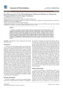
exposed via a midline subperiosteal approach (Figure 1A). A retractor with 2 cm-width blades was used. Under microscopic visualization, decompression was performed at the indicated spinal segment (s) using chisels and Kerisson rongeurs. The decompression procedure included unilateral total facetectomy, partial laminectoy and contralateral medical facetectomy. Subsequently a nearly complete discectomy and endplate decortication were carried out through a portal created at the total facetectomy site. The cartilaginous material was removed from the endplates both above and below the disk space using Cobb elevators, pituitary rongeures and end plate scrapers. Care was taken not to injure the bony endplates. An appropriately sized cage was then selected on the basis of the preoperative templating and intraoperative evaluation using a trial cage. Bones harvested from the lamina and facet joints were morcellized (Figure 1B) and inserted into the anterior and lateral parts of the intervertebral disc space. A single titanium cage filled with local morcellized bone was then inserted (Figure 1C). Fluoroscopy was used to ensure satisfactory placement of the cage. Gender
Female 33, Male 21
Age
Average 65.2 (range: 31-84)
Diagnosis
Degenerative spondylolisthesis: 31 Spinal stenosis with instability: 20 Spondylolytic spondylolisthesis: 3
Fusion segment
L2-L3: 2 L3-L4: 12 L4-L5: 30 L5-S1: 5 Two-level fusion: 5
Methods Patients Between March 2006 and November 2007, 63 patients underwent instrumented TLIF with local bone graft for degenerative lumbar or lumbosacral diseases in the authors’ hospitals. Operations were carried out by two authors (SK and HO). All patients suffered from both low back pain and neurological symptoms (i.e. radicular pain, intermittent claudication, and bowel and bladder dysfunction). All underwent at least 6 months of conservative treatment before surgery. Of these, 54 patients (86 %) completed clinical and radiological assessments both at 1 year and 2 years. There were 33 female and 21 male patients with the average age of 65.2 years. Thirty-one patients were 65 years or older. Table 1 depicts demographic information of the patients.
Surgical technique All patients underwent surgery in the prone position. A longitudinal median skin incision, 4 cm in a single-level fusion and 6cm in a two-level fusion, was made under fluoroscopic guidance. For decompression, the spinal processes, laminae, and facet joints were J Spine ISSN: 2165-7939 JSP an open access journal
One-level fusion: 49
L3-L5: 3 L4-S1: 2 Interbody cage
Novel ACC (Alphatec Spine Inc): 23 CAPSTONE (Medtronic Inc): 18
KIDNEY BEAN MESH (Medtronic Inc): 13 Table 1: Demographic information of 54 patients.
*Corresponding author: Satoshi Kawaguchi, Department of Orthopaedic Surgery, Asahikawa Kosei Hospital, Asahikawa, Japan, Tel: +81-166-33-7171; Fax: +81166-33-6075; E-mail:
[email protected] Received December 07, 2011; Accepted December 14, 2011; Published December 17, 2011 Citation: Kawaguchi S, Oguma H, Yamamura M, Akatsuka T, Matsuo S, et al. (2012) Fusion Status and Clinical Outcomes at One Year and Two Years of Instrumented, Local Bone Transforaminal Lumbar Interbody Fusion. J Spine 1:103. doi:10.4172/2165-7939.1000103 Copyright: © 2012 Kawaguchi S, et al. This is an open-access article distributed under the terms of the Creative Commons Attribution License, which permits unrestricted use, distribution, and reproduction in any medium, provided the original author and source are credited.
Volume 1 • Issue 1 • 1000103
Citation: Kawaguchi S, Oguma H, Yamamura M, Akatsuka T, Matsuo S, et al. (2012) Fusion Status and Clinical Outcomes at One Year and Two Years of Instrumented, Local Bone Transforaminal Lumbar Interbody Fusion. J Spine 1:103. doi:10.4172/2165-7939.1000103 Page 2 of 4
Two paramedian fascial incisions were then made over the cleft between the multifudus and longissimus muscles. Via a Wiltze intermuscular paraspinal approach [16,17], pedicle screws were placed bilaterally (Figure 1D). Two rods were applied to the pedicle screws and tightened under compressive force. A total of three drainage tubes were inserted through three subcutaneous incisions (a midline incision for decompression and two paramedian incisions for screwing), respectively. Postoperatively all patients wore a soft lumbar corset for a period of three months.
Assessment Data on operation time, intraoperative blood loss, and operationrelated complications including deep venous thrombosis, pulmonary embolism, surgical site infection and adjacent segment disease, were gathered. Clinical and radiographic evaluations were made preoperatively and again at 1 year and 2 years postoperatively. Clinical outcomes were determined by using the Japanese Orthopaedic Association (JOA) scores for the management of low-back pain (minimum score-6, maximum score 29, Appendix 1). Radiographic evaluations included (i) mobility of the fusion segment on flexion/ extension lateral radiographs, (ii) continuous trabecular bone formation through the cage and (iii) cage migration on both coronal and sagittal reconstruction images of CT scans. Union refers to the case showing (i) absence of mobility of the fusion segment on flexion/ extension lateral radiographs, and (ii) presence of trabecular bone formation through the cage [18] (Figure 2). When a case showed union with cage subsidence of 2 mm or deeper, it is referred to as “collapsed union” (Figure 3). All radiographs and CT images were analyzed by two authors (SK and HO) working in consensus.
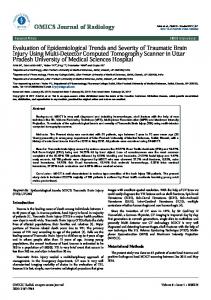
Figure 2: Representative radiographs and CT scan images of union. A 56-year-old man underwent instrumented TLIF with local autogenous bone graft of L4-L5 using a novel ACC cage. Radiographs (A: anteroposterior image, B: lateral image in flexion, C: lateral image in extension) at 2 years postoperatively show no mobility of the fusion segment. CT scans at 2 years postoperatively show continuous trabecular bone formation through the cage to both upper and lower endplates on both coronal (D) and sagittal (E) images. There is no migration of the cage. A transverse image (F) represents the location of the grafted bone.
Statistical significance was determined by using the Mann-Whitney test and the Fisher’s exact probability test. A probability of less than 0.05 was considered statistically significant.
Results The operation time averaged 200 min (range: 180-240 min) in 49 patients with a one-level fusion, 267 min (range: 240-33 min) in
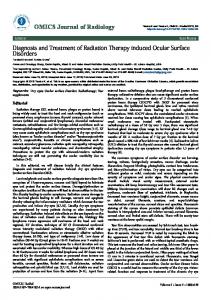
Figure 3: Representative radiographs and CT scan images of collapsed union.An 84-year-old woman underwent instrumented TLIF with local autogenous bone graft of L4-L5 using a novel ACC cage. Radiographs (A: anteroposterior image, B: lateral image in flexion, C: lateral image in extension) at 2 years postoperatively show no mobility of the segment. CT scans (D: coronal, E: sagittal) revealed continuous trabecular bone formation through the cage to both upper and lower endplates, consistent with union. However, there is sinking of the cage and a radiolucent zone around screws (arrows) (D). The case is referred to as “collapsed union”.
five patients with a two-level fusion. Intraoperative bleeding averaged 169 ml (range: 70-400 ml) in patients with a one-level fusion, 250 ml (range: 200-300 ml) in those with a two-level fusion.
Figure 1: Intraoperative photographs. (A) Through a 4-cm longitudinal skin incision, the spinal processes, laminae, and facet joints were exposed. (B) Local bones were harvested and morcellized. (C) Following decompression and discectomy, a single titanium cage with local morcellized bone was inserted into the intervertebral space. (D) Pedicle screws were inserted via the Wiltse intermuscular paraspinal approach.
J Spine ISSN: 2165-7939 JSP an open access journal
The JOA scores were 13.4 +/-3.1 (average +/- standard deviation) preoperatively, 24.0 +/- 3.5 at 1 year and 24.5 +/- 3.4 at 2 years. Improvement of the JOA scores was statistically significant. Mobility of the fusion segment in flexion/extension radiographs was less than 4 degrees in all 54 but one patient at 1 year and in all patients at 2 years. In reconstruction CT scans at 1 year, 47 patients (87%) showed union, including all five patients with a two-level fusion. At 2 years, 53
Volume 1 • Issue 1 • 1000103
Citation: Kawaguchi S, Oguma H, Yamamura M, Akatsuka T, Matsuo S, et al. (2012) Fusion Status and Clinical Outcomes at One Year and Two Years of Instrumented, Local Bone Transforaminal Lumbar Interbody Fusion. J Spine 1:103. doi:10.4172/2165-7939.1000103 Page 3 of 4
patients (98%) showed union (Figure 2). Two of them were referred to as “collapsed union” (Figure 3). These two patients were female of 78 years and 84 years, respectively. Complications included infection in one patient, which healed by surgical debridement without removal of the implant. Fracture of the fusion segment occurred in one patient. There were no wound complications. The 47 patients with union and seven patients without union at 1 year were compared (Table 2). There was no significant difference between the groups with regard to age, gender, preoperative score, and type of the cage used. Despite the difference in the union status between the groups, the JOA total scores and low back scores at 1 year were not significantly different, respectively.
Discussion In the present study, we evaluated the clinical and radiographic outcomes at 1 year and 2 years for 54 patients who had undergone instrumented TLIF with a single cage and local bone graft. More than half of the study population was 65 years of age or older. We discovered at 1 year that 53 patients (98%) exhibited biomechanical stability and 47 patients (87%) showed complete union. The JOA scores improved significantly. The scores were indistinguishable between the 47 patients with union and the remaining seven patients without union. At 2 years, the number of patients with union increased to 98% even though two patients were referred to collapsed union. These findings indicated that biomechanical fixation offered by instrumented TLIF with a single cage leads to symptomatic relief and union of the grafted local bones during a postoperative period of 2 years in the population with the average age of 65 years. In the literature, four studies [12-15] have to date addressed TLIF with local bone graft (Table 3). As in line with our procedures, each one used one cage per fusion segment. Union was achieved in all
cases in the study by Peng et al. [13] and Zhuo et al. [15] and all cases but one in the remaining two studies [12,14] within 2 years. No cage subsidence was reported in these studies. As with our own report, these other studies collectively support the efficacy of TLIF with local bone in achieving segmental fusion at 2 years postoperatively. Collapsed union and screw loosening are of particular concern with the interbody fusion procedure in the elderly who tend to have osteoporotic bones. We observed sinking of the cage with screw loosening in two patients (4%). Okuda et al. [9] reported collapsed union in 10% of the 31 patients who were over 70 years and underwent PLIF using two cages with local bone graft. To avoid cage sinking, it would be of technical importance not to injure bony endplates and pack the disk space with bone graft both inside and around the cage. Concern for the use of local bone graft in fusion surgery is its limited quantity and quality. Regarding the limited quantity of local bone graft, bone graft expanders such as hydroxyapatite [19,20] and beta tricalcium phosphate [21] have been used in PLF. In our experience, the amount of local bone collected from the laminae and the facet joints was sufficient for TLIF when chisels and rongeurs were properly used. We are of the opinion that the use of bone graft expanders is less advantageous in lumbar interbody fusion than in PLF. The quality of local bone graft can be improved by supplementation of biological modulators including bone marrow aspirate (stroma cells), platelet glue, and bone morphogenetic protein [3,4]. Efficacy and safety of these materials remain controversial and are not fully approved in many countries. The limitations of the present study include the small number of patients and lack of independent examiners for the clinical and radiographic evaluations. Due to the small number of patients, a comparative analysis was not made between the young patient group (less than 65 years) and the older patient group (65 years or older). These limitations should be considered when interpreting the results.
with union (n=47)
without union (n=7)
Age
65.1+/- 10.1
66.4+/- 7.8
Male gender
18 (38%)
3 (43%)
Degenerative spondylolisthesis
24 (51%)
5 (71%)
Two-segment fusion
5 (11%)
0 (0%)
Preoperative JOA score
13.4 +/- 3.0
13.3 +/- 4.0
Novel ACC
22 (47%)
1 (14%)
CAPSTONE
10 (21%)
3 (43%)
KIDNEY BEAN MESH
15 (32%)
3 (43%)
Cage
JOA score at 1 year Total
24.4 +/- 3.0
Low back pain
2.4 +/- 0.6
21.4 +/- 5.0
2.1 +/- 0.7
Table 2: Comparison of 47 cases with union and 7 cases without union. Authors
Average
Number of
Number of
Image
Follow-up
Union rate
Reference
age (years)
participant
cage
(months)
(%)
Dhall et al
54.0
15
One
XR
24
93
12
Peng et al
52.5
29
One
XR
24
100
13
Xiao et al
49.5
52
One
XR, CT
18
97
14
Xhou et al
50.9
76
One
XR, CT
22
100
15
Present study
65.2
54
One
XR, CT
24
98
XR: X ray Table 3: Union rate of transforaminal lumbar interbody fusion surgery with local bone graft.
J Spine ISSN: 2165-7939 JSP an open access journal
Volume 1 • Issue 1 • 1000103
Citation: Kawaguchi S, Oguma H, Yamamura M, Akatsuka T, Matsuo S, et al. (2012) Fusion Status and Clinical Outcomes at One Year and Two Years of Instrumented, Local Bone Transforaminal Lumbar Interbody Fusion. J Spine 1:103. doi:10.4172/2165-7939.1000103 Page 4 of 4
In conclusion, biomechanical fixation offered by instrumented TLIF with a single cage led to symptomatic relief, union of the grafted local bones, and a low rate of complications during a postoperative period of 2 years in patients with the average age of 65 years. References 1. Sasso RC, LeHuec JC, Shaffrey (2005) Iliac crest bone graft donor site pain after anterior lumbar interbody fusion: a prospective patient satisfaction outcome assessment. J Spinal Disord Tech 18 Suppl: S77-S81. 2. Kim DH, Rhim R, Li L, Martha J, Swaim BH, et al. (2009) Prospective study of iliac crest bone graft harvest site pain and morbidity. Spine J 9: 886-892. 3. Agarwal R, Williams K, Umscheid CA, Welch WC (2009) Osteoinductive bone graft substitutes for lumbar fusion: a systematic review. J Neurosurg Spine 11: 729-740. 4. Rihn JA, Kirkpatrick K, Albert TJ (2010) Graft options in posterolateral and posterior interbody lumbar fusion. Spine (Phila Pa 1976) 35: 1629-1639. 5. Sengupta DK, Truumees E, Patel CK, Kazmierczak C, Hughes B, et al. (2006) Outcome of local bone versus autogenous iliac crest bone graft in the instrumented posterolateral fusion of the lumbar spine. Spine (Phila Pa 1976) 31: 985-991. 6. Lee SC, Chen JF, Wu CT, Lee ST (2009) In situ local autograft for instrumented lower lumbar or lumbosacral posterolateral fusion. J Clin Neurosci 16: 37-43. 7. Ohtori S, Suzuki M, Koshi T, Takaso M, Yamashita M, et al. (2011) Single-level instrumented posterolateral fusion of the lumbar spine with a local bone graft versus an iliac crest bone graft: a prospective, randomized study with a 2-year follow-up. Eur Spine J 20: 635-639. 8. Miura Y, Imagama S, Yoda M, Mitsuguchi H, Kachi H (2003) Is local bone viable as a source of bone graft in posterior lumbar interbody fusion? Spine (Phila Pa 1976) 28: 2386-2389. 9. Okuda S, Oda T, Miyauchi A, Haku T, Yamamoto T, et al. (2006) Surgical outcomes of posterior lumbar interbody fusion in elderly patients. J Bone Joint Surg Am 88: 2714-2720. 10. Tsutsumimoto T, Shimogata M, Ohta H, Misawa H (2009) Mini-open versus conventional open posterior lumbar interbody fusion for the treatment of lumbar degenerative spondylolisthesis: comparison of paraspinal muscle damage and slip reduction. Spine (Phila Pa 1976) 34: 1923-1928.
11. Ito Z, Matsuyama Y, Sakai Y, Imagama S, Wakao N, et al. (2010) Bone union rate with autologous iliac bone versus local bone graft in posterior lumbar interbody fusion. Spine (Phila Pa 1976) 35: E1101-E1105. 12. Dhall SS, Wang MY, Mummaneni PV (2008) Clinical and radiographic comparison of mini-open transforaminal lumbar interbody fusion with open transforaminal lumbar interbody fusion in 42 patients with long-term follow-up. J Neurosurg Spine 9: 560-565. 13. Peng CW, Yue WM, Poh SY, Yeo W, Tan SB (2009) Clinical and radiological outcomes of minimally invasive versus open transforaminal lumbar interbody fusion. Spine (Phila Pa 1976) 34: 1385-1389. 14. Xiao Y, Li F, Chen Q (2010) Transforaminal lumbar interbody fusion with one cage and excised local bone. Arch Orthop Trauma Surg 130: 591-597. 15. Zhou J, Wang B, Dong J, Li X, Zhou X, et al. (2011) Instrumented transforaminal lumbar interbody fusion with single cage for the treatment of degenerative lumbar disease. Arch Orthop Trauma Surg 131: 1239-1245. 16. Wiltse LL, Spencer CW (1988) New uses and refinements of the paraspinal approach to the lumbar spine. Spine (Phila Pa 1976) 13: 696-706. 17. Taneichi H, Suda K, Kajino T, Matsumura A, Moridaira H, et al. (2006) Unilateral transforaminal lumbar interbody fusion and bilateral anterior-column fixation with two Brantigan I/F cages per level: clinical outcomes during a minimum 2-year follow-up period. J Neurosurg Spine 4: 198-205. 18. Lee DY, Jung TG, Lee SH (2008) Single-level instrumented mini-open transforaminal lumbar interbody fusion in elderly patients. J Neurosurg Spine 9: 137-144. 19. Chen WJ, Tsai TT, Chen LH, Niu CC, Lai PL, et al.(2005) The fusion rate of calcium sulfate with local autograft bone compared with autologous iliac bone graft for instrumented short-segment spinal fusion. Spine (Phila Pa 1976) 30: 2293-2297. 20. Lee JH, Hwang CJ, Song BW, Koo KH, Chang BS, et al. (2009) A prospective consecutive study of instrumented posterolateral lumbar fusion using synthetic hydroxyapatite (Bongros-HA) as a bone graft extender. J Biomed Mater Res A 90: 804-810. 21. Epstein NE, Silvergleid RS, Hollingsworth R (2009) Increased postoperative cervical myelopathy and cord compression resulting from the use of Gelfoam. Spine J 9: e19-e21.
Submit your next manuscript and get advantages of OMICS Group submissions Unique features: • • •
User friendly/feasible website-translation of your paper to 50 world’s leading languages Audio Version of published paper Digital articles to share and explore
Special features: • • • • • • • •
200 Open Access Journals 15,000 editorial team 21 days rapid review process Quality and quick editorial, review and publication processing Indexing at PubMed (partial), Scopus, DOAJ, EBSCO, Index Copernicus and Google Scholar etc Sharing Option: Social Networking Enabled Authors, Reviewers and Editors rewarded with online Scientific Credits Better discount for your subsequent articles
Submit your manuscript at: http://www.omicsgroup.info/editorialtracking/spine/
J Spine ISSN: 2165-7939 JSP an open access journal
Volume 1 • Issue 1 • 1000103