Keloids: Pathogenesis, Clinical Features, and Management Chuma J. Chike-Obi, M.D.,1 Patrick D. Cole, M.D.,1 and Anthony E. Brissett, M.D., F.A.C.S.2
ABSTRACT
Cutaneous wound healing is a complex response to skin injury. Deregulation of this process can lead to excessive scar formation, as seen in keloids. Keloids are common skin lesions that are difficult to treat and are associated with high recurrence rates despite the large number of available treatment options. With increased knowledge of the disease process and further scientific advancements, future approaches will hopefully improve keloid treatment. In this article, we review the epidemiology, genetic basis, etiology, clinical features, pathogenesis, and management of keloids. KEYWORDS: Keloid, hypertrophic scar, treatment
Cutaneous wound healing represents an inter-
play between cellular processes that act to restore skin integrity and function after injury. In the adult human, these processes end in scar formation. Keloids form when excessive scar tissue is deposited within and beyond the boundaries of the wound. In addition to the aesthetic problems they pose, keloids are often pruritic, painful, and psychologically debilitating. In this review, we discuss the epidemiology, pathophysiology, clinical features, and management of keloids.
PATHOGENESIS The process by which keloids develop is poorly understood. Because they have only been observed in humans, research efforts have been hindered by a lack of reliable animal models.1 The process is known to be induced by skin trauma in predisposed individuals. Skin trauma can be secondary to acne, folliculitis, body piercings, burns, lacerations, and surgical wounds. Whereas most keloids develop within 3 months of the injury, some may occur 1 Division of Plastic Surgery, Baylor College of Medicine, Houston, Texas; 2Bobby R. Alford Department of Otorhinolaryngology, Baylor College of Medicine, Houston, Texas. Address for correspondence and reprint requests: Chuma J. Chike-Obi, M.D., Division of Plastic Surgery, Baylor College of Medicine, 6701 Fannin, CC610.00, Houston, TX 77030-2399 (e-mail:
[email protected]).
178
up to 1 year after skin trauma.2 There are several theories of keloid etiology, most of which are related to fibroblast dysfunction. Keloid fibroblasts, when compared with fibroblasts isolated from a normal wound, overproduce type I procollagen and express higher levels of certain growth factors including vascular endothelial growth factor, transforming growth factor b1 and b2, and platelet-derived growth factor.3 In addition, these cells have lower rates of apoptosis and demonstrate a downregulation of apoptosis-related genes, including p53.4–6 Abnormalities in connexins, which are specialized proteins that are important for gap junction formation, have been implicated in keloid formation. In one study, fibroblasts harvested from keloids were found to have decreased connexin expression and less gap junctional intercellular communication than that of those harvested from normal, nonkeloid tissue.7 The investigators concluded that this difference may contribute to keloid development as adjacent cells cannot exchange inhibitory signals and as a result may undergo programmed cell death at a slower rate than normal. Interestingly, there is Cosmetic Surgery in the Ethnic Population: Special Considerations and Procedures; Guest Editor, Jamal M. Bullocks, M.D. Semin Plast Surg 2009;23:178–184. Copyright # 2009 by Thieme Medical Publishers, Inc., 333 Seventh Avenue, New York, NY 10001, USA. Tel: +1(212) 584-4662. DOI 10.1055/s-0029-1224797. ISSN 1535-2188.
KELOIDS/CHIKE-OBI ET AL
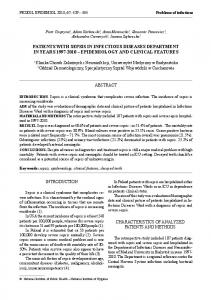
some evidence suggesting that whereas fibroblasts may play a central role in keloid formation, these cells may not be abnormal when compared with fibroblasts involved in normal scar tissue. Instead, keloids may contain normal fibroblasts that are responding to abnormal extracellular signals in predisposed individuals.8 Histologically, keloids demonstrate increased collagen and glycosaminoglycan content with whorls of thickened hyalinized collagen bundles.9 In contrast, collagen bundle orientation in normal scar tissue is parallel to the epidermis. Keloid tissue has been shown to be more metabolically active and to use more oxygen than normal scar tissue. This leads to a relative state of hypoxia in keloid fibroblasts. This high oxygen-consuming potential and low oxygen diffusion may contribute to the pathophysiology of keloid formation.10 Several investigators have reported the presence of mast cells and histamine in keloid tissue, especially early in the clinical course, which explains the pruritus associated with these lesions.11,12 The etiology of keloidrelated pain is unclear, but evidence suggests a small nerve fiber neuropathy affecting the perikeloidal skin as a possible explanation.13 Figure 1 A 53-year-old man with presternal keloids after trauma.
EPIDEMIOLOGY AND GENETICS Although keloids have been documented in virtually all major ethnic groups, they are most commonly seen in individuals of African, Asian, and, to a lesser degree, Hispanic and Mediterranean descent. Dark-skinned individuals form keloids 15 times more frequently than do their lighter-skinned counterparts.2 In both black and Hispanic populations, the incidence of keloid formation is as high as 16%, with higher frequencies during puberty and pregnancy.14 A slight female predominance is also noted, but this could be related to the higher rate of earlobe piercing in females.15 Although keloids can occur at any age, they are most likely to occur between the ages of 11 and 30 years.16 The increased risk of keloid formation in certain ethnic groups suggests a genetic component to the disease. There are documented familial cases that suggest an autosomal dominant pattern of inheritance with incomplete penetrance and variable expression.17 Keloid susceptibility loci were identified on chromosomes 2 and 7 in one large familial study; individuals with mutations at these loci developed keloids.18 However, most cases are sporadic and do not follow any clear inheritance patterns. Therefore, it is unlikely that a single candidate gene is responsible for the majority of keloids.
CLINICAL FEATURES A keloid is a growth of dense fibrous tissue that extends beyond the boundaries of the original wound (Figs. 1, 2).
This is different from a hypertrophic scar, which is typically confined to the original wound borders and tends to flatten with time. Keloids are raised, firm, frequently pruritic and painful, and generally do not spontaneously regress (Table 1). In a review of 28 keloid patients, Lee et al found that 86% and 46% of patients reported associated pruritus and pain, respectively.13 In the Caucasian patient, keloids tend to be erythematous and telangiectatic; they are often hyperpigmented in darker-skinned individuals. Keloids most commonly occur on the chest, shoulders, upper back, posterior
Figure 2 A 46-year-old woman with postauricular and cervical keloids after ventriculoperitoneal shunt removal.
179
180
SEMINARS IN PLASTIC SURGERY/VOLUME 23, NUMBER 3
2009
Table 1 Common Keloid Characteristics Age distribution
10 to 30 years
Ethnic groups
African, Asian, Hispanic,
Location
Chest, shoulders, neck, ears
Appearance
Raised, firm, hyperpigmented
Signs, symptoms
Pain, pruritus, ulceration, bleeding
Mediterranean ancestry
neck, and earlobes.19 In a retrospective review of 1000 keloid patients over an 8-year period, Ramakrishnan et al found that 34% (336 of 1000) of the keloids in the study population occurred in the presternal region. Other common sites included the deltoids (17%), upper limbs (13%), lower limbs (10%), and ears (9%).16 The reason for this site specificity is unclear, however it is generally accepted that keloids tend to form in areas of high skin tension and mechanical stress.2 Exceptions to this theory, such as earlobe keloids, suggest other contributing factors. We hypothesize that keloids form in areas of minimal tension because of the proliferation of trapped dermal elements in predisposed individuals after injury.
MANAGEMENT Current approaches to keloid management are based on an algorithm including surgical excision, intralesional steroids, oral antihistamines, cryotherapy, laser removal, radiotherapy, occlusive dressings, and immunomodulators.
Surgical Excision Sharp excision of a keloid allows the healing process to start anew; the idea is that the cooperative efforts of surgeon and patient will lead to an improved scar. A well-executed keloid excision requires complete removal of all abnormal tissue, which may increase the final scar length. Thus keloid recurrence in the setting of a longer scar may lead to a larger keloid.20 Because excessive wound tension has been proposed as a causative factor of keloid formation, wound edges should be approximated with just enough tension to close the wound. The surgeon should make incisions along skin tension lines and evert the wound edges during closure.21 The importance of meticulous technique must be emphasized. We have found that the use of a minimal number of deep resorbable stitches and permanent sutures for skin closure minimizes the inflammatory response during the early healing period. Surgical excision alone, however, is rarely curative with recurrence rates ranging between 45% and 100%.22 This high rate of recurrence suggests that surgical excision without adjuvant therapy should be undertaken with caution. For this reason, adjuvant therapies such as corticosteroids and immunomodulators are commonly started at the time of skin closure.23
Corticosteroids Corticosteroids are first-line agents for keloid treatment. When administered intralesionally, they decrease inflammation, increase vasoconstriction, and inhibit fibroblast proliferation in a dose-dependent fashion. The most commonly used corticosteroid is triamcinolone, and response rates vary from 50% to 100%, with recurrence rates between 9% and 50%.24 Commonly reported side effects include hypopigmentation, skin atrophy, and telangiectasia.25 The longer the course of treatment, the more likely it is that these side effects will occur. Corticosteroids can be administered concurrently with lidocaine to decrease injection pain and improve patient comfort. When used as an adjunct to surgical excision, triamcinolone is best initiated in the preoperative period followed by weekly injections for at least 2 weeks, then monthly injections for up to 3 months (Fig. 3). Weekly corticosteroid injections are not necessary if topical imiquimod is included in the treatment regimen (Table 2). Topical corticosteroid application is of limited benefit as the drug is poorly absorbed through an intact epidermis.
Immunomodulators IMIQUIMOD
Approved for clinical use in 1997, imiquimod is an immunomodulating agent that was first used for treating external genital and perianal warts.26 It acts as a potent inducer of interferon and cytokine release at the site of skin application, specifically IFN-a, TNF-a, IL-1, IL-6, and IL-8.27 These immunomodulators act to decrease excessive collagen production by keloid fibroblasts. Additionally, topical application of imiquimod has been shown to upregulate certain apoptosis-related genes in keloid fibroblasts.28 In one study, the investigators concluded that imiquimod application after surgical excision of earlobe and presternal keloid lesions decreased recurrence rates.29 In the same report, however, all lesions recurred after the termination of imiquimod therapy. When formulated as a 5% cream, imiquimod is a safe and well-tolerated drug. Side effects include skin erosion, excoriation, flaking, and edema at the site of application.30 Although more rigorous studies are needed to elucidate the exact mechanism of action of imiquimod for keloid management, this therapy appears to be of clinical benefit when used judiciously. INTERFERON-a-2b
Interferons are naturally occurring cytokines with antiviral, antiproliferative, and immunomodulating properties. Interferon-a-2b (IFN-a2b) specifically has undergone extensive study for keloid therapy. It has been shown in experimental models to deactivate
KELOIDS/CHIKE-OBI ET AL
Figure 3 (A) A 27-year-old woman with recurrent right earlobe keloid after two attempts at excision. (B) Sixteen months after treatment using protocol shown in Table 2.
Table 2 Keloid Treatment Protocol Time*
Treatment Regimen
0
Injection 1: Intralesional triamcinolone
1 month
Injection 2: Same as injection 1
1 month
Injection 3: Same as injection 1
acetonide, 40 mg/mL
1 month
Surgical excision Injection 4: Triamcinolone acetonide, 10 mg/mL, intraoperatively
5–7 days 1–3 weeks
Suture removal Apply silicone gel sheeting
5-FLUOROURACIL
Injection 5: Triamcinolone acetonide,
5-Fluorouracil (5-FU) is a pyrimidine analogue that is used widely for its antimetabolite and chemotherapeutic properties. It has been shown to interfere with TGF-b signaling and to decrease type I collagen gene expression in keloid fibroblasts in vitro.36 Several investigators have reported some clinical benefit with 5-FU therapy. In one randomized prospective trial, 28 keloid patients were treated with between 25 mg and 100 mg of intralesional 5-FU at weekly intervals for 12 weeks. The authors reported a response to therapy in all patients including eight patients who had failed intralesional steroid treatment, and no recurrences.37 In another study involving 24 patients with a similar protocol, the authors reported keloid flattening in all patients, with a greater response
10 mg/mL, to 3 parts Apply topical 5% imiquimod cream twice weekly 4–6 weeks
fibroblasts and to inhibit collagen production in a dosedependent fashion.31,32 It is typically injected intralesionally in recombinant form either alone or after surgical excision. Results from intralesional IFN-a2b therapy have not been favorable. Davison et al reported the results of a prospective controlled trial that demonstrated a higher recurrence rate (54%) associated with postoperative intralesional IFN-a2b than with intralesional triamcinolone (15%).33 Other investigators have reported similarly disappointing clinical results.34,35
Injection 6: Same as injection 5 Begin tapering silicone gel sheeting
4–6 weeks
Injection 7: Same as injection 5
4 weeks to 9 months
Continue injections as needed to prevent recurrence Consider silicone gel sheeting at signs of recurrence
*Each time given is counted from the previous step of the protocol. Source: Modified from Brissett AE, Sherris DA. Scar contractures, hypertrophic scars, and keloids. Facial Plast Surg 2001;17(4): 263–272.
181
182
SEMINARS IN PLASTIC SURGERY/VOLUME 23, NUMBER 3
2009
to treatment in keloids that had been present for 5 years or fewer.38 Uppal et al report that a single dose of 5-FU after extralesional excision led to decreased contractility of keloid fibroblasts 1 month after treatment, and a significant reduction in keloid scar height compared with the control population was observed.39 Common side effects of 5-FU therapy include pain, burning, hyperpigmentation, and ulceration at the injection site. MITOMYCIN C
Mitomycin C is an antitumor antibiotic isolated from Streptomyces caespitosus that has been shown to have antifibroblastic effect without inhibiting epithelialization.40 However, this has not translated into clinical efficacy in recent studies. In a prospective controlled study by Sanders et al, 15 patients with multiple keloids underwent postexcision application of topical mitomycin C (0.4 mg/mL) for 5 minutes postoperatively. After a follow-up period of 6 months, the investigators noted no difference in recurrence rate between the mitomycin C–treated lesions and the control (untreated) lesions.41 Because mitomycin C is typically applied after excision, the common side effect of poor wound healing limits its widespread use.42
transformation of the surrounding tissues. We recommend the use of radiation therapy for only the most refractory keloids.
Laser Therapy Laser therapy involves the use of light energy of a specific wavelength and pulse duration to ablate targeted tissues. Keloids can be treated with any of several different lasers including carbon dioxide (CO2), argon, and neodymium:yttrium-aluminum-garnet (Nd:YAG) lasers.50 Argon laser therapy is associated with a high recurrence rate, and carbon dioxide lasers have not been shown to be highly effective.51,52 In one study of 23 patients who underwent CO2 laser treatment for keloids, 13 patients were considered failures, and 9 patients required steroids to treat recurrent lesions.53 In addition to their high failure rate, carbon dioxide lasers vaporize tissue, which can aerosolize infectious agents such as the hepatitis and human immune deficiency viruses and endanger health care personnel.54 Laser technology continues to improve, with promises of higher cure rates and fewer postoperative complications. Still, these devices should be used with caution until long-term outcome data are available.
RETINOIDS
Retinoids are a class of compounds chemically related to vitamin A that are used to treat various skin conditions including photoaging, acne, and psoriasis. These compounds have been shown to inhibit fibroblast proliferation, enhance epidermal growth, and promote normal regeneration.43,44 Since the early 1980s, their application to keloid therapy has been attempted by several investigators, with limited efficacy.45–47 More recent studies on the effects of retinoids on keloids are needed to further assess the efficacy of these agents in keloid management.
Radiotherapy Radiotherapy can be used as monotherapy or as an adjunct to surgical excision. The most commonly used modality is external beam radiotherapy, specifically electron beam radiotherapy .When administered after surgery, it is commonly initiated within 48 hours of keloid excision. Satisfactory results have been reported with 10 to 20 Gy over 2 to 4 days in the early postoperative period.48 Ogawa et al report a recurrence rate of 32.7% after postoperative electron beam irradiation (15 Gy over a 3-day period) after 24 months of follow-up in a series of 129 patients.49 Although radiotherapy technology and equipment continue to improve and lower effective doses are being reported, this treatment modality still confers serious risks that must be discussed with the patient before treatment. These include postoperative fibrosis, impaired wound healing, and the potential for malignant
Cryotherapy Cryotherapy involves the use of a freezing medium to ablate targeted lesions. In one study, cryotherapy modified collagen synthesis and facilitated fibroblast differentiation in keloids.55 It has been used on keloids for decades, and there is evidence that small lesions may respond better than do larger lesions. However, many patients report considerable pain from the procedure and fail to return for additional treatments.15 Multiple treatment sessions are typically necessary, and hypopigmentation in darker-skinned patients is common.56 Cryotherapy continues to decline in popularity due to the associated pain and poor patient compliance.
Occlusive Dressings SILICONE THERAPY
Silicone gel products have become popular for scar therapy. Recent studies show keloid prevention in as many as 80% of cases after excision.57 Whereas the exact mechanism of action is not known, effective silicone products must be able to provide occlusion, hydration, and increased temperature to the scar.24 There are many commercial silicone-containing products, however only the gel sheet and self-drying tube formulations have been associated with improved final scar appearance. The constant, water-tight occlusion and hydration applied by the gel sheets soften scars and decrease scar size,
KELOIDS/CHIKE-OBI ET AL
erythema, pain, and itching.58 Based on these findings, an international panel of experts recommended silicone gel sheet therapy for at least 1 month after excision.22 Silicone gel sheeting should be applied as soon as reepithelialization is complete and should be worn for at least 12 hours daily.2 These sheets are expensive, but each one can be washed and reapplied to last up to 12 days.59 They need to be worn constantly for good effect, so patient compliance can be an issue especially when the scar is located in a highly visible area. Furthermore, they are difficult to keep in place around joints and areas such as the face. Silicone gel in a tube, constituted in a self-drying formulation, has been shown to be at least as effective as silicone gel sheeting in one clinical study.60 A comparison of silicone versus nonsilicone gel sheets revealed that nonsilicone gel sheets were just as effective in reducing scar size, induration, and symptoms.58 This supports the theory that the occlusion and hydration of a scar play a prominent role in affecting cytokine-mediated keratinocyte-fibroblast signaling, perhaps a more important role than the silicone itself.61 Moreover, nonocclusive silicone-based products such as creams containing silicone oil have yielded disappointing results.62 Similarly, one would expect newer formulations such as silicone sprays or foams to be ineffective because they do not provide occlusion.
CONCLUSION Keloids are difficult problems to treat. Many therapeutic options are available, with varying degrees of proven clinical success. No optimal treatment modality has been identified to date. Surgical excision, perioperative intralesional steroids, topical imiquimod, and occlusive silicone therapy are among the more efficacious options that are currently available. The surgeon must ensure adequate patient education and informed consent, including an indepth discussion of the high recurrence rate and limitations of current options before initiating treatment.
REFERENCES 1. Bayat A, Bock O, Mrowietz U, Ollier WE, Ferguson MW. Genetic susceptibility to keloid disease and hypertrophic scarring: transforming growth factor beta1 common polymorphisms and plasma levels. Plast Reconstr Surg 2003;111: 535–543; discussion 544–546 2. Brissett AE, Sherris DA. Scar contractures, hypertrophic scars, and keloids. Facial Plast Surg 2001;17:263–272 3. Marneros AG, Krieg T. Keloids—clinical diagnosis, pathogenesis, and treatment options. J Dtsch Dermatol Ges 2004; 2:905–913 4. Messadi DV, Le A, Berg S, Huang G, Zhuang W, Bertolami CN. Effect of TGF-beta 1 on PDGF receptors expression in human scar fibroblasts. Front Biosci 1998;3:a16–a22
5. Sayah DN, Soo C, Shaw WW, et al. Downregulation of apoptosis-related genes in keloid tissues. J Surg Res 1999; 87:209–216 6. De Felice B, Ciarmiello LF, Mondola P, et al. Differential p63 and p53 expression in human keloid fibroblasts and hypertrophic scar fibroblasts. DNA Cell Biol 2007;26:541– 547 7. Lu F, Gao J, Ogawa R, Hyakusoku H. Variations in gap junctional intercellular communication and connexin expression in fibroblasts derived from keloid and hypertrophic scars. Plast Reconstr Surg 2007;119:844–851 8. Chevray PM, Manson PN. Keloid scars are formed by polyclonal fibroblasts. Ann Plast Surg 2004;52:605– 608 9. Berman B, Flores F. The treatment of hypertrophic scars and keloids. Eur J Dermatol 1998;8:591–595 10. Ichioka S, Ando T, Shibata M, Sekiya N, Nakatsuka T. Oxygen consumption of keloids and hypertrophic scars. Ann Plast Surg 2008;60:194–197 11. Sasaki A, Mueller RV, Xi G, Sipe R, Buck D, Hollinger J. Mast cells: an unexpected finding in the modulation of cutaneous wound repair by charged beads. Plast Reconstr Surg 2003;111:1446–1453 12. Sharquie KE, Al-Dhalimi MA. Keloid in Iraqi patients: a clinicohistopathologic study. Dermatol Surg 2003;29: 847–851 13. Lee SS, Yosipovitch G, Chan YH, Goh CL. Pruritus, pain, and small nerve fiber function in keloids: a controlled study. J Am Acad Dermatol 2004;51:1002–1006 14. Ako¨z T, Giderog˘lu K, Akan M. Combination of different techniques for the treatment of earlobe keloids. Aesthetic Plast Surg 2002;26:184–188 15. Kelly AP. Medical and surgical therapies for keloids. Dermatol Ther 2004;17:212–218 16. Ramakrishnan KM, Thomas KP, Sundararajan CR. Study of 1,000 patients with keloids in South India. Plast Reconstr Surg 1974;53:276–280 17. Marneros AG, Norris JE, Olsen BR, Reichenberger E. Clinical genetics of familial keloids. Arch Dermatol 2001;137:1429–1434 18. Marneros AG, Norris JE, Watanabe S, Reichenberger E, Olsen BR. Genome scans provide evidence for keloid susceptibility loci on chromosomes 2q23 and 7p11. J Invest Dermatol 2004;122:1126–1132 19. Bayat A, Arscott G, Ollier WE, Ferguson MW, Mc Grouther DA. Description of site-specific morphology of keloid phenotypes in an Afrocaribbean population. Br J Plast Surg 2004;57:122–133 20. Poochareon VN, Berman B. New therapies for the management of keloids. J Craniofac Surg 2003;14:654–657 21. Chen MA, Davidson TM. Scar management: prevention and treatment strategies. Curr Opin Otolaryngol Head Neck Surg 2005;13:242–247 22. Mustoe TA, Cooter RD, Gold MH, et al. International clinical recommendations on scar management. Plast Reconstr Surg 2002;110:560–571 23. Berman B, Kaufman J. Pilot study of the effect of postoperative imiquimod 5% cream on the recurrence rate of excised keloids. J Am Acad Dermatol 2002;47(4, Suppl): S209–S211 24. Niessen FB, Spauwen PH, Schalkwijk J, Kon M. On the nature of hypertrophic scars and keloids: a review. Plast Reconstr Surg 1999;104:1435–1458
183
184
SEMINARS IN PLASTIC SURGERY/VOLUME 23, NUMBER 3
2009
25. Alster TS, Tanzi EL. Hypertrophic scars and keloids: etiology and management. Am J Clin Dermatol 2003;4: 235–243 26. Rudy SJ. Imiquimod (Aldara): modifying the immune response. Dermatol Nurs 2002;14:268–270 27. Weeks CE, Gibson SJ. Induction of interferon and other cytokines by imiquimod and its hydroxylated metabolite R842 in human blood cells in vitro. J Interferon Res 1994; 14:81–85 28. Jacob SE, Berman B, Nassiri M, Vincek V. Topical application of imiquimod 5% cream to keloids alters expression genes associated with apoptosis. Br J Dermatol 2003;149(Suppl 66):62–65 29. Malhotra AK, Gupta S, Khaitan BK, Sharma VK. Imiquimod 5% cream for the prevention of recurrence after excision of presternal keloids. Dermatology 2007;215:63–65 30. Bilu D, Sauder DN. Imiquimod: modes of action. Br J Dermatol 2003;(149 Suppl 66):5–8 31. Berman B, Duncan MR. Short-term keloid treatment in vivo with human interferon alfa-2b results in a selective and persistent normalization of keloidal fibroblast collagen, glycosaminoglycan, and collagenase production in vitro. J Am Acad Dermatol 1989;21(4 Pt 1):694–702 32. Duncan MR, Berman B. Persistence of a reduced-collagenproducing phenotype in cultured scleroderma fibroblasts after short-term exposure to interferons. J Clin Invest 1987;79: 1318–1324 33. Davison SP, Mess S, Kauffman LC, Al-Attar A. Ineffective treatment of keloids with interferon alpha-2b. Plast Reconstr Surg 2006;117:247–252 34. al-Khawajah MM. Failure of interferon-alpha 2b in the treatment of mature keloids. Int J Dermatol 1996;35:515– 517 35. Wong TW, Chiu HC, Yip KM. Intralesional interferon alpha-2b has no effect in the treatment of keloids. Br J Dermatol 1994;130:683–685 36. Wendling J, Marchand A, Mauviel A, Verrecchia F. 5Fluorouracil blocks transforming growth factor-beta-induced alpha 2 type I collagen gene (COL1A2) expression in human fibroblasts via c-Jun NH2-terminal kinase/activator protein-1 activation. Mol Pharmacol 2003;64:707–713 37. Nanda S, Reddy BS. Intralesional 5-fluorouracil as a treatment modality of keloids. Dermatol Surg 2004;30:54– 56, discussion 56–57 38. Gupta S, Kalra A. Efficacy and safety of intralesional 5fluorouracil in the treatment of keloids. Dermatology 2002; 204:130–132 39. Uppal RS, Khan U, Kakar S, Talas G, Chapman P, McGrouther AD. The effects of a single dose of 5fluorouracil on keloid scars: a clinical trial of timed wound irrigation after extralesional excision. Plast Reconstr Surg 2001;108:1218–1224 40. Lee DA. Antifibrosis agents and glaucoma surgery. Invest Ophthalmol Vis Sci 1994;35:3789–3791 41. Sanders KW, Gage-White L, Stucker FJ. Topical mitomycin C in the prevention of keloid scar recurrence. Arch Facial Plast Surg 2005;7:172–175 42. Sewall GK, Robertson KM, Connor NP, Heisey DM, Hartig GK. Effect of topical mitomycin on skin wound contraction. Arch Facial Plast Surg 2003;5:59–62 43. Prutkin L. Wound healing and vitamin A acid. Acta Derm Venereol 1972;52:489–492
44. Christophers E. Growth stimulation of cultured postembryonic epidermal cells by vitamin A acid. J Invest Dermatol 1974;63:450–455 45. Janssen de Limpens AM. The local treatment of hypertrophic scars and keloids with topical retinoic acid. Br J Dermatol 1980;103:319–323 46. Daly TJ, Weston WL. Retinoid effects on fibroblast proliferation and collagen synthesis in vitro and on fibrotic disease in vivo. J Am Acad Dermatol 1986;15(4 Pt 2):900– 902 47. Al-Attar A, Mess S, Thomassen JM, Kauffman CL, Davison SP. Keloid pathogenesis and treatment. Plast Reconstr Surg 2006;117:286–300 48. Ogawa R, Miyashita T, Hyakusoku H, Akaishi S, Kuribayashi S, Tateno A. Postoperative radiation protocol for keloids and hypertrophic scars: statistical analysis of 370 sites followed for over 18 months. Ann Plast Surg 2007;59:688– 691 49. Ogawa R, Mitsuhashi K, Hyakusoku H, Miyashita T. Postoperative electron-beam irradiation therapy for keloids and hypertrophic scars: retrospective study of 147 cases followed for more than 18 months. Plast Reconstr Surg 2003;111:547–553, discussion 554–555 50. Nouri K, Vidulich K, Rivas MP. Lasers for scars: a review. J Cosmet Dermatol 2006;5:14–22 51. Apfelberg DB, Maser MR, Lash H, White D, Weston J. Preliminary results of argon and carbon dioxide laser treatment of keloid scars. Lasers Surg Med 1984;4:283– 290 52. Apfelberg DB, Maser MR, White DN, Lash H. Failure of carbon dioxide laser excision of keloids. Lasers Surg Med 1989;9:382–388 53. Norris JE. The effect of carbon dioxide laser surgery on the recurrence of keloids. Plast Reconstr Surg 1991;87:44–49, discussion 50–53 54. Atiyeh BS. Nonsurgical management of hypertrophic scars: evidence-based therapies, standard practices, and emerging methods. Aesthetic Plast Surg 2007;31:468–492; discussion 493–494 55. Rusciani L, Paradisi A, Alfano C, Chiummariello S, Rusciani A. Cryotherapy in the treatment of keloids. J Drugs Dermatol 2006;5:591–595 56. Har-Shai Y, Amar M, Sabo E. Intralesional cryotherapy for enhancing the involution of hypertrophic scars and keloids. Plast Reconstr Surg 2003;111:1841–1852 57. Borgognoni L. Biological effects of silicone gel sheeting. Wound Repair Regen 2002;10:118–121 58. de Oliveira GV, Nunes TA, Magna LA, et al. Silicone versus nonsilicone gel dressings: a controlled trial. Dermatol Surg 2001;27:721–726 59. Chang CW, Ries WR. Nonoperative techniques for scar management and revision. Facial Plast Surg 2001;17:283–288 60. Chernoff WG, Cramer H, Su-Huang S. The efficacy of topical silicone gel elastomers in the treatment of hypertrophic scars, keloid scars, and post-laser exfoliation erythema. Aesthetic Plast Surg 2007;31:495–500 61. Mustoe TA. Evolution of silicone therapy and mechanism of action in scar management. Aesthetic Plast Surg 2008;32:82– 92 62. Sawada Y, Sone K. Treatment of scars and keloids with a cream containing silicone oil. Br J Plast Surg 1990;43:683– 688