Chronic Wounds: Combating an “Axis of Evil” with the “Forces of Healing” This is a war which must be won. Ischemia Ischemia in extremities with open wounds prolongs or prevents healing and is a risk factor for amputation. Ischemia must be recognized and iding the healing of chronic treated. The history, clinical exam, wounds and preventing and imaging are all used to make consequences such as the diagnosis. Patients should be amputation can, in a sense, be likened questioned about to the “War on intermittent Terror.” The claudication “terror” that A patient with a lower and rest pain. is present in Frequently, these patients extremity wound and a patient is the fear of with diabetic amputation contributing peripheral neuropathy resulting in loss may lack the of independence. ischemia should have a true symptoms We already of intermittent know that 84% consult for a vascular claudication, of amputations but rather have are preceded by intervention. fatigue after a foot wound.1 walking, which We know that an is relieved infected wound is by rest. The American Diabetes a significant risk factor for limb loss.2 Association has published guidelines Directing resources toward keeping for screening of peripheral arterial wounds from getting infected, and disease in persons with diabetes assisting them to close in a timely recommending intervals of 5 years.3 fashion, can maximize the potential for limb preservation. The “Axis of A patient with a lower extremity Evil” consists of ischemia, infection, wound and contributing peripheral and pressure. ischemia should have a consult for By Lee C. Rogers, DPM, Nicholas J. Bevilacqua, DPM, and David G. Armstrong, DPM, PhD.
A
NOVEMBER/DECEMBER 2007 • PODIATRY MANAGEMENT • www.pediatrym.com
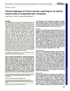
Figure 1: The VersaJet (Smith & Nephew) hydroscalpel debriding a full-thickness ulcer.
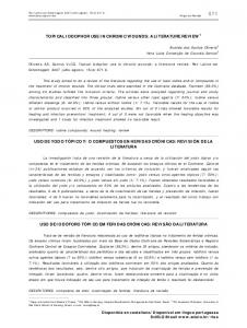
Figure 2: Maggot Debridement Therapy used to convert a fibrotic or necrotic wound, to a granular one.
1
of muscle/tendon/joint/bone AND the patient is systemically well and metabolically stable.
a vascular intervention. Although distal bypass is considered the “gold standard” for limb revascularization, an endovascular procedure may restore enough flow to the limb to allow the wound to heal—achieving the desired endpoint. Infection Infection is a clinical diagnosis. A culture should only be taken if the wound is clinically infected and is used to direct therapy, not to make the diagnosis of infection. The Infectious Diseases Society of America (IDSA) classifies diabetic foot infections as uninfected, mild, moderate, and severe. Uninfected wounds have no purulence or manifestations of inflammation. Mild infections have >2 manifestations of inflammation (purulence, erythema, pain, tenderness, warmth, induration);
Figure 4: Puracol (Medline) is 100% collagen and is moistened with a few drops of saline and placed on the wound.
Figure 3: The Wound V. A. C. (KCI, San Antonio) produces topical negative pressure and promotes granulation tissue.
AND any erythema extends less than 2 cm from the wound, AND infection is limited to skin/superficial subcutaneous tissue without systemic illness. Moderate infections have one or more of the following descriptions: cellulitis extending greater than 2 cm from the wound, lymphangitis, deep tissue abscess, gangrene, involvement
Figure 5: A split thickness skin graft (STSG) placed on the dorsum of the foot, after 1 week the interstices are healing.
Figure 6A & 6B: The use of GraftJacket (Wright Medical) to replace “like with like” tissue on the plantar foot.
NOVEMBER/DECEMBER 2007 • PODIATRY MANAGEMENT • www.pediatrym.com
Severe infections have the characteristics of a moderate infection, but the patient has additional signs/ symptoms of systemic toxicity such as fever, chills, tachycardia, hypotension, confusion, vomiting, leukocytosis, acidosis, hyperglycemia or azotemia.2 Our group has since validated this classification system, noting a substantial increase in hospitalization and amputation with increased infection severity.4 Pressure Pressure is probably the most commonly overlooked component of wound chronicity. High foot pressures are a major etiologic factor in wound development5 and can keep a foot ulcer from healing. Foot deformities are predictive of elevated pressures, such as hallux valgus, hammered digits, and Charcot foot. Some deformities are not visible to inspection. Limited joint mobility (LJM) is as important for prediction of foot ulcerations as deformity.6 The joints in the foot that should be checked for restricted range of motion are the first metatarsophalangeal joint and the ankle joint. The hallux should have at least 50 degrees of passive dorsiflexion.7 Less than 10 degrees of dorsiflexion at the ankle is an equinus deformity. Ankle equinus increases plantar forefoot pressures and the risk for ulceration.8 Likewise, a tendo- Achilles lengthening has been shown to reduce plantar forefoot pressures and is beneficial to heal or prevent forefoot ulcerations. Lin, et al. reported rapid healing of previously recalcitrant plantar wounds with an Achilles tendon lengthening procedure.9 Mueller, et al. subsequently reported a similar trend toward lower ulcer recurrence when performing an Achilles tendon lengthening.10 The “Forces of Healing” are debridement, promotion of granulation 2
tissue, and wound closure. These steps must be completed in order. It is assumed that before one begins the healing treatments below, the “axis of evil” components (vascular, infection, and pressure) all have been addressed. Debridement Debridement is a key component to the management of foot ulcerations. Debridement removes senescent cells and reduces bioburden. Armstrong, et al. found that diabetic foot ulcers that were debrided on each visit had a 5.3times greater chance of healing in 12 weeks than ulcers that were debrided less often.11 There are different forms of debridement: sharp/surgical, enzymatic, mechanical, autolytic, and biosurgical. When debriding a wound, it is important to remove all necrotic/non-viable tissue, and any undermining. The surgeon must not be concerned with the defect caused by the initial debridement, since removing this tissue is important to attain closure. Most wounds require serial debridements. New tools are available to make debridement easier and can help discriminate between healthy and nonviable tissue. The VersaJet (Smith & Nephew) is a “hydroscapel” which uses a thin spray of saline at pressures greater than 10,000 P.S.I. to debride tissue, layer by layer (Figure 1). The SonicOne (Misonix) is a device that uses ultrasonic energy to create cavitation which separates non-viable tissue. Maggot debridement therapy (MonarchLabs com) is the only FDA-approved device that is a living organism. Maggots arrive in a sterile vial containing 250-500 larvae. They are placed inside a fibrotic wound and covered with a mesh dressing for three days (Figure 2). The species Phaenicia sericata (green blowfly) is unique in that it only consumes fibrotic/necrotic tissue and will not digest living granulation. Occasionally, multiple applications are required. Promotion of Granulation After a wound has been debrided,
Figure 3: The Wound V. A. C. (KCI, San Antonio) produces topical negative pressure and promotes granulation tissue.
via any modality granulation tissue must be promoted. Multiple methods exist, but the Wound VAC (KCI, San Antonio) is by far the most superior method (Figure 3). With an acute surgical debridement, we wait 24 hours to allow hemorrhage to stop; then, we apply the Wound VAC. Most often the wound bed can be prepared for closure within four weeks. Granulation tissue can also be promoted by using collagen dressings (Puracol or Promogran), which act as a substrate for MMPs and elastase, preventing breakdown of native collagen in the wound (Figure 4). Growth factors, such as PDGF (Regranex), can be useful in a minority of cases. The living skin equivalent, Apligraf, which contains live cells, acts like a “growth factor factory”. We have found it more useful at promoting granulation tissue than providing wound epithelialization. We have shown that marrow-derived stem cells are potent, rapid stimulators of granulation tissue, providing wound bed preparation in an average of 16 days.12 The marrow aspirate can be harvested from the distal tibial metaphysis and reinjected into the periwound area. We have placed the Wound VAC on these wounds after 24-48 hours. Wound Closure The best method of wound closure depends on the location of the ulcer and the stresses that will be present during and after wound closure. Dorsal foot wounds are quite amenable to split thickness skin
NOVEMBER/DECEMBER 2007 • PODIATRY MANAGEMENT • www.pediatrym.com
grafting (Figure 5). We use the Wound VAC as a bolster dressing at 125 mmHg, continuous for three to five days post-grafting. It is important to place a non-adherent dressing, such as the silicone Mepitel (Molnlycke), between the foam and the graft, as not to avulse it during the VAC removal. The VAC helps to reduce the two main factors responsible for graft failure, shearing force and hematoma/seroma. Dorsal wounds can also be closed with skin stretching devices. Medial, lateral, and plantar wounds require more resilient treatments, given the pressures that these surfaces are subjected to during ambulation. Plantar wounds have been responsive to GraftJacket (Wright Medical) and this seems to replace “like tissue with like tissue” (Figure 6). The subsequent healed plantar skin is of good quality. GraftJacket undergoes a phase during which it almost liquefies, but we have found that leaving it alone and keeping it covered with a nonadherent dressing allows it to respond as desired. Plantar wounds can also be closed with a fasciocutaneous flap. A medial plantar artery rotational flap allows the plantar arch skin to be rotated in the midfoot area. A split thickness skin graft can then be placed in the donor arch area, which is not subjected to much weight-bearing force (Figure 7). Medial and lateral wounds can respond to a variety of treatments. Unite (Pegasus) is an equine pericardium graft used for tendon repair (Figure 8). It is extremely tough and resistant to breakdown since the processing does not destroy the crosslinking of collagen.
Figure 8: The xenograft Unite (Pegasus) is made from equine pericardium and can be used as a skin substitute.
3
“Mission Accomplished— Victory Over Wounds” Once treatment has begun, clinicians should not be satisfied with delayed healing in wounds. We recommend using a wound healing prediction model to track progress, such as described by Sheehan, et al., which states that a wound which does not reduce in area (simple length x width) by at least 50% in four weeks, has a 91% chance of not healing in 12 weeks.13 This means that the wound should be re-evaluated and an alternate treatment should be considered. The best offense is a good defense. The post-ulcerative patient should be closely monitored, as re-ulceration rates are very high. The etiology of the wound should be managed, with close attention given to pressure. Prescriptive footwear and prophylactic surgery should be considered. Neuropathic patients should be encouraged to purchase a dermal thermometer (TempTouch, Xilas) and monitor their foot temperatures daily. A spike in foot temperature can predict ulcerations days in advance, when an intervention might prevent the wound.14 ■
References 1. Pecoraro, R.E., G.E. Reiber, and E.M. Burgess, Pathways to diabetic limb amputation: basis for prevention. Diabetes Care, 1990. 13: p. 513-521. 2. Lavery, L.A., et al., Validation of the Infectious Diseases Society of America’s diabetic foot infection classification system. Clin Infect Dis, 2007. 44(4): p. 562-5. 3. Association”, A.D., Peripheral arterial disease in people with diabetes. Diabetes Care, 2003. 26: p. 3333-3341. 4. Lavery, L.A., et al., Validation of the Infectious Disease Society of America’s Diabetic Foot Infection Classification System. Clinical Infectious Diseases, 2006(In Review). 5. Frykberg, R.G., et al., Role of neuropathy and high foot pressures in diabetic foot ulceration. Diabetes Care, 1998. 21(10): p. 1714-9. 6. Fernando, D.J.S., et al., Relationship of limited joint mobility to abnormal foot pressures and diabetic foot ulceration. Diabetes Care, 1991. 14: p. 8-11. 7. Birke, J., M.A. Cornwall, and M. Jackson, Relationship between hallux limitus and ulceration of the great toe. Sports Phys Ther. J. Orthop., 1988. 10: p. 172-176. 8. Lavery, L.A., D.G. Armstrong, and A.J.M. Boulton, Ankle equinus deformity and its relationship to high plantar pressure in a large population with diabetes mellitus. J Am Podiatr Med Assoc, 2002. 92(9): p. 479-482. 9. Lin, S.S., T.H. Lee, and K.L. Wapner, Plantar forefoot ulceration with equinus deformity of the ankle in diabetic patients: the effect of tendo-achilles lengthening and total contact casting. Orthopaedics, 1996. 19(5): p. 465-475. 10. Mueller, M.J., et al., Effect of Achilles tendon lengthening on neuropathic plantar ulcers. A randomized clinical trial. J Bone Joint Surg, 2003. 85A(8): p. 1436-1445. 11. Armstrong, D.G. Serial surgical debridement increases healing rates in chronic lower extremity wounds. in American Podiatric Medical Association Oral Abstracts. 2007. Philadelphia, PA. 12. Rogers, L.C. Marrow-derived stem cells to augment chronic wound healing. in American Podiatric Medical Association Oral Abstracts. 2007. Philadelphia, PA. 13. Sheehan, P., et al., Percent Change in Wound Area of Diabetic Foot Ulcers Over a 4-Week Period Is a Robust Predictor of Complete Healing in a 12-Week Prospective Trial. Diabetes Care, 2003. 26(6): p. 1879-1882.
Dr. Rogers is the Director of the Amputation Prevention Center at Broadlawns Medical Center in Des Moines, IA. He completed the CLEAR Fellowship in Diabetic Limb Preservation and Research in 2007 and is an active CLEAR collaborator (www.broadlawns.org and www.diabetic-foot.net). Email
[email protected]. Dr. Bevilacqua is a staff surgeon in the Division of Foot and Ankle Surgery at Broadlawns Medical Center in Des Moines, IA. He is a 2007 alumnus of the CLEAR Fellowship in Diabetic Limb Preservation and Research and is an active CLEAR collaborator (www. diabeticfoot.net). Email Nicholas.
[email protected]. Dr. Armstrong is the Director of Scholl’s Center for Lower Extremity Ambulatory Research (CLEAR) at Rosalind Franklin University of Medicine and Science in Chicago, IL. He is the co-chair of the Diabetic Foot Global Conference (DFCon) held annually in Hollywood, CA (www.dfcon.com and www. diabetic-foot.net). E-mail Armstrong@ USA.net.
14. Armstrong, D.G., et al., Skin temperature monitoring reduces the risk for diabetic foot ulceration in high-risk patients. Am J. Med, 2007: p. In Press.
NOVEMBER/DECEMBER 2007 • PODIATRY MANAGEMENT • www.pediatrym.com
VJ-0063-0408
4