KISEP
LABORATORY INVESTIGATION J Korean Neurosurg Soc 40 : 267-272, 2006
Motor Function Recovery after Adipose Tissue Derived Mesenchymal Stem Cell Therapy in Rats with Cerebral Infarction Chang-Hwan Kim, M.D.,1 Yong-Woon Kim, M.D.,2 Sung-Ho Jang, M.D.,3 Chul-Hoon Chang, M.D.,1 Jae-Ho Jung, M.D.,4 Seong Ho Kim, M.D.1 Departments of Neurosurgery,1 Physiology,2 Rehabilitation Medicine,3 Plastic Surgery,4 College of Medicine Yeungnam University, Daegu, Korea
Objective : There have been recent reports that mesenchymal stromal cells that are harvested from adipose tissue are able to differentiate into neurons. In the present study, we administered adipose tissue derived stem cells in rats with cerebral infarction in order to determine whether those stem cells could enhance the recovery of motor function. Methods : Cerebral infarction was induced by intraluminal occlusion of middle cerebral artery in rats. The adipose tissue-derived mesenchymal stem cells were harvested from inguinal fat pad and proliferated for 2 weeks in DMEM media. Approximately 1x10⁶ cells were injected intravenously or into subdural space of the peri-lesional area. The rotor rod test was performed at preoperative state(before MCA occlusion), and 1, 2, 3, 4, 6, 8 and 10 weeks after the cell therapy. Results : The motor functions that were assessed by rotor rod test at 1 week of the cell therapy were nearly zero among the experimental groups. However, there was apparent motor function recovery after 2 weeks and 4 weeks of cell injection in intravenously treated rats and peri-lesionaly treated rats, respectively, while there was no significant improvement till 8 weeks in vehicle treated rats. Conclusion : These results demonstrate that the adipose derived stem cell treatment improves motor function recovery in rats with cerebral infarction.
KEY WORDS : Cerebral infarction·Adipose tissue derived mesenchymal stem cell· Motor Function recovery.
Introduction
M
ature nerve cell has a limited capacity for self repair. Thus, various stem cell transplantations, such as embryonic stem cells, neural stem cells, and bone marrow stromal cells, have been applied to experimental animals with cerebral ischemia, because stem cells have the capacity for self-renewal and differentiation into various cell types. There have been recent reports that mesenchymal stromal cells that were harvested from adipose tissue were able to differentiate into neurons. Thus, adipose tissue derived stem cells(ADSC) therapy has emerged as a excellent candidate for stem cell therapy in ischemic brain disease because ADSCs have some advantages, such as their abundancy and less effort required for tissue harvesting, compared with bone marrow derived stem cell
(BDSC). Kang et al.10) reported that intracerebral transplantation of human ADSC improved the neurological deficits in the cerebral ischemia in rats. However, there are no research whether intravenous and peri-ischemic regional injection of autologous ADSC improve the motor function recovery to cerebral infarction. In this study, we evaluated the effect of autologous ADSC treatment improves the motor function in rats with cerebral infarction.
Materials and Methods Animal care
Adult male Sprague-Dawley rats (250~300g) were used in this study. Animals were divided randomly into three groups : middle cerebral artery(MCA) occlusion+intrave-
Received:April 26, 2006 Accepted:May 16, 2006 Address for reprints : Seong Ho Kim, M.D., Department of Neurosurgery, College of Medicine, Yeung-nam University, 317-1 Daemyeong-dong, Nam-gu, Daegu 705-717, Korea Tel:+82-53-620-3790, Fax:+82-53-620-3770, E-mail :
[email protected]
267
J Korean Neurosurg Soc 40|October 2006
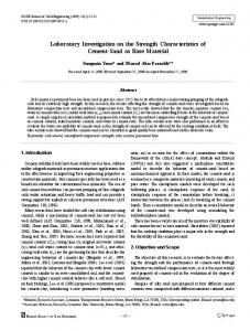
Rotor rod test, sec
nous buffer treatment (n=9), MCA occlusion+intravenous ADSC treatment (n=7), MCA occlusion +peri-lesional ADSC treatment (n=8). The cerebral infarction was induced by MCA occlusion. The left common carotid artery, external carotid artery(ECA), and internal carotid artery(ICA) were exposed under anesthesia with xylazine hydrochloride (8mg/kg) and ketamine (90mg/kg, i.p). A length of 4-0 monofilament nylon suture (18.5~19.5mm) with its tip rounded by heating, was adv- Fig. 1. Photomicrograph showing the adipose tissue-derived stem cell. (Prussian blue stain, X400). anced from the ECA into the lumen of the ICA until it blocked the origin of the MCA. The 16 nylon suture was then tied with the stump of ECA. Control 14 Isolation and culture of adipose tissue derived stem cells(ADSC)
Adipose tissue was excised from inguinal area at the same time as the MCA occlusion surgery for isolation of ADSC in the rats. Adipose tissue was washed extensively with equal volumes of phosphate-buffered saline to remove contaminating red blood cells and debris. Sequently, the extracellular matrix was digested with 0.075% collagenase for 30 minutes at 37。 C. Enzyme activity was neutralized with 10% fetal bovine serum (Hyclone, Logan, Utah), and the cells were centrifuged at 1200×g for 5 minutes. The resulting pellet, which contained ADSC cells, was resuspended in complete culture medium that consisted of Dulbecco’s Modified Eagle Medium (Gibco BRL, Rockville, MD, USA.), 10% fetal bovine serum, and 1% antibiotic/antimycotic. All the cells were distributed in 100×20mm tissue culture dishes and incubated at 37。 C with 5% humidified carbon dioxide. The cells were washed thoroughly with phosphatebuffered saline after 24 hours of incubation, and all the nonadherent cells were discarded. Fresh complete culture medium was added every 3 days. Treatment of the ADSCs
The proliferated ADSCs (approximately 1×10⁶cells) were injected intravenously or perilesionally into the MCA occluded rats. It was 2-weeks after MCA occlusion. For perilesional treatment, the rats were anesthesized with xylazine hydrochloride (8mg/kg) and ketamine (90mg/kg, i.p). The rats were then transferred to a stereotaxic instrument in a clean field. A small incision (1cm) was made over the midline of the skull to expose the landmarks of the cranium (bregma
IV Perilesional
12 10 8 6 4 2 0
Initial
1
2
3
4
6
8
10
Weeks after MCA occlusion Fig. 2. Functional recovery after treatment of adipose-derived stem cells in the rats with cerebral infarction. Values are means ± S.E.. Arrow means the time of cell treatment.
and lambda). A burr hole was made in the bone 6mm posterior to bregma and 3mm lateral to midline with a dental drill. An incision was made over the dura, and then approximately 100ul of ADSC suspension was loaded slowly into the hole. Rotor rod test
In all animals rotor rod tests were assessed at preoperative state, and 1, 2, 3, 4, 6, 8, and 10 weeks after MCA occlusion, with and without cell treatment. In order to measure the motor function, the rats were placed at a rotoring rod (frequency, 6 per minute; rotor diameter, 20cm). The time, in second, that the rat was stayed there was recorded. Statistical analysis
The data were presented as means+SE, and were analyzed by a T-test between the individual groups. A value of p<0.05 was considered significant.
268
Mesenchymal Stem Cell Therapy|CH Kim, et al.
A
B
C
Fig. 3. Microphotograph taken from the rat's excised brain tissue after adipose-derived stem cell treatment. The blue spots are stained adipose-derived stem cell and they are seen at the basal area of the brain. (Prussian blue stain, A : X10, B : X40, C : X100).
Results
T
he features of cultured ADSCs demonstrated a spindlelike shape (Fig. 1). The results of the preoperative motor function of the three groups was 8 to 13 seconds. This variation in the results can be considered as the difference in basal motor function. After the 1 week of the occlusion of left middle cerebral artery, most rats were not tolerated on the RotaRod machine. A majority had minimal recovery of motor function. The experimental groups had a fast recovery after treatment of stem cells. In particular, the first experimental group had the fastest recovery of results, which was statistically significant compared with the control group (Table 1, Fig. 2, P=0.0029). The results of the second experimental group, which received injections into the peri-ischemic area after the burr-hole operation, recovered more slowly recovery than the first experimental group; however, 4 weeks later after operation, motor function recovery was faster than the previous time. That also was statistically significant compared with the control group (Table 1, Fig. 2, P=0.0064). The control group had a natural recovery of the motor function, but there was difference in the results compared with the experimental groups, especially after 4 weeks after operation, and there was no recovery of motor function at 6 weeks later after operation (Table 1, Fig. 2).
And ADSCs were performed by the rat’s brain histology (Fig. 3). ADSCs were detected at the basal area of the brain and were taken by the prussian blue stain as the morphology of blue spot. By the these images, we can confirm the living of the ADSCs in brain tissue of the ischemic rats.
Discussion
S
tem cells are premordial, self-renewal and potentially can differentiate into multi-lineage cells. Numerous studies on stem cell treatment have been reported for cerebral infarction4,8), myocardiac infarction15), type 1 diabetes3), renal disease17), and spinal cord disease5). Gorio et al.6) reported about the fate of autologous dermal stem cells that were transplanted into the spinal cord after traumatic injury. There are two types of stem cells : embryonal stem cells and adult stem cells. Embryonal stem cells are derived from the one embryo, so these present the ethical dilemma with respect to when they actually differentiate into a human life. Adult stem cells present fewer ethical problems compared with embryonal stem cells. However, adult stem cells do not have a precise mechanism for differentiation and can be differentiate to cancer cells, so there are more problems with respect their clinical application2). The majority of studies on cerebral infarction have proposed the intravenous transplantation of human BDSC into rats with cerebral infarctions. Chu et Table 1. Means of motor function according to the groups al.4) performed research about the Preoperative distribution and in situ proliferaWeek AT#1 AT#2 AT#3 AT#4 AT#6 AT#8 AT#10 state tion patterns of intravenously inGroup I jected immortalized human neu12.43±1.58 0±0 1.43±0.35 4.86±1.06 9.86±1.87 12.00±1.45 13.00±1.29 13.57*±1.20 (n=7) ral stem-like cells in rats with foGroup II 17.75±0.73 0±0 0.50±0.26 1.13±0.39 4.38±0.52 10.75±1.48 12.87±0.83 13.25†±0.66 cal cerebral ischemia. And Hon(n=8) ma et al.8) estimated the reduced Control 11.00±1.40 0±0 0.78±0.36 3.00±0.65 3.67±0.53 5.00±0.47 15.67±0.29 †15.89±0.26 cerebral infarction volume in a (n=9) Values are means ± S.E. of the number of cases. Group I : stem cell treatment group through the intravenous route (tail vein). cerebral ischemia model in adult Group II : stem cell treatment group through the perilesional route (cerebral cortex). Preoperative state : before MCA rat using magnetic resonance imagocclusion. AT: After ADSC treatment. Stem cells treatment is performed at 2 week after MCA occlusion. * P value = 0.0029 ing, spectroscopy, and histological (