| www.fao.org/ag/empres.html. Laboratory protocols and algorithms.
June 2013
[email protected] | www.fao.org/ag/empres.html
Addressing avian influenza A(H7N9) Laboratory protocols and algorithms
Contents Introduction 1. Overview of primers and probes 2. Validated RT-PCR protocols 3. Surveillance laboratory flow chart
1 2 2 6
Introduction In response to the emergence of the novel avian influenza A(H7N9) virus in China in March 2013, public and veterinary health sectors have collaborated to make diagnostic protocols available to governments and research institutions. Chinese authorities have been sharing H7N9 virus isolates and their sequences and other phenotypic and genotypic data. This has enabled international laboratories to evaluate the biological properties of the virus and improve their own diagnostic protocols and procedures. The Food and Agriculture Organization of the United Nations (FAO), the World Organisation for Animal Health (OIE), the World Health Organization (WHO) and other partners have been working with the Government of the People’s Republic of China to help respond to the situation. The OIE/FAO network of expertise on animal influenza (OFFLU) has compiled laboratory algorithms, protocols and validation data for the detection and characterization of H7N9. This guidance has been validated by OFFLU reference centres and WHO collaborating centres. FAO recommends member countries apply OFFLU guidance for H7N9 diagnosis. All protocols are fit for purpose, since they are
highly sensitive and specific in detecting H7 viruses in the field, including H7N9. Early during the H7N9 event, FAO in collaboration with its reference centres chose to proceed with the Friedrich Löffler Institut (FLI) protocols as preferred procedures for the detection of H7N9. FAO made this choice on the predicted reaction of these primers and probes with the sequences that became available. There were no mismatches in sequences when compared to A/Anhui/1/2013. Information on this and other validated protocols for the detection of H7N9 are publicly available on the OFFLU website: www.offlu.net. The Australian Animal Health Laboratories (AAHL) in Geelong developed the previous diagnostic algorithms for the detection of avian influenza viruses (AIVs) in the Southeast Asia region. AAHL developed these algorithms in consultation with the Association of Southeast Asian Nations (ASEAN) regional animal health laboratory network. FAO updated the algorithms to accommodate H7N9 screening in addition to H5N1 highly pathogenic avian influenza which continues to circulate in the region. This document describes the protocols from FLI and the updated laboratory algorithms from AAHL and ASEAN. It also provides links to other reference documents. These include the WHO N9 protocol, alternative H7 conventional and reversetranscriptase (RT) polymerase chain reaction (PCR) protocols and validation data provided by OFFLU contributing laboratories. FAO has provided three categories of guidance for the laboratory testing of animal samples for the presence of avian influenza A(H7N9) virus: 1. Overview of primers and probes 2. Validated RT-PCR protocols 3. Surveillance laboratory flow chart
JUNE 2013
|
Laboratory protocols and algorithms
1. Overview of primers and probes 1.1 Primers and probes for the detection of H7N9 in real-time RT-PCR assays (see table below) H7 ASSAYS
Prediction for H7N9 detection
Primer forward
Primer reverse
Probe
Dye
Quencher BHQ1
CNIC H7(N9) rRT-PCR
VALIDATED
AGAAA TGAAATGGCT CCTGTCAA
GGTTTTTTCTT GTATTTTTATA TGACTTAG
AGATAATGCT GCATTCCCGC AGATG
FAM
rRT_PCR_CODA
VALIDATED
GYAGYGGYTACAAA GATGTG
GAAGACAAG GCCCATTGC AA
TGGTTTAGCTT CGGGGC
not specified
rRT-PCR_IZSVe
VALIDATED
TTTGGTTTAGCTTCGGG
GAAGAMAAG GCYCATTG
CATCATGTTTC RTACTTCT
VIC
MGB
Eurasian H7 HA2 rRT-PCR-AHVLA
VALIDATED
GGCCAGTA TTAGAAACAAC ACCTATGA
GCCCCGAAGC TAAACCAAAG TAT
CCGCTGCTTA GTTTGACTGG GTCAATCT
FAM
BHQ-1
rRT-PCR H7.2-FLI
VALIDATED
AYA GAA TAC AGA TWG ACC CAGT
TAG TGC ACY GCA TGT TTC CA
TGG TTT AGC TTC GGG GCA TCA TG
FAM
BHQ-1
Eurasian H7 rRTPCR assay 2-HKU
PREDICTED to work
ATAGATAGCAG GGCAGTTGG
GATCWATTGC HGAYTGRGTG
CCYTCYCCYTG TGCRTTYTG
FAM
3BHQ1
Eurasian H7 rRTPCR assay 1-HKU
PREDICTED to work
ATMAATAGCAG RGCARTRGG
GATCWATTGC HGAYTGRGTG
CCYTCYCCYTG TGCRTTYTG
FAM
3BHQ1
H7 CS rRT-PCRAHVLA
PREDICTED to work
CGTGCAAGTTTT CTGAGAGG
GACCTTCCCAT CCATTTTCA
AAC CCG CTA TGG CAC CAA ATA GGC CTC
FAM
BHQ-1
N9 assays
Prediction for H7N9 detection
Primer forward
Primer reverse
Probe
Dye
Quencher
CNIC (H7)N9 rRT-PCR
VALIDATED Detection of N9
TGGCAATGACA CACACTAGTCA GT
ATTACCTGGA TAAGGGTCGT TACACT
AGACAATCCC CGACCGAATG ACCC
FAM
BHQ1
Eurasian N9 rRTPCR-SEPRL
VALIDATED Detection of N9
GCA TTG ACA GAT GAT AGA TCA AA
AGC CCA ATA GTC CAT GAA AGA
CTG ACT GGA / ZEN/GTG GTT ACA GTG G
FAM
IABkFQ
M assays
Prediction for H7N9 detection
Primer forward
Primer reverse
Probe
Dye
Quencher
GACCRATCCTGT CACCTCTGA C
AGGGCATTYT GGACAAAKCG TCTA
TGC AGT CCT CGC TCA CTG GGC ACG
FAM
BHQ1
InfA rRT-PCR (by CDC/WHO)
VALIDATED Detection of H7N9 as FluA positive
1.2 Additional information RT-PCR assays: http://www.offlu.net/fileadmin/home/en/guidance/pdf/OFFLU_real-time_diagnostics_of_H7N9_Dv07.pdf Conventional PCR assays: http://www.offlu.net/fileadmin/home/en/guidance/pdf/OFFLU_conventional_diagnostics_of_H7N9_Dv05.pdf
2. Validated RT-PCR protocols The below mentioned protocols describe recommended real-time RT-PCR procedure 1(rRT-PCR) for the detection of influenza A virus of subtype H7
2.1 Purpose • This assay can be used to detect viral ribonucleic acid (RNA) of AIV subtype H7 strains including the novel avian H7N9 (A/Anhui/1/2013-like). • RNA isolated from various sample types (e.g. blood, tissues and swabs) can be used. • The AIV-H7.2 method was developed by FLI and validated for Eurasian and some North American AIV H7 strains as well as novel avian H7N9. • The assay will detect a broad variety of H7 strains. A positive result does not prove H7N9 presence in the sample. Further testing is required through sequencing or N9 RT-PCR. • Weak cross-reaction with H10 strains is possible.
2.2 RNA extraction • This assay is to be used with sample RNA that has already been tested positive for AIV (by M-gene RT-PCR). 1 Based upon ‘FLI Method AIV-H7.2; Code: NRLAI_PCR_4V1‘, courtesy of Timm Harder, Friedrich Löeffler Institut, Insel Riems, Germany
2
Addressing avian influenza (H7N9)
|
JUNE 2013
2.3 Real-time RT-PCR procedure 2.3.1 Kits and controls • Validation was based on SuperScript™ III One-Step RT-PCR System with Platinum® Taq DNA Polymerase (#12574-026; Invitrogen). Different One-Step RT-PCR kits are expected to work as well, but procedures likely need to be adjusted to accommodate for different settings required by other kits. • Run controls: a ‘no template’ control (NTC), positive control (PC). • In order to prevent/minimize cross contamination, the concentration of the positive control should be low (with a Cq-threshold of approximately 33 in the M-gene assay). • After preparation of the mastermix (see table below), 20 µl will be used for each reaction and 5 µl template RNA are to be added for 25 µl total volume. The volume can be adjusted to accommodate other detection equipment. 2.3.2 Preparation of primer/probe mix AIV-H7.2-Mix-FAM (10 pmol Primer/µl + 1,25 pmol probe/µl) Primer/probe name
Sequence
Volume
AIV-HA7_1593-F
5´-AYA GAA TAC AGA TWG ACC CAG T -3´
20 µl
AIV-HA7_1740-R
5´-TAG TGC ACY GCA TGT TTC CA -3´
20 µl
AIV-HA7_1649-FAM
5´-FAM- TGG TTT AGC TTC GGG GCA TCA TG-BHQ2-3´
2,5 µl
TE buffer (pH 8,0)
-----
157,5 µl
Total volume
200 µl AIV-H7-2-Mix-FAM
(100 pmol/µl)
(100 pmol/µl)
2.3.3 Preparation of the mastermix Mastermix components
Volume
RNase free water
4,5 µl
2x reaction mix
12,5 µl
Enzyme mix
1,0 µl
Primer-probe mix:
2,0 µl
AIV-H7.2-mix-FAM Total volume mastermix
20 µl/ reaction tube or well
Addition of template RNA
Volume
Samples
5 µl
NTC PC Total volume of reaction mixture
25 µl
2.3.4 Programming the PCR cycler • This can be adjusted to accommodate other One-Step kits. Setting
Temperature
Time
Reverse transcription
50°C
30 min
Inactivation RT /activation Taq
94°C
2 min
30 sec
Denaturing
94°C
Annealing *
56°C
30 sec
Elongation
68°C
30 sec
*
Cycles
42-45 cycles
Fluorescence is recorded in the FAM-channel during annealing
2.3.5 Analysis • Analyse results as for M-gene or H5 rRT-PCR • Failure to detect the positive control means the run is invalid. • If negative controls are “positive” reagents may be RNA-contaminated. The run is invalid. 3
JUNE 2013
|
Laboratory protocols and algorithms
2.4 Example laboratory chart
Real-time RT-PCR run #:
Date:
Processed by:
Time:
Remarks: When added, tick box
AIV -H7.2
Mastermix
1x
RNase free water
x
4,5 µl
2x Reaction mix
12,5 µl
Enzyme mix
1,0 µl
AIV -H7.2 -Mix -FAM
2 µl
Total volume mastermix per reaction
20 µl 5 µL each
Addition of template -RNA Field samples PC, NTC Total volume per reaction
25 µl
96 -well -plate: SAMPLE -IDs
1 cycle: RT ActivationTaq 42 cycles: Denat. Annealing 30sec Elongation 30sec
4
30min 2min
50°C 94°C
30sec 56°C 68°C
94°C
Name: Date: Valid □ Yes Results:
□ No
Addressing avian influenza (H7N9)
|
JUNE 2013
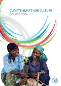
2.5 Algorithm for the detection of AIV through PCR and/or virus isolation
1
Healthy animal surveillance
Sick animal or in-contact flock
Prepare samples
2 PCR (M)
Virus isolation
3
NEG
4
POS
POS
NEG Report
Report
a
Typing (H5. H7)
PCR (H5,H7)
NEG
POS
POS
NEG
Report
b
c
6
5.1
Report
d
PCR (N1, N9)
5.2
REFERENCE LAB
H & N typing sequencing
7
e
Notes 1
Diagnosis: Use PCR or virus isolation (preferably PCR); Surveillance: Start with PCR, then isolate virus from positive sample.
2
Avoid pooling samples in the field whenever possible. When required for testing purposes, conduct pooling at the laboratory by combining a maximum of five similar samples per pool from the same sample species, epidemiological unit and type (i.e. either oropharyngeal/tracheal or cloacal, but not mixed cloacal and oropharyngeal).
3
Screening flocks for all influenza viruses using PCR (M) is recommended where possible. Specific virus PCR can be used first (e.g. H5 or H7) where a diagnosis is required for a specific virus in an emergency.
4
For isolation use up to three passages in eggs and hemagglutinin assay (HA).
5.1 & 5.2
Use recommended regional PCR primers and probes.
6
Use hemagglutinin inhibition (HI) assays for subtyping of isolates and/or PCR and/or sequencing. HI antigens and antisera must match the circulating H5 or H7 clade.
7
Confirmation of H and N type requires sequencing of the isolate.
a
Not influenza A. Differential diagnosis (Newcastle disease, infectious bursal disease, duck viral enteritis; optional).
b
Report as influenza A, not H5 and not H7. Optional: further subtyping to determine H and N.
c
Not influenza A by virus isolation.
d
Report as influenza A, not H5, not H7. Optional: further subtyping to determine H and N.
e
Whole genome sequencing (optional).
5
JUNE 2013
|
Laboratory protocols and algorithms
2.6 Additional information OFFLU protocols: available at http://www.offlu.net/index.php?id=267 WHO protocols (H7 and N9) available at http://www.who.int/influenza/gisrs_laboratory/cnic_realtime_rt_pcr_protocol_a_h7n9.pdf
3. Surveillance laboratory flow chart 3.1 Serological considerations for the detection of poultry exposure to avian influenza A(H7N9) In vivo infectivity studies in poultry with H7N9 virus have shown that the virus is shed for a limited period of time following infection. This significantly reduces the opportunity to detect the virus in poultry populations during targeted surveillance. Therefore serological surveillance may be an appropriate method of choice for detection of H7N9 infection when conducting an active surveillance programme. Laboratory experiments with H7N9 in various animal species (e.g. chickens, quails, turkeys, pigeons, and ducks) have shown that HI testing can be used to detect exposure of animals to H7N9 infections. Data show that poultry become seropositive at 5 to 7 days after infection; the longevity of the antibody response is currently being investigated, but it will wane over time. AAHL and the Animal Health and Veterinary Laboratories Agency (AHVLA) have found high levels of cross-reactivity in HI assay results using various antisera against H7N9 as an antigen. The AAHL and AHVLA results have been posted by OFFLU. 2 Therefore, standard HI assays using the Chinese H7N9 or Eurasian H7 antigens can be used for the serosurveillance for H7N9. These methods have been described in the OIE Manual of Diagnostic Tests and Vaccines for Terrestrial Animals 2012. 3 However, when performing serosurveillance for H7N9, it is essential to consider that: • Antibodies to the avian influenza A(H7N9) virus and antibodies to other Eurasian-lineage H7Nx influenza viruses are highly crossreactive. • Anti-H7 antibodies do not unequivocally proof infection with H7N9 as the hemagglutinin inhibition assay cannot distinguish between antibodies to H7N9 and antibodies to other Eurasian-lineage H7Nx influenza viruses. • In infected areas where H7N9 virus has been detected, the presence of anti-H7 antibodies in sera should be considered highly indicative of previous H7N9 infection. • Neuraminidase inhibitor (NAI) assays using HxN9 antigens should be utilized to determine presence of anti-N9 antibodies, however, other HxN9 viruses will also cross-react with novel H7N9. 2. OFFLU results: i) http://www.offlu.net/fileadmin/home/en/guidance/pdf/HI_and_NI_results_AHVLA_7May13.pdf; and ii) http://www.offlu.net/fileadmin/home/en/resource-centre/pdf/AAHL_H7N9_Experiment_17May.pdf 3. ttp://www.oie.int/fileadmin/Home/eng/Health_standards/tahm/2.03.04_AI.pdf
© FAO/Stephanie Sonnberg
6
Addressing avian influenza (H7N9)
|
JUNE 2013
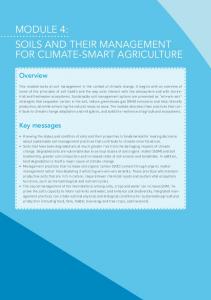
3.2 Laboratory diagnostic algorithm for the serosurveillance of AIV
Sampling frame
Healthy flock (Serum)
1
3.1
Screening Test (ELISA, AGID)
2
N E G
P O S
Report
HI (H5, H7)
3.2
N typing
a
4
P O S
N E G
Report
b
Report
c
Notes 1
Investigate vaccination history of flock
2
Avoid pooling of serum samples
3
If flock has been previously vaccinated against H5N1 or H7Nx: • 3.1 Use homologous antigen to determine vaccination coverage; • 3.2 Screen for H5 or H7 antibodies using appropriate antigens for these subtypes
4
To determine the NA type: use NAI assays
a
No influenza A antibodies present. Therefore; there is no evidence of previous infection, though infection with H5 or H7 viruses can fail to cause seroconversion. In chickens seroconversion requires approximately 5—7 days after infection and antibodies wane over time.
b
Report as influenza A antibodies present, but not anti-H5, not anti-H7; optional: further subtyping of HA-specific antibodies using full HI panel (H1-H16).
c
Report either as H5- or H7-specific antibodies; optional: also include NA subtype.
7
CONTACT The Emergency Prevention System (EMPRES) is an FAO programme, founded in 1994, with the goal of enhancing world food security, fighting transboundary animal and plant pests and diseases and reducing the adverse impact of food safety threats. EMPRES-Animal Health is the component dealing with the prevention and control of transboundary animal diseases (TADs). To subscribe or to ask for information about EMPRES-Animal Health send an e-mail to:
These guidelines are based on the information available to date and will be reviewed as new information becomes available.
[email protected] or a fax to (+39) 06 57053023 For more information visit us at http://www.fao.org/ag/empres.html EMPRES-Animal Health can assist countries in the shipment of samples for TAD diagnostic testing at a FAO reference laboratory and reference centre. Please contact
[email protected] for information prior to sampling or shipment. Please note that sending samples out of a country requires an export permit from the Chief Veterinarian’s Office of the country and an import permit from the receiving country.
8
The views expressed in this information product are those of the author(s) and do not necessarily reflect the views or policies of FAO.
Photo cover&back cover: ©FAO/Stephanie Sonnberg
acknowledgement of FAO as the source and copyright holder is given and that FAO’s endorsement of users’ views, products or services is not implied in any way.
© FAO, 2013 FAO encourages the use, reproduction and dissemination of material in this information product. Except where otherwise indicated, material may be copied, downloaded and printed for private study, research and teaching purposes, or for use in non-commercial products or services, provided that appropriate
All requests for translation and adaptation rights, and for resale and other commercial use rights should be made via www.fao.org/contact-us/ licence-request or addressed to
[email protected]. FAO information products are available on the FAO website (www.fao.org/publications) and can be purchased through
[email protected].
I3596E
The designations employed and the presentation of material in this information product do not imply the expression of any opinion whatsoever on the part of the Food and Agriculture Organization of the United Nations (FAO) concerning the legal or development status of any country, territory, city or area or of its authorities, or concerning the delimitation of its frontiers or boundaries. The mention of specific companies or products of manufacturers, whether or not these have been patented, does not imply that these have been endorsed or recommended by FAO in preference to others of a similar nature that are not mentioned.
Recommended citation FAO. 2013. Laboratory protocols and algorithms. Addressing avian influenza A(H7N9). Rome