RESEARCH ARTICLE
Lack of serological and molecular evidence of arbovirus infections in bats from Brazil Cı´ntia Bittar1☯, Rafael R. G. Machado ID1☯*, Manuela T. Comelis2, Larissa M. Bueno2, Eliana Morielle-Versute2, Matheus R. Beguelini2,3, Renato P. de Souza4, Maurı´cio L. Nogueira5, Paula Rahal1
a1111111111 a1111111111 a1111111111 a1111111111 a1111111111
1 Laborato´rio de Estudos Genoˆmicos, Instituto de Biociências, Letras e Ciências Exatas, UNESP— Universidade Estadual Paulista “Ju´lio de Mesquita Filho”, São Jose´ do Rio Preto, São Paulo, Brazil, 2 Laborato´rio de Chiroptera, Instituto de Biociências, Letras e Ciências Exatas, Universidade Estadual Paulista “Ju´lio de Mesquita Filho”, São Jose´ do Rio Preto, São Paulo, Brazil, 3 Centro das Ciências Biolo´gicas e da Sau´de, UFOB—Universidade Federal do Oeste da Bahia, Barreiras, Bahia, Brazil, 4 Nu´cleo de Doenc¸as de Transmissão Vetorial, Instituto Adolfo Lutz, São Paulo, Brazil, 5 Laborato´rio de Pesquisa em Virologia, FAMERP-Faculdade de Medicina de São Jose´ do Rio Preto, São Jose´ do Rio Preto, São Paulo, Brazil ☯ These authors contributed equally to this work. *
[email protected]
Abstract OPEN ACCESS Citation: Bittar C, Machado RRG, Comelis MT, Bueno LM, Morielle-Versute E, Beguelini MR, et al. (2018) Lack of serological and molecular evidence of arbovirus infections in bats from Brazil. PLoS ONE 13(11): e0207010. https://doi.org/10.1371/ journal.pone.0207010 Editor: Naomi Forrester, Keele University Faculty of Natural Sciences, UNITED KINGDOM Received: May 16, 2018 Accepted: October 23, 2018 Published: November 7, 2018 Copyright: © 2018 Bittar et al. This is an open access article distributed under the terms of the Creative Commons Attribution License, which permits unrestricted use, distribution, and reproduction in any medium, provided the original author and source are credited.
Viruses are important agents of emerging zoonoses and are a substantial public health issue. Among emerging viruses, an important group are arboviruses, which are characterized by being maintained in nature in cycles involving hematophagous arthropod vectors and a wide range of vertebrate hosts. Recently, bats have received increasing attention as an important source for the emergence of zoonoses and as possible viral reservoirs. Among the arboviruses, there are many representatives of the genera Flavivirus and Alphavirus, which are responsible for important epidemics such as Dengue virus, Zika virus and Chikungunya virus. Due to the importance of analyzing potential viral reservoirs for zoonosis control and expanding our knowledge of bat viruses, this study aimed to investigate the presence of viruses of the Alphavirus and Flavivirus genera in bats. We analyzed serum, liver, lungs and intestine from 103 bats sampled in northeast and southern Brazil via Nested-PCR and the hemagglutination inhibition test. All samples tested in this study were negative for arboviruses, suggesting that no active or past infection was present in the captured bats. These data indicate that the bats examined herein probably do not constitute a reservoir for these viruses in the studied areas. Further studies are needed to clarify the role of bats as reservoirs and sources of infection of these viral zoonoses.
Data Availability Statement: All relevant data are within the paper and its Supporting Information files. Funding: This work was financially supported by FAPESP (São Paulo Research Foundation - http:// www.fapesp.br/) grant number 2015/09704-6, received by RRGM and by CNPq (National Council for Scientific and Technological Development http://cnpq.br/) grant number 165802/2015-4, received by CB.
Introduction Among emerging viruses, an important group are arboviruses (arthropod-borne viruses), which are characterized by being maintained in nature in cycles involving hematophagous arthropod vectors and a wide range of hosts [1,2]. These hosts are often vertebrates, especially mammals and birds.
PLOS ONE | https://doi.org/10.1371/journal.pone.0207010 November 7, 2018
1 / 10
Investigation of arbovirus in bats
Competing interests: The authors have declared that no competing interests exist.
Bats are mammals belonging to the order Chiroptera [3], and they are considered to be one of the most abundant, diverse and geographically distributed vertebrates in the world [4]. In Brazil, there is a great diversity of bats, with approximately 179 species (10 of which are endemic) and 68 subspecies belonging to 68 genera documented [5,6]. They present a broad geographic distribution, being able to fly long distances and often coming into direct or indirect contact with humans [3]. Recently, these vertebrates have received increasing attention as an important source for the emergence of zoonoses and possibly as viral reservoirs [7–9]. The infection of bats by arboviruses has long been reported by several authors [8,10–14]. Although the possibility of them acting as reservoirs has been raised, it is not clear yet if they play this role in the ecological cycle [15,16]. Being a large highly populated tropical country with one third of its territory covered by forests, Brazil presents ideal conditions for the existence of many arboviruses [17]. More than 200 arboviruses have been isolated in the country, and approximately 40 of these viruses cause diseases in humans [17,18]. The country presents a constant risk of emergence and re-emergence of arboviruses due to the existence of densely populated cities infested by mosquitoes of the genera Culex and Aedes, which are important vectors for arboviruses [1,7,19,20]. Focusing on the importance of the analysis of potential sources of zoonoses and viral reservoirs for the control of emerging viruses, the aim of this study was to investigate arbovirus infections, of the Alphavirus and Flavivirus genera, in bats from southeast and northeast Brazil. In addition, we aim to provide relevant information that may contribute to the epidemiological surveillance of diseases of great public health impact.
Materials and methods Ethics statement The animals were collected under environmental licenses from the Brazilian Institute of Environment and Natural Renewable Resources (IBAMA), SISBIO n˚: 21707–1 (Process: 02027.001957 / 2006–02); 46190–1 and handled according to the Guide for the Care and Use of Laboratory Animals of the Institute for Laboratory Animal Resources Commission on Life Sciences National Research Council [21]. In addition, it should be noted that this project was approved by the Ethics Committee on the Use of Animals of the Institute of Biosciences, Letters and Exact Sciences (CEUA / IBILCE—Protocol 135/2016).
Study areas Sampling was performed between 2014 and 2017 at ten different sites in urban and peri-urban areas of two cities from two different states of Brazil: São Jose´ do Rio Preto, state of São Paulo (SP) and Barreiras, state of Bahia (BA). The complete geographical coordinates address and ecological characteristics of all sampling sites are available as supplementary information (S1 Table). Additionally, climatological characteristics (temperature and rainfall) are given in S1 Fig (“A” for São Jose´ do Rio Preto—SP and “B” for Barreiras-BA).
Samples Bats were collected using mist nets, and the sex and species of the animals were determined [22–24]. Following euthanasia, which was performed by subcutaneous anesthesia with 80 mg/ kg ketamine (Dopalen-Vertebrands, Paulı´nia, SP, Brazil) and 20 mg/kg xylazine (RompunBayer S.A., São Paulo, SP, Brazil), the liver, intestines and lungs from each specimen were removed and stored at -150˚C. Blood was also collected, and serum was separated and stored at -150˚C. Finally, the animals were fixed in 10% formaldehyde for 24 hours and deposited in
PLOS ONE | https://doi.org/10.1371/journal.pone.0207010 November 7, 2018
2 / 10
Investigation of arbovirus in bats
70% alcohol in the Chiroptera Collection of the Department of Zoology and Botany—IBILCE / UNESP, where they are available for taxonomic studies.
RNA extraction and cDNA synthesis The RNA of the lungs, intestines and liver was extracted in order to test for the presence of Alphavirus and Flavivirus RNA. Tissues were homogenized in a Turrax-MA102 (Marconi, Piracicaba, SP, Brazil), and pellets were used for RNA extractions in TRIzol (Thermo Fisher Scientific, Waltham, MA, USA), according to manufacturer’s instructions. Finally, the RNA was resuspended in 100 μl of water treated with DEPC (Sigma Aldrich, St. Louis, Missouri, USA) and stored at -150˚C. Quantification of samples was performed on the NanoDrop 2000 spectrophotometer (Thermo Fisher Scientific, Waltham, MA, USA). Nucleic acids extracted from the organs of individual bats were subjected to cDNA synthesis using the High-Capacity cDNA Reverse Transcription Kit (Applied Biosystems, Foster City, CA, USA).
Endogenous control amplification After cDNA synthesis, in order to check the RNA quality, PCR was performed for the endogenous control, β-actin, as described previously [25].
Molecular detection of Alphavirus and Flavivirus Detection of viral RNA in the tissue samples was carried out by Nested-PCR assay. We designed a set of primers targeting the nsp4 region of Alphavirus based on 23 viruses from this genus (S1 File). To test for Flavivirus, we used PCR primers external to Flav100F and Flav200R, published by Maher-Sturgess et al., 2008 [26], for the Nested-PCR reaction, which targets the NS5 region. The sets of primers used for each reaction is described in Table 1. Additional information on primer design and molecular tests are available in S1 and S2 Files. The PCR reaction was carried out using Long PCR Enzyme Mix (Thermo Fisher Scientific, Waltham, MA, USA). Amplification conditions were 3 min at 94˚C, 35 cycles at 94˚C for 1 min, annealing at 50˚C (for Alphavirus) and 48˚C (for Flavivirus) for 45 seconds, and extension at 72˚C for 1 min followed by a final extension at 72˚C for 10 minutes. The Nested-PCR reaction was performed as described above except for the annealing temperature, 45˚C (for Alphavirus) and 42˚C (for Flavivirus). Samples were resolved in 1% agarose gels. The expected sizes of the amplicons were 803 bp for Flavivirus and 913 bp for Alphavirus.
Alphavirus and Flavivirus antibody detection The presence of antibodies for Alphavirus and Flavivirus in the serum samples were tested by hemagglutination inhibition (HI), as described previously [27]. Additional information about Table 1. Sequences 5’-3’ of Alphavirus and Flavivirus specific primers used for Nested-PCR reactions. PCR reaction Alphavirus Flavivirus
�
Nested-PCR reaction
Name
Sequence 5’-3’
Name
Sequence 5’-3’
α.nsP4.PF
TAYYTDGAYATGGTIGABGG
α.nsP4.NF
TGYAAYGTIACICARATGMG
α.nsP4.PR
GGYTTICCIADYTTVAAIAG
α.nsP4.NR
AAICCICCRCARAARTAIGG
F.NS5P-F
ACIMTBITSTGTGACATHGGIGA
Flav100F�
AAYTCIACICAIGARATGTAY
F.NS5P-R
GTBAHIGTGTTIARGGCRTAIGT
Flav200R�
CCIARCCACATRWACCA
Flav100F and Flav200R primers were published by Maher-Sturgess et al., 2008 [26].
https://doi.org/10.1371/journal.pone.0207010.t001
PLOS ONE | https://doi.org/10.1371/journal.pone.0207010 November 7, 2018
3 / 10
Investigation of arbovirus in bats
the antigens and the positive controls used in the HI, are given in S2 Table. The tests were performed by the Adolfo Lutz Institute–SP.
Results A total of 39 animals were collected in Barreiras-BA and 64 in the region of São Jose´ do Rio Preto-SP, totaling 103 bats. These were distributed in four families: Molossidae, Phyllostomidae, Emballonuridae and Vespertilionidae (Fig 1 and Table 2). The diversity of species and the amount of male/female per species are reported in Table 2. The livers and intestines were collected from 28 specimens from Barreiras–BA and only the livers from 11 specimens. From the region of São Jose´ do Rio Preto-SP liver, lungs and intestines were collected from all bats. Detailed information on species, sex, organs tested, site of sampling and month of collection for each specimen are presented in S3 Table. The necropsies did not show any morphological changes that indicated a pathological condition. RNA from all tissue samples was extracted and quantified. The quality of the RNA was confirmed by the amplification of the endogenous gene, β-actin, from all tested samples. The presence of Alphavirus and Flavivirus RNA was investigated by Nested-PCR in all available tissue samples. The results revealed that none of the 103 samples were infected by Alphavirus and Flavivirus at the time they were collected. We also tested for serological evidence of previous infections by viruses from this groups using HI. A total of 73 serum samples (46 from São Jose´ do Rio Preto–SP and 27 from Barreiras–BA) were tested for anti-DENV-1, anti-DENV-2, anti-DENV-3, anti-DENV-4, anti-
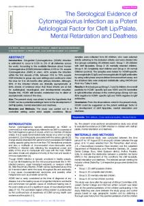
Fig 1. Locations of bat collections and the families found in each sampling site. On the left is the map of Latin America, highlighting the two Brazilian states, Bahia (in blue) and São Paulo (in green) in which bats were collected. At greater magnification, the collection sites within the regions of São Jose´ do Rio Preto–SP and Barreiras–BA are shown. The families of bats collected are represented by colored circles. The image was designed using QGIS 2.18.15 software (http://www.qgis.org/en/ site/about/index.html). https://doi.org/10.1371/journal.pone.0207010.g001
PLOS ONE | https://doi.org/10.1371/journal.pone.0207010 November 7, 2018
4 / 10
Investigation of arbovirus in bats
Table 2. Diversity of the species collected in relation to the sex of each specimen. Family Molossidae
Vespertilionidae
Phyllostomidae
Emballonuridae
Species
Number of specimens Male
Female
Eumops glaucinus
-
1
Molossops temminckii
1
-
Molossus molossus
6
18
Molossus rufus
14
4
Eptesicus furinalis
1
2
Eptesicus sp
-
1
Myotis nigricans
1
-
Artibeus lituratus
17
5
Artibeus planirostris
9
3
Carollia perspicillata
1
-
Desmodus rotundus
1
-
Diphylla ecaudata
2
-
Glossophaga soricina
2
1
Phyllostomus discolor
3
2
Phyllostomus hastatus
1
-
Platyrrhinus lineatus
1
-
Sturnira lilium
1
-
Vampyressa pussilla
-
1
Peropteryx leucoptera
1
-
https://doi.org/10.1371/journal.pone.0207010.t002
MAYV, anti-WEEV, anti-EEEV, anti-YFV, anti-SLEV, anti-ROCV, anti-ILHV, anti-MUCV and anti-IGUV antibodies. No HI was detected in any of the bats’ serum or in the negative serum used as a control, indicating no previous infection by any of these viruses.
Discussion The role of bats in the transmission and ecology of arboviruses, such as Flavivirus and Alphavirus, is not fully elucidated; however, several studies over the past 40 years have demonstrated that bats are susceptible to infection by viruses of these genera. For example, evidence of nucleic acids and antibodies from the four DENV serotypes has been reported in bats in Central and South America [28]. In addition, CHIKV was detected in bats from Asia [29]. In this study, we analyzed the presence of RNA from Alphavirus and Flavivirus and antibodies from viruses from these genera in animals collected in São Jose´ do Rio Preto (São Paulo State) and Barreiras (Bahia State). The first city is in the northwest region of São Paulo state and comprises an area with a high incidence of arbovirus infection in humans. Several studies have shown that this is a region of YFV transmission [30,31], is hyper-endemic for DENV [32–34], has confirmed cases of MAYV [35,36], Saint Louis encephalitis virus (SLEV) [34,37] and ZIKV [38–40]. Additionally, some cases of DENV intra-serotype co-infection [32] and coinfection with other Flaviviruses [34,41] have been reported. In the Barreiras region, epidemiological reports from 2014 to 2017 also show the occurrence of DENV, CHIKV, ZIKV and YFV [42–44]. In addition, all animals were collected in densely populated urban or peri-urban areas where the main vector of these arboviruses, Aedes aegypti, has extensive circulation. Our results suggest that no active or past infections by the arboviruses in this study were present in the captured bats. These facts indicate that although the bats collected are in close contact with these viruses, that they are not being infected and that they probably do not constitute a reservoir in the studied regions.
PLOS ONE | https://doi.org/10.1371/journal.pone.0207010 November 7, 2018
5 / 10
Investigation of arbovirus in bats
Our study sampled bats from the Molossidae, Vespertilionidae, Phyllostomidae and Emballonuridae families. The few previous studies that sought to identify arboviruses in these bat families show that Molossidae and Phyllostomidae have the highest incidence of arbovirus infection. As an example of Molossidae infection, St. Louis encephalitis virus (SLEV) was isolated from Tadarida brasiliensis mexicana in Texas [45]. Additionally, in east Africa, antibodies reactive to ZIKV were detected at a high seroprevalence in Mops condylurus using the indirect hemagglutination test (HAI) [10]. In the Phyllostomidae family, there is a single work that detected the viral RNA of DENV-1 in Carollia perspicillata in French Guiana [46]. Additionally, specific antibodies against West Nile Virus (WNV), SLEV and DENV 1–4 were detected by the plaque reduction neutralization test (PRNT) in three bat species from Mexico: Glossophaga soricina, Artibeus jamaicensis and Artibeus lituratus, with a Flavivirus antibody prevalence of 33%, 24%, and 9%, respectively [47]. For the Emballonuridae and Vespertilionidae families, there are no records of viral RNA or antibodies against the Flavivirus and Alphavirus genera [46]. Even with the absence of studies that reported the lack of active and/or past arbovirus infection in bats, some studies with DENV corroborate the results obtained in this work. In a study carried out in Mexico, which investigated the presence of the 4 serotypes of DENV in serum, lung and liver of 240 bats, no active or past infection in these animals was found [48]. Additionally, other studies with DENV, demonstrate that some serotypes of this virus did not replicate efficiently in cell lines derived from neotropical bat species and indicate that some species are incapable of sustaining Dengue virus replication and are unlikely to act as reservoirs for this virus [49–51]. Moreover, a recent work showed that bats sampled from households in Costa Rican urban environments do not sustain DENV amplification, since they do not support sufficient virus replication. These findings excluded them as potential hosts or reservoirs with no role in the transmission cycle and more likely are functioning as epidemiological dead-end hosts for this virus [52]. To date, few studies have aimed to identify arbovirus in bats, even with all the recognition of the importance of these animals in the emergence of zoonotic viruses. Some studies have demonstrated that ecological, behavioral and phylogenetic characteristics can influence and diversify the immunological response to viral infections in different species of bats [53,54]. For example, some evidence suggests that large colonies and higher species richness were significantly positively associated with European Bat lyssavirus 1 (EBLV-1) seroprevalence [54]. Additionally, they observed that EBLV-1 seroprevalence in bats from the Vespertilionidae and Rhinolophidae families were different. This difference is likely due to differences between bat species in the immune response and the lifespan of immunity to a virus infection [53,55]. Therefore, it is more likely to find seropositive bats in species with long lifespan immunity [55]. However, no study aimed to investigate differences in immunological responses between bat species to arbovirus infection. Even though there is no serological and molecular evidence of arbovirus infections in Brazilian bats from the studied regions, we emphasize the importance of continuing studies in other locations in order to evaluate the importance of bats as arbovirus reservoirs and to determine if these animals are an important part of its enzootic cycle.
Supporting information S1 Fig. Climatological characteristics of the sampling sites. A. Average monthly rainfall and temperature in the São Jose´ do Rio Preto region, from 2014 to 2017. The data were provided by the Integrated agrometeorological information center (CIIAGRO-Brazil: http://www. ciiagro.sp.gov.br). B. Average monthly rainfall and temperatures in the Barreiras region, from
PLOS ONE | https://doi.org/10.1371/journal.pone.0207010 November 7, 2018
6 / 10
Investigation of arbovirus in bats
2014–2015. The data were provided by the National Institute of Meteorology (INMET-Brazil: www.inmet.gov.br). (TIF) S1 Table. Sampling sites information. Additional information, geographic coordinates and ecological characteristics of sampling sites. The ecological characteristics were obtained by the Brazilian company of agricultural research—Ministry of Agriculture, Livestock and Supply (EMBRAPA-Brazil: https://www.embrapa.br/) and by the Forestry Institute—Sao Paulo’s State Government—Secretariat of Environment (IF-Brazil: http://iflorestal.sp.gov.br/). (XLSX) S2 Table. HI Antigens and Positive Controls. Description of the antigens (strain, virus and sample) and the positive controls used in HI. The positive controls were pools of mouse hyperimmune sera for each subgroup of arboviruses (Pool A—Alphavirus and B—Flavivirus). (XLSX) S3 Table. Characteristics of bats collected. Characteristics of bats collected (identification number—ID, family, species, sex, type of sample collected of each specimen and sampling site and month/year of collection). (XLSX) S1 File. Alphavirus primers design and molecular tests. (PDF) S2 File. Flavivirus primers design and molecular tests. (PDF)
Acknowledgments The authors would also like to thank all members of the Laboratory of Genomics Studies (IBILCE-UNESP, BR) for their helpful suggestions and discussions. This work was financially supported by FAPESP (São Paulo Research Foundation) grant number 2015/09704-6 and by CNPq (National Council for Scientific and Technological Development) grant number 165802/2015-4.
Author Contributions Conceptualization: Cı´ntia Bittar, Paula Rahal. Data curation: Cı´ntia Bittar, Rafael R. G. Machado. Formal analysis: Cı´ntia Bittar, Rafael R. G. Machado, Paula Rahal. Funding acquisition: Maurı´cio L. Nogueira, Paula Rahal. Investigation: Cı´ntia Bittar, Rafael R. G. Machado. Methodology: Cı´ntia Bittar, Rafael R. G. Machado, Manuela T. Comelis, Larissa M. Bueno, Eliana Morielle-Versute, Matheus R. Beguelini, Renato P. de Souza. Project administration: Cı´ntia Bittar, Rafael R. G. Machado. Resources: Paula Rahal. Supervision: Maurı´cio L. Nogueira, Paula Rahal. Validation: Cı´ntia Bittar, Rafael R. G. Machado.
PLOS ONE | https://doi.org/10.1371/journal.pone.0207010 November 7, 2018
7 / 10
Investigation of arbovirus in bats
Visualization: Cı´ntia Bittar, Rafael R. G. Machado. Writing – original draft: Cı´ntia Bittar, Rafael R. G. Machado. Writing – review & editing: Cı´ntia Bittar, Rafael R. G. Machado, Paula Rahal.
References 1.
Weaver SC, Barrett ADT. Transmission cycles, host range, evolution and emergence of arboviral disease. Nat Rev Microbiol. 2004; 2: 789–801. https://doi.org/10.1038/nrmicro1006 PMID: 15378043
2.
Coffey LL, Vasilakis N, Brault AC, Powers AM, Tripet F, Weaver SC. Arbovirus evolution in vivo is constrained by host alternation. Proc Natl Acad Sci U S A. 2008; 105: 6970–6975. https://doi.org/10.1073/ pnas.0712130105 PMID: 18458341
3.
Hill JE, Smith JD. Bats: a natural history. London: British Museum (Natural History); 1984.
4.
Feldhamer GA, Drickamer LC, Vessey SH, Merritt JF, Krajewski C. Mammalogy: Adaptation, Diversity, Ecology. 4th ed. Baltimore: Johns Hopkins University Press; 2015.
5.
Nogueira MR, Lima IP De, Moratelli R, Cunha V, Gregorin R, Peracchi AL. Checklist of Brazilian bats, with comments on original records. J species List Distrib. 2014; 10: 808–821. https://doi.org/10.15560/ 10.4.808
6.
Gregorim R. Cata´logo Taxonoˆmico da Fauna do Brasil. In: Nogueira MR, Isaac P. Lima, editors. Boletim da Sociedade Brasileira para o Estudo de Quiro´pteros. 2nd ed. Rio de Janeiro; 2016. p. 28.
7.
Olival KJ, Hosseini PR, Zambrana-Torrelio C, Ross N, Bogich TL, Daszak P. Host and viral traits predict zoonotic spillover from mammals. Nature. Nature Publishing Group; 2017; 546: 646–650. https://doi. org/10.1038/nature22975 PMID: 28636590
8.
Calisher CH, Childs JE, Field HE, Holmes K V., Schountz T. Bats: Important reservoir hosts of emerging viruses. Clin Microbiol Rev. 2006; 19: 531–545. https://doi.org/10.1128/CMR.00017-06 PMID: 16847084
9.
Wang L-F, Cowled C. Bats and Viruses: A New Frontier of Emerging Infectious Diseases. Hoboken, New Jersey: Wiley-Blackwell; 2015.
10.
Shephard RC WM. Studies on viruses in east African bats (Chiroptera). 1. Hemagglutination inhibition and circulation of arboviruses. Zoonoses Res. 1964; 3: 125–139. PMID: 5895899
11.
Thompson NN, Auguste AJ, Travassos da Rosa APA, Carrington CVF, Blitvich BJ, Chadee DD, et al. Seroepidemiology of selected alphaviruses and flaviviruses in bats in Trinidad. Zoonoses Public Health. 2015; 62: 53–60. https://doi.org/10.1111/zph.12118 PMID: 24751420
12.
Luis AD, Hayman DTS, O’Shea TJ, Cryan PM, Gilbert AT, Pulliam JRC, et al. A comparison of bats and rodents as reservoirs of zoonotic viruses: are bats special? Proc Biol Sci. 2013; 280: 20122753. https:// doi.org/10.1098/rspb.2012.2753 PMID: 23378666
13.
Williams MC, Simpson DI SR. Studies on viruses in East African bats. (Chiroptera). 2. Virus isolation. Zoonoses Res. 1964; 3: 141–53. PMID: 5895900
14.
Williams MC, Simpson DIH, Shepherd RC. Bats and Arboviruses in East Africa. Nature. 1964; 203: 670.
15.
Kuno G, Chang GJJG-JJ, Kuno G, Chang GJJG-JJ. Biological Transmission of Arboviruses: Reexamination of and New Insights into Components, Mechanisms, and Unique Traits as Well as Their Evolutionary Trends Biological Transmission of Arboviruses: Reexamination of and New Insights into Components, M. Clin Microbiol Rev. 2005; 18: 608–637. https://doi.org/10.1128/CMR.18.4.608-637. 2005 PMID: 16223950
16.
Kuno G, Mackenzie JS, Junglen S, Huba´lek Z, Plyusnin A, Gubler DJ. Vertebrate reservoirs of arboviruses: Myth, synonym of amplifier, or reality? Viruses. 2017; 9: 1–28. https://doi.org/10.3390/v9070185 PMID: 28703771
17.
Figueiredo LTM. Emergent arboviruses in Brazil. Rev Soc Bras Med Trop. 2007; 40: 224–229. https:// doi.org/10.1590/S0037-86822007000200016 PMID: 17568894
18.
Casseb ADR, Casseb LMN, Silva SP Da, Vasconcelos PFDC. Arbovı´rus: Importante Zoonose Na Amazoˆnia Brasileira. Veterina´ria e Zootec. 2013; 20: 391–403. Available: http://www.fmvz.unesp.br/rvz/ index.php/rvz/article/view/191
19.
Woolhouse MEJ, Gowtage-Sequeria S. Host range and emerging and reemerging infectious diseases. Emerg Infect Dis. 2005; 11: 1842–1847. https://doi.org/10.3201/eid1112.050997 PMID: 16485468
20.
Kuno G, Chang GJ, Tsuchiya KR, Karabatsos N, Cropp CB. Phylogeny of the genus Flavivirus. J Virol. 1998; 72: 73–83. PMID: 9420202
PLOS ONE | https://doi.org/10.1371/journal.pone.0207010 November 7, 2018
8 / 10
Investigation of arbovirus in bats
21.
Institute for Laboratory Animal Resources Comission on Life Sciences National Research Council. Guide for the Care and Use of Laboratory Animals [Internet]. 8th ed. Washington, D.C.: The National Academies Press; 2011. https://doi.org/10.1163/1573-3912_islam_DUM_3825
22.
Vizotto LD, Taddei VA. Chave para determinac¸ão de quiro´pteros brasileiros. São Jose´ do Rio Preto, SP: Gra´fica Francal; 1973.
23.
Miranda JMD, Bernardi IP, Passos FC. A new species of Eptesicus (Mammalia: Chiroptera: Vespertilionidae) from the Atlantic Forest, Brazil. Zootaxa. 2006; 1383: 57–68.
24.
Laval RK. A revision of the neotropical bats of the genus Myotis. Sci Bull Nat Hist Museum Los Angeles Cty. 1973; 15: 1–53.
25.
Spandidos A, Wang X, Wang H, Seed B. PrimerBank: A resource of human and mouse PCR primer pairs for gene expression detection and quantification. Nucleic Acids Res. 2009; 38: 792–799. https:// doi.org/10.1093/nar/gkp1005 PMID: 19906719
26.
Maher-Sturgess SL, Forrester NL, Wayper PJ, Gould E a, Hall R a, Barnard RT, et al. Universal primers that amplify RNA from all three flavivirus subgroups. Virol J. 2008; 5: 16. https://doi.org/10.1186/1743422X-5-16 PMID: 18218114
27.
Clarke D, Casals J. Techniques for hemagglutination and hemagglutination-inhibition with arthropodborne viruses. Am J Trop Med Hyg. 1958; 7: 561–573. PMID: 13571577
28.
Aguilar-Setie´n a, Romero-Almaraz ML, Sa´nchez-Herna´ndez C, Figueroa R, Jua´rez-Palma LP, Garcı´aFlores MM, et al. Dengue virus in Mexican bats. Epidemiol Infect. 2008; 136: 1678–1683. https://doi. org/10.1017/S0950268808000460 PMID: 18325131
29.
Dash a. P, Bhatia R, Sunyoto T, Mourya DT. Emerging and re-emerging arboviral diseases in Southeast Asia. J Vector Borne Dis. 2013; 50: 77–84. PMID: 23995308
30.
Moreno ES, Rocco IM, Bergo ES, Brasil RA, Siciliano MM, Suzuki A, et al. Reemergência de febre amarela: Detecc¸ão de transmissão no estado de São Paulo, Brasil, 2008. Rev Soc Bras Med Trop. 2011; 44: 290–296. https://doi.org/10.1590/S0037-86822011005000041 PMID: 21739073
31.
Saad LDC, Barata RB, Saad LDC, Barata RB. Surtos de febre amarela no estado de São Paulo, 2000– 2010*. Epidemiol e Servic¸os Sau´de. 2016; 25: 531–540. https://doi.org/10.5123/S167949742016000300009 PMID: 27869924
32.
Colombo TE, Vedovello D, Mondini A, Reis AFN, Cury AAF, Oliveira FH de, et al. Co-infecc¸ão por vı´rus dengue, sorotipos 1 e 4, em paciente de cidade de porte me´dio no Brasil. Rev Inst Med Trop Sao Paulo. 2013; 55: 275–281. https://doi.org/10.1590/S0036-46652013000400009
33.
Colombo TE, Vedovello D, Pacca-Mazaro CC, Mondini A, Arau´jo JP, Cabrera E, et al. Dengue virus surveillance: Detection of DENV-4 in the city of São Jose´ do Rio Preto, SP, Brazil. Acta Trop. Elsevier B.V.; 2016; 164: 84–89. https://doi.org/10.1016/j.actatropica.2016.09.004 PMID: 27609639
34.
Mondini A, Bronzoni RV de M, Cardeal ILS, Santos TMIL dos, La´zaro E, Nunes SHP, et al. Simultaneous infection by DENV-3 and SLEV in Brazil. J Clin Virol. 2007; 40: 84–86. https://doi.org/10.1016/j. jcv.2007.06.007 PMID: 17658293
35.
Mota MTO, Vedovello D, Estofolete C, Malossi CD, Arau´jo JP, Nogueira ML. Complete Genome Sequence of Mayaro Virus Imported from the Amazon Basin to São Paulo State, Brazil. Genome Announc. 2015; 3: e01341–15. https://doi.org/10.1128/genomeA.01341-15 PMID: 26607884
36.
Estofolete CF, Mota MTO, Vedovello D, de Go´ngora DVN, Maia IL, Nogueira ML. Mayaro fever in an HIV-infected patient suspected of having chikungunya fever. Rev Soc Bras Med Trop. 2016; 49: 648– 652. https://doi.org/10.1590/0037-8682-0093-2016 PMID: 27812665
37.
Mondini A, Cardeal ILS, La´zaro E, Nunes SH, Moreira CC, Rahal P, et al. Saint Louis encephalitis virus, Brazil. Emerg Infect Dis. 2007; 13: 176–178. https://doi.org/10.3201/eid1301.060905 PMID: 17370543
38.
Colombo TE, Estofolete CF, Reis AFN, da Silva NS, Aguiar ML, Cabrera EMS, et al. Clinical, laboratory and virological data from suspected ZIKV patients in an endemic arbovirus area. J Clin Virol. Elsevier; 2017; 96: 20–25. https://doi.org/10.1016/j.jcv.2017.09.002 PMID: 28918127
39.
Fernanda Estofolete C, Terzian ACB, Parreira R, Esteves A, Hardman L, Greque GV, et al. Clinical and laboratory profile of Zika virus infection in dengue suspected patients: A case series. J Clin Virol. Elsevier B.V.; 2016; 81: 25–30. https://doi.org/10.1016/j.jcv.2016.05.012 PMID: 27289428
40.
Nogueira ML, Rocha Nery Ju´nior NR, Estofolete CF, Bernardes Terzian AC, de Freitas Guimarães G, Zini N, et al. Adverse birth outcomes associated with Zika virus exposure during pregnancy in São Jose´ do Rio Preto, Brazil [Internet]. Clinical Microbiology and Infection. European Society of Clinical Microbiology and Infectious Diseases; 2017. https://doi.org/10.1016/j.cmi.2017.11.004 PMID: 29133154
41.
Terzian ACB, Mondini A, de Moraes Bronzoni RV, Drumond BP, Ferro BP, Cabrera EMS, et al. Detection of Saint Louis Encephalitis Virus in Dengue-Suspected Cases During a Dengue 3 Outbreak. Vector-Borne Zoonotic Dis. 2011; 11: 291–300. https://doi.org/10.1089/vbz.2009.0200 PMID: 20645866
PLOS ONE | https://doi.org/10.1371/journal.pone.0207010 November 7, 2018
9 / 10
Investigation of arbovirus in bats
42.
Costa IMP, Calado DC, Costa IMP, Calado DC. Incidência dos casos de dengue (2007–2013) e distribuic¸ão sazonal de culicı´deos (2012–2013) em Barreiras, Bahia*. Epidemiol e Servic¸os Sau´de. 2016; 25: 735–744. https://doi.org/10.5123/S1679-49742016000400007 PMID: 27869967
43.
Bahia S da S do E da. Boletim Epidemiolo´gico da Febre Amarela. 2017; 3. Available: http://www.saude. mg.gov.br/component/gmg/story/9282-informe-epidemiologico-da-febre-amarela-12-04
44.
Sau´de S de V em S− M da. Boletim Epidemiolo´gico. 2017;48: 1–10.
45.
Sulkin SE, Sims RA, Allen R. Isolation of St. Louis Encephalitis Virus from Bats (Tadarida b. mexicana) in Texas. Science (80-). 1966;Vol. 152: 223–225.
46.
Chen L, Liu B, Yang J, Jin Q. DBatVir: The database of bat-associated viruses. Database. 2014; 2014: 1–7. https://doi.org/10.1093/database/bau021 PMID: 24647629
47.
Machain-williams C, Lo´pez-uribe M, Talavera-aguilar L, Vera-escalante L, Puerto-manzano F, Ulloa A, et al. Serologic Evidence of Flavivirus Infection in Bats in the Yucatan Peninsula of Mexico. J Wildl Dis. 2014; 49: 1–8. https://doi.org/10.7589/2012-12-318.Serologic Cabrera-Romo S, Max Ramirez C, Recio-To´toro B, Tolentino-Chi J, Lanz H, del A´ngel RM, et al. No Evidence of Dengue Virus Infections in Several Species of Bats Captured in Central and Southern Mexico. Zoonoses Public Health. 2016; 63: 579–583. https://doi.org/10.1111/zph.12276 PMID: 27357156
48.
49.
Moreira-Soto A, Soto-Garita C, Corrales-Aguilar E. Neotropical primary bat cell lines show restricted dengue virus replication. Comp Immunol Microbiol Infect Dis. Elsevier Ltd; 2017; 50: 101–105. https:// doi.org/10.1016/j.cimid.2016.12.004 PMID: 28131369
50.
Perea-Martı´nez L, Moreno-Sandoval HN, Moreno-Altamirano MM, Salas-Rojas M, Garcı´a-Flores MM, Are´chiga-Ceballos N, et al. Experimental infection of Artibeus intermedius bats with serotype-2 dengue virus. Comp Immunol Microbiol Infect Dis. Elsevier Ltd; 2013; 36: 193–198. https://doi.org/10.1016/j. cimid.2012.12.002 PMID: 23312108
51.
Cabrera-Romo S, Recio-Totoro B, Alcala´ AC, Lanz H, Del Angel RM, Sa´nchez-Cordero V, et al. Experimental inoculation of artibeus jamaicensis bats with dengue virus serotypes 1 or 4 showed no evidence of sustained replication. Am J Trop Med Hyg. 2014; 91: 1227–1234. https://doi.org/10.4269/ajtmh.140361 PMID: 25311698
52.
Vicente-Santos A, Moreira-Soto A, Soto-Garita C, Chaverri LG, Chaves A, Drexler JF, et al. Neotropical bats that co-habit with humans function as dead-end hosts for dengue virus. PLoS Negl Trop Dis. 2017; 11: 1–18. https://doi.org/10.1371/journal.pntd.0005537 PMID: 28545090
53.
Lo´pez-Roig M, Bourhy H, Lavenir R, Serra-Cobo J. Seroprevalence dynamics of European bat Lyssavirus type 1 in a multispecies bat colony. Viruses. 2014; 6: 3386–3399. https://doi.org/10.3390/ v6093386 PMID: 25192547
54.
Serra-Cobo J, Lo´pez-Roig M, Seguı´ M, Sa´nchez LP, Nadal J, Borra´s M, et al. Ecological Factors Associated with European Bat Lyssavirus Seroprevalence in Spanish Bats. PLoS One. 2013; 8. https://doi. org/10.1371/journal.pone.0064467 PMID: 23700480
55.
Serra-Cobo J, Lopez-Roig M. Bats and Emerging Infections: An Ecological and Virological Puzzle. In: Giuseppe R, Ippolito G, editors. Emerging and Re-emerging Viral Infections: Advances in Microbiology, Infectious Diseases and Public Health. 2017. pp. 35–49.
PLOS ONE | https://doi.org/10.1371/journal.pone.0207010 November 7, 2018
10 / 10