Published 17 August 2009 Cite this as: BMJ Case Reports 2009 [doi:10.1136/bcr.04.2009.1764] Copyright © 2009 by the BMJ Publishing Group Ltd.
Learning from errors Pulmonary oedema and hyponatraemia after an ironman triathlon Georgia Stefanko1, Bill Lancashire2, Jeff S Coombes3, Robert G Fassett4 1
Prince of Wales Hospital, Sydney, New South Wales, 2000, Australia 2 Port Macquarie Base Hospital, Port Macquarie, New South Wales, 2000, Australia 3 The University of Queensland, St Lucia, Brisbane, Queensland, 4000, Australia 4 Royal Brisbane and Women’s Hospital and The University of Queensland, Renal Medicine, Level 9 Ned Hanlon Building, Brisbane, Queensland, 4029, Australia Correspondence to: Robert G Fassett,
[email protected]
SUMMARY A 36-year-old man presented with symptoms of acute pulmonary oedema at the conclusion of the Australian ironman triathlon. He was alert, orientated, with an oxygen saturation of 75% on room air. Chest examination revealed bilateral basal crepitations. Serum sodium was 120 mmol/L and chest x ray revealed bilateral basal opacities. He was treated for acute pulmonary oedema with prompt improvement and given 200 ml of intravenous hypertonic saline followed by normal saline. Serum sodium decreased to 117 mmol/L and 30 hours after presentation he had a seizure. He fully recovered and was discharged 5 days after admission. This case highlights that exercise-associated hyponatraemia and pulmonary oedema are still not widely understood and there is still a reluctance to treat hyponatraemia aggressively with ongoing hypertonic saline.
BACKGROUND This case illustrates that inadequately treated exercise-associated hyponatraemia (EAH) can result in seizure. In addition, the coexistence of the clinically more
apparent exercise-induced pulmonary oedema may distract attention from the underlying electrolyte disturbance. We report the case to draw attention to these clinical issues and to emphasise the importance of correctly treating the exerciseassociated hyponatremia with hypertonic saline.
CASE PRESENTATION A 36-year-old man presented to the medical tent immediately following completion of an ironman triathlon (3.8 km swim, 180 km bike and 42.2 km run) in 11 hours 50 minutes. He had a preceding 3-week history of a viral upper respiratory tract infection for which he had completed a course of oral antibiotics and took ibuprofen 2 days prior to the event. There was no other medical history and he did not take any medications. There was a family history of coronary artery disease. He did not feel "100%" after the swim, and at the conclusion of the event his symptoms included chest tightness, shortness of breath and a productive cough. He recalls consuming approximately 9 L of fluid over the course of the race comprised of both water and an electrolyte-containing drink and did not pass urine during the run. In the post-race medical tent his heart rate was 96 bpm, respiratory rate 32 bpm, systolic blood pressure 115 mmHg and chest examination revealed vesicular breath sounds only. During transfer to hospital, he developed rapidly progressive respiratory distress with a cough productive of pink frothy sputum. On arrival, he was alert, orientated, afebrile, pale and diaphoretic, with heart rate 89 bpm, blood pressure 134/89, respiratory rate 47 bpm and oxygen saturation 75% breathing room air that improved to 80% on 6 L/min of oxygen. Chest examination revealed diffuse coarse crepitations and two normal heart sounds. The patient’s weight was not recorded.
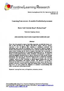
INVESTIGATIONS The chest x ray showed bilateral diffuse infiltrates (fig 1) and the electrocardiogram was normal. Laboratory findings showed a serum sodium 120 mmol/L, and plasma and urine osmolality 249 and 265 mmol/kg, respectively. Spot urine sodium and potassium were 101 and 20 mmom/L, respectively. His haematocrit was 0.38, white cell count 20.5x10⁹/L, neutrophils 17.6x10⁹/L and Bnatriuretic peptide (BNP) 1950 pg/mL. Arterial blood gas results following initiation of bi-level positive airway pressure (BiPAP) were pH 7.50, pCO2 27 mmHg, pO2
247 mmHg, bicarbonate 21.1 mmol/L, base excess 0.6 mmol/L and lactate 1.5 mmol/L.
View this figure (120K): in this window | in a new window | PowerPoint for Teaching Figure 1 Admission chest x ray showing pulmonary oedema.
Subsequent investigations found a troponin I peak of 0.33 ug/L and an increase in C reactive protein to 95.8 mg/L. Blood and sputum cultures were negative. An echocardiogram was performed the day after presentation that showed there was suspicion of mild hypokinesis of the inferior wall of the left ventricle with trivial mitral regurgitation and estimated systolic pulmonary artery pressure of 23 mmHg. Heart size, wall thickness and global left and right ventricular systolic function were normal. Renal artery stenosis was not evident on a doppler ultrasound study.
DIFFERENTIAL DIAGNOSIS A clinical diagnosis of acute pulmonary oedema (APO) was made, although initially respiratory infection was also considered based on his recent upper respiratory tract infection. The diagnosis of EAH was made based on the serum electrolytes.
TREATMENT The patient was treated with a glyceryl trinitrate infusion for 5 hours, a stat dose of intravenous frusemide and BiPAP. The patient improved rapidly and BiPAP was not required again after the initial 90 minutes in hospital. In addition, aspirin, clopidogrel, captopril and intravenous ceftriaxone and azithromycin were administered. In view of the hyponatraemia, 200 mL of 3% sodium chloride was infused intravenously over 30 minutes, which was followed by a normal saline infusion. Four hours after presentation the serum sodium was measured at 117 mmol/L.
OUTCOME AND FOLLOW-UP This patient did not experience any further episodes of respiratory compromise throughout the rest of his hospital admission. Subsequent chest x ray showed widespread bilateral infiltration. Thirty hours after presentation, the patient had a seizure at which time the serum sodium was 124 mmol/L. The Glasgow coma scale was 15/15, but he remained amnesic of his participation in the sporting event and ensuing hospital admission for several hours. A CT head scan with intravenous contrast was normal and no further seizures occurred. The serum sodium reached 134 mmol/L 60 hours after admission. A coronary angiogram was performed, which was reported as normal. He was discharged without further problems. Subsequent exercise stress testing 6 months later was normal.
DISCUSSION APO has previously been reported in endurance athletes both with normal serum electrolyte values1,2 and in those with EAH in conjunction with other evidence of cerebral encephalopathy.3–5 To the authors’ knowledge, there has only been one report of APO as the primary manifestation of EAH in the absence of any symptoms of cerebral encephalopathy.3 Therefore, this particular case is noteworthy in that it highlights the need to be mindful that severe hyponatraemia could present solely as life-threatening APO. In addition, the prompt and resolute response to initial short-lived treatment in spite of ongoing hyponatraemia suggests alternative or multifactorial causes, and emphasises our lack of understanding of the mechanisms by which exercise-associated APO occurs. This case and the management of EAH in endurance events are discussed below. EAH is becoming an increasingly well-recognised syndrome in endurance athletes. It develops in susceptible individuals from excess oral intake of hypotonic fluid,6–8 sometimes in the setting of a concurrent variant of the syndrome of inappropriate secretion of anti-diuretic hormone (SIADH).8,9 Potential mechanisms for ADH secretion in endurance athletes are hypovolaemia, as well as pain and nausea.6 Muscle derived interleukin-6 from exertional rhabdomyolysis has also been suggested as a cause.9,10 Impaired renal diluting ability and metabolic water production from the breakdown of
glycogen may have varying contributory roles.11 Due to the above factors, and possibly augmented by failure to mobilise osmotically inactive sodium stores or osmotically inactivate circulating sodium,12 hyponatraemia results (serum sodium 30 mmol/L) occurs9,10 promoted by atrial natriuretic peptide secreted in response to atrial stretch induced by plasma volume expansion.6 All these features were present in ours patient. Hyponatraemia in this athlete was a result of over-zealous hypotonic fluid consumption with an underlying SIADH. Failure to pass urine during the final leg of the race supports this notion, while his plasma and urinary electrolyte abnormalities confirm it. The ensuing fall in serum sodium from 120 mmol/L to 117 mmol/L following both hypertonic saline and normal saline reflect an intact renin-angiotensin-aldosterone axis in the face of an expanded plasma volume from impaired free water excretion. As well as the use of normal saline, the return of normal gastro-intestinal function after ceasing exercise and subsequent absorption of fluid ingested during the race may have contributed to the exacerbation. Neurological symptoms follow the development of severe hyponatraemia when cerebral oedema occurs as water moves from the extracellular to the intracellular compartment according to osmotic gradients. Common manifestations include nausea, weakness, dizziness and headache,6–8 although these are not uncommon following prolonged exercise in the absence of hyponatraemia.7,8 However, vomiting was found to be a discriminating symptom in one observational retrospective study,7 but was absent in our patient. Current treatment recommendations for acute symptomatic EAH state that administration of hypertonic saline should be promptly employed to reduce brain oedema.8 A variety of protocols exist including infusing 3% sodium chloride (NaCl) at a rate of 1–2 mL/kg/h with hourly serum sodium measurements until symptomatic improvement and the patient is stable or "until the patient regains consciousness". The use of isotonic or hypotonic intravenous fluids is contraindicated, as circulating ADH will facilitate the retention of free water, possibly aggravating the situation. Hyponatraemia in this patient was initially treated with intravenous 3% NaCl. However, fluid administration was subsequently changed to isotonic 0.9% normal saline due to concern regarding the potential for development of central pontine myelinolysis. This concern was unfounded as there are no reports of this complication occurring in those patients with rapidly corrected symptomatic EAH.
Notably, the patient’s serum sodium level decreased further to 117 mmol/L and a seizure resulted. Interestingly, this patient’s severe hyponatraemia occurred in the absence of neurological symptoms. It was not immediately apparent to the treating clinicians that the patient’s respiratory distress was possibly the heralding symptom. Moreover, the APO resolved swiftly and absolutely, even though the severe hyponatraemia actually worsened. These facts stimulate an examination as to the exact mechanisms of exercise associated pulmonary oedema. The precise mechanism by which APO develops in association with hyponatraemia is unclear and is further complicated by conflicting evidence in the literature.13 Clearly, for APO to occur there must either be a disturbance in Starling’s forces—changes in hydrostatic or oncotic pulmonary capillary or interstitial pressures—or a change in permeability of the blood-gas barrier. Experimental evidence suggests that compression and distortion of the medulla and cervical spinal cord from raised intracranial pressure—and, therefore, hyponatraemia—results in profound sympathetic activation.14 How, exactly, this increased sympathetic outflow manifests as pulmonary oedema is debatable. Pulmonary oedema fluid analysis described in retrospective reviews of patients with neurogenic oedema has both been used as evidence of increased permeability as the primary mechanism15 and, contrarily, in support of a hydrostatic one.16 Neither of these mechanisms, which are commonly linked by the inciting event of the development of cerebral encephalopathy, can account for the reports of oedema occurring in long distance athletes with normal electrolytes. In these situations, exercise itself may have a facilitatory role. For example, high pulmonary venous pressures are necessary to facilitate left ventricular filling in order to meet demands for increased cardiac output. Inevitably, raised pulmonary capillary hydrostatic pressures are also present, and in combination with increased longitudinal wall tension from the larger volumes during exercise, mechanical disruption of the blood-gas barrier could result. Alternative causes of a cardiac nature for APO in this endurance athelete and others could include non-ST elevation myocardial infarction, Takotsubo’s cardiomyopathy and acute myocarditis. An elevated BNP is in keeping with a cardiogenic origin—although equally this could reflect left ventricular stretch secondary to increased end diastolic volumes associated with exercise. So too, a raised troponin is not uncommon following endurance events and of dubious significance.17 Takotsubo cardiomyopathy, otherwise known as left ventricular apical ballooning syndrome, presents similar to ST-segment elevation myocardial
infarction with chest pain and dyspnoea but can be associated with heart failure in 15.9% of patients.18 It tends to follow an emotionally or physiologically stressful event with over 90% of cases being in women.18 Neither our patient, nor other reported cases, have exhibited the characteristic appearance of apical ballooning of the left ventricle with hyperdynamic basal segments on echocardiogram, and in this particular case it is highly unlikely that this abnormality would have entirely resolved prior to performing this investigation the day following admission, had this been the cause.18 More likely, contributing cardiac factors may have included depression of left ventricular function associated with prolonged exercise itself19 and possible subclinical viral myocarditis in view of a recent viral upper respiratory tract infection. Irrespective of the cause, APO in endurance athletes can be life-threatening and requires prompt action. So too, symptomatic EAH should be actively managed in accordance with current guidelines.
LEARNING POINTS
This case highlights that knowledge of the recommended treatment of exercise-associated hyponatraemia (EAH) is still not widely disseminated nor necessarily accepted and because of this seizures can result. Appropriate and adequate treatment with hypertonic saline is required EAH and acute pulmonary oedema may co-exist with one condition being more clinically apparent. Both conditions must be treated adequately
Competing interests: none.
Patient consent: Patient/guardian consent was obtained for publication.
REFERENCES Luks, AM, Robertson, HT, & Swenson, ER. An ultracyclist with pulmonary edema during the Bicycle Race Across America. Med Sci Sports Exerc 2007; 39: 8–12.[CrossRef][Medline] McKechnie, JK, Leary, WP, Noakes, TD, et al. Acute pulmonary oedema in two athletes during a 90-km running race. S Afr Med J 1979; 56: 261– 5.[Medline] Noakes, TD, Goodwin, N, Rayner, BL, et al. Water intoxication: a possible complication during endurance exercise. Med Sci Sports Exerc 1985; 17: 370–5.[Medline] Young, M, Sciurba, F, & Rinaldo, J. Delirium and pulmonary edema after completing a marathon. Am Rev Respir Dis 1987; 136: 737–9.[Medline] Ayus, JC, Varon, J, & Arieff, AI. Hyponatremia, cerebral edema, and noncardiogenic pulmonary edema in marathon runners. Ann Intern Med 2000; 132: 711–4.[Abstract/Free Full Text] Davis, DP, Videen, JS, Marino, A, et al. Exercise-associated hyponatremia in marathon runners: a two-year experience. J Emerg Med 2001; 21: 47– 57.[CrossRef][Medline] Hew, TD, Chorley, JN, Cianca, JC, et al. The incidence, risk factors, and clinical manifestations of hyponatremia in marathon runners. Clin J Sport Med 2003; 13: 41–7.[CrossRef][Medline] Hew-Butler, T, Almond, C, Ayus, JC, et al. Consensus statement of the 1st International Exercise-Associated Hyponatremia Consensus Development Conference, Cape Town, South Africa 2005. Clin J Sport Med 2005; 15: 208–13.[CrossRef][Medline] Siegel, AJ, Verbalis, JG, Clement, S, et al. Hyponatremia in marathon runners due to inappropriate arginine vasopressin secretion. Am J Med 2007; 120: 461 e11–7. Siegel, AJ. Exercise-associated hyponatremia: role of cytokines. Am J Med
2006; 119: S74–8.[Medline] Rosner, MH, & Kirven, J. Exercise-associated hyponatremia. Clin J Am Soc Nephrol 2007; 2: 151–61.[Abstract/Free Full Text] Noakes, TD, Sharwood, K, Speedy, D, et al. Three independent biological mechanisms cause exercise-associated hyponatremia: evidence from 2,135 weighed competitive athletic performances. Proc Natl Acad Sci U S A 2005; 102: 18550–5.[Abstract/Free Full Text] Colice, GL, Matthay, MA, Bass, E, et al. Neurogenic pulmonary edema. Am Rev Respir Dis 1984; 130: 941–8.[Medline] Chen, HI, Sun, SC, & Chai, CY. Pulmonary edema and hemorrhage resulting from cerebral compression. Am J Physiol 1973; 224: 223– 9.[Free Full Text] Mackersie, RC, Christensen, JM, Pitts, LH, et al. Pulmonary extravascular fluid accumulation following intracranial injury. J Trauma 1983; 23: 968– 75.[Medline] Smith, WS, & Matthay, MA. Evidence for a hydrostatic mechanism in human neurogenic pulmonary edema. Chest 1997; 111: 1326– 33.[CrossRef][Medline] Koller, A. Exercise-induced increases in cardiac troponins and prothrombotic markers. Med Sci Sports Exerc 2003; 35: 444–8.[CrossRef][Medline] Pilgrim, TM, & Wyss, TR. Takotsubo cardiomyopathy or transient left ventricular apical ballooning syndrome: A systematic review. Int J Cardiol 2008; 124: 283–92.[CrossRef][Medline] Niemela, KO, Palatsi, IJ, Ikaheimo, MJ, et al. Evidence of impaired left ventricular performance after an uninterrupted competitive 24 hour run. !
Circulation 1984; 70: 350–6.[Abstract/Free Full Text]