Research Article
2747
Alterations in the lens capsule contribute to cataractogenesis in SPARC-null mice Qi Yan1, John I. Clark2, Thomas N. Wight1 and E. Helene Sage1,* 1Department of Vascular Biology, The Hope Heart Institute, Seattle, WA 98104-2046, USA 2Departments of Biological Structure and Ophthalmology, University of Washington, Seattle,
WA, USA
*Author for correspondence (e-mail:
[email protected])
Accepted 23 April 2002 Journal of Cell Science 115, 2747-2756 (2002) © The Company of Biologists Ltd
Summary The lens capsule, which is also called the lens basement membrane, is a specialized extracellular matrix produced anteriorly by the lens epithelium and posteriorly by newly differentiated fiber cells. SPARC (secreted protein, acidic and rich in cysteine) is a matricellular glycoprotein that regulates cell-cell and cell-matrix interactions, cellular proliferation and differentiation, and the expression of genes encoding extracellular matrix components. SPARCnull mice exhibit lens opacity 1 month after birth and mature cataract and capsular rupture at 5-7 months. In this study, we report disruption of the structural integrity of the lens capsule in mice lacking SPARC. The major structural protein of basement membrane, collagen type IV, in the lens capsule was substantially altered in the absence of SPARC. The lens cells immediately beneath the capsule showed aberrant morphology, with numerous protrusions into the lens basement membrane. SPARC-null lenses at 1 month of age exhibited an increased penetration
Introduction Targeted disruption of the SPARC (secreted protein, acidic and rich in cysteine) gene has been performed in mice from three different genetic backgrounds (Gilmour et al., 1998; Norose et al., 1998). The mice are viable and fertile and appear grossly similar to their wild-type (wt, +/+) counterparts. Unexpectedly, a predominant and consistent phenotype of all SPARC-null (–/–) mice is the presence of cataracts. The mechanism of cataractogenesis in SPARC-null mice is not known. In this report, we provide a partial explanation for this pathology. SPARC, which is also termed osteonectin or BM-40, belongs to the matricellular class of secreted glycoproteins (thrombospondins 1 and 2, osteopontin, tenascins C and X) that function as modulators of cell-cell and cell-matrix interactions. SPARC mediates these interactions by (1) binding to extracellular matrix (ECM) proteins, (2) regulating ECM and growth factor production/efficacy and/or 3) modulating matrix metalloproteinase expression (Lane and Sage, 1994; Tremble et al., 1993; Yan and Sage, 1999; Sage, 1997). Both counteradhesive and antiproliferative properties in vitro have been attributed to SPARC, which induces rounding of cultured cells and disassembly of focal adhesions (Lane and Sage, 1990; Murphy-Ullrich et al., 1995). SPARC participates in morphogenesis, tissue repair and differentiation by virtue of its regulation of cell cycle, cell shape change, migration, adhesion
of dye or radioactive tracer through the capsule, as well as a higher content of water than their wild-type counterparts. Moreover, SPARC-null fibers exhibited swelling as early as 1 month of age; by 3 months, all the fiber cells appeared swollen to a marked degree. By contrast, the absence of SPARC had no apparent morphological effect on the early stages of lens formation, cell proliferation or fiber cell differentiation. Degradation of crystallins and MIP 26, or changes in the levels of these proteins, were not detected. These results underscore the importance of the capsular extracellular matrix in the maintenance of lens transparency and indicate that SPARC participates in the synthesis, assembly and/or stabilization of the lens basement membrane. Key words: SPARC, Lens capsule, Basement membrane, Extracellular matrix, Epithelial cells, Fiber cells, Collagen IV, Cataract, Permeability
and ECM production (Lane and Sage, 1994; Yan and Sage, 1999). Therefore, SPARC does not appear to serve a structural role in the ECM but is a functional modulator of various activities attributed to protein/proteoglycan networks (Sage and Bornstein, 1991; Bornstein, 1995). Despite its expression as a consequence of development or injury-related remodeling (Brekken and Sage, 2001), the production of SPARC in normal adult tissues is rather limited. Interestingly, SPARC is expressed in lenticular epithelium in both developing and adult mammals (Yan et al., 1998; Yan et al., 2000). The phenotype of early cataract formation in SPARC-null mice indicates that SPARC participates significantly in the function and homeostasis of the normal lens. The lens is a cellular structure without blood vessels, lymphatics and nerves. It is enclosed by an avascular thick capsule and is nourished by diffusion from the aqueous humor through the lens capsule and epithelium. The capsule, also called the lens basement membrane (BM), is a substantial, acellular and structurally complex ECM, consisting of an orderly meshwork of various glycoproteins and proteoglycans (for example, collagen type IV, laminin, perlecan, nidogen, and fibronectin) (Timpl and Dziadek, 1986; Bosman et al., 1989; Cammarata et al., 1986). BM produced by different cells has been correlated with functions such as proliferation, differentiation, adhesion and permeability (Yurchenco and
2748
Journal of Cell Science 115 (13)
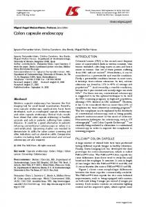
Fig. 1. Absence of SPARC results in cortical cataract. Slit-lamp photographs of wt mice (+/+) at 1, 3 and 7 months of age and SPARC-null mice (–/–) at 1, 3 and 7 months are shown. The SPARCnull lens at 1 month shows anterior subcapsular cortical opacity (arrow). SPARC-null lens at 3 months displayed anterior and posterior subcapsular opacity (arrows), whereas the nucleus is transparent. The SPARC-null lens at 7 months has a mature cataract (arrow). Arrowheads indicate the cornea, and arrows indicate lens opacity. mo, month. Bar, 240 µm.
Schittny, 1990). Thus, the composition of the ECM can be expected to be of particular importance for the maintenance of normal morphological and functional properties of the lens. Other important functions of the lens capsule that are dependent upon the organization of its constituent proteins and proteoglycans are filtration and permeability (Fisher, 1977; Lee et al., 1997; Winkler et al., 2001). The capsular ECM of the lens is not a static structure; rather, it is continually produced and remodeled anteriorly by the lens epithelial cells and posteriorly by newly differentiated fiber cells (Johnson and Beebe, 1984). SPARC regulates the production of certain ECM proteins, in addition to its interaction with collagens (Sasaki et al., 1998; Sage et al., 1989; Mayer et al., 1991; Maurer et al., 1995) and growth factors (Raines et al., 1992; Kupprion et al., 1998). In this study we have asked whether SPARC plays a significant role in the organization and deposition of ECM proteins in the lens capsule and in its structural integrity. This report demonstrates alterations in the structure of the lens capsule, increased dye or radioactive tracer penetration through the capsule and swelling of the lens fiber cells in lenses from mice with a targeted disruption of the SPARC gene. The absence of SPARC disturbed the normal relationship between the capsular ECM and the underlying cells. We propose that damaged capsular integrity contributes significantly to cataractogenesis in SPARC-null mice and that cell-matrix
Fig. 2. Histology of SPARC-null lens at E14 and E18. Sections of wt and SPARC-null lenses were stained with hematoxylin and eosin and were examined by light microscopy. The sizes of the SPARC+/+ and SPARC-null (–/–) lenses at E14 and E18 are highly similar. The primary lens fibers have elongated to contact the anterior epithelium, and the lens vesicle lumen has disappeared at E14. The –/– lens fiber elongation is complete, and there are no vacuoles in the –/– lenses. Bar (A,B), 270 µm; bar (C,D), 400 µm.
interactions that are sensitive to the presence (or diminution) of SPARC are a major component of cataractogenesis. The SPARC-null mouse appears to be an opportune model for understanding the role of SPARC in the modulation of ECM organization and function. Materials and Methods Mice Targeted disruption of the SPARC gene was performed in embryonic stem cells from 129SvEv mice (Norose et al., 1998), and these cells were injected into C57BL/6J blastocysts to generate chimeras. F1 SPARC heterozygous mice were produced by the mating of male chimeras with C57BL/6J females (Norose et al., 1998). Crosses between heterozygotes produced a Mendelian distribution of homozygous (–/–), heterozygous (+/–) and wt animals (+/+). The homozygous mutants expressed neither SPARC mRNA nor protein. A polymerase chain reaction (PCR) method, as described previously (Bassuk et al., 1999), was used to genotype the transgenic mice. The presence of the vaginal plug was counted as day 0 of gestation (E0). All the mice used in this study were maintained under pathogen-free conditions. The treatment and use of mice followed the guidelines for the Care and Use of Laboratory Animals by the National Institutes of Health and the Association for Research in Vision and Ophthalmology Statement for the use of Animals in Ophthalmology and Vision Research. Slit lamp examination Lenses of both wt and mutant mice were examined and photographed by slit-lamp photomicroscopy (Nikon FS-2). The pupils of unanesthetized mice were dilated with 0.1% Mydriacyl tropicanide
Lens capsule in SPARC-null mice
2749
Fig. 3. Histology of SPARC-null lens at 1 month of age. Sections of wt and SPARC-null lenses were stained with hematoxylin and eosin. Comparable regions of wt (A,C,E) and SPARC-null lenses (B,D,F) are shown. (C,D) center anterior region of cortex; (E,F) equatorial (bow) region of lens. Note that +/+ and SPARC-null (–/–) lenses are grossly indistinguishable, with the lens nucleus centrally located (A,B). A few of the fiber cells in the SPARC-null (–/–) lens show altered morphology (arrows in D and F). Bar (A,B), 540 µm; bar (C-F), 36 µm.
Fig. 4. Histology of SPARC-null lens at 3 months of age. Sections of wt and SPARC-null lenses were stained with hematoxylin and eosin. Loss of normal shape of secondary fiber cells in SPARC-null lenses is shown. Comparable center cortical anterior regions from +/+ (A,C,E,G) and SPARC-null (–/–) lenses (B,D,F,H) are shown. Note that in the SPARC-null lens, most of the secondary fiber cells have become rounded and swollen, and the nucleus has been displaced toward the posterior capsule (B). Bar (A,B), 540 µm; bar (C-H), 18 µm.
ophthalmic solution and 10% phenylephrine hydrochloride ophthalmic solution (1:1 by volume). Slit views were taken at a 30° angle to the optic axis with a Nikon electronic flash power supply at a maximum setting.
for each time point. All counts were performed without knowing the identity of the animals. Wild-type and transgenic mice were compared within the same age group. Significant differences were determined by Student’s paired t-test for comparison of two sample means. Lenses from embryos and postnatal mice were prepared by fixation with methyl Carnoy’s solution (60% methanol, 30% chloroform and 10% glacial acidic acid) for 4 hours. The eyeballs were dehydrated in a solution of ethanol and were embedded in paraffin for staining with hematoxylin and eosin and for immunofluorescence (anti-mouse collagen IV(α1/α2) IgG, Collaborative Biomedical Research, Bedford, MA). For reaction with the anti-MIP26 antibody, the eyeballs were fixed with 4% paraformaldehyde for 20 minutes, washed with PBS and soaked for 4 hours in 30% sucrose in PBS. Frozen sections were processed and exposed to anti-MIP (major intrinsic protein) IgG, followed by a secondary antibody conjugated with fluorescein isothiocyanate. For electron microscopy (EM), lenses were fixed in 2.5% glutaradehyde in 0.1M sodium cacodylate buffer and were processed and photographed as described (Wight et al., 1997; Norose et al., 2000).
BrdU delivery, histology, immunohistochemistry and EM Pregnant mice were given 100 µg BrdU (5-bromo-2′-deoxyuridine; Sigma, St Louis, MO) suspended in phosphate-buffered saline (PBS) by intraperitoneal injection. One hour after the injection, embryonic eyes were collected. Postnatal mice younger than 2 months of age and weighing less than 20 g were injected with 500 µg BrdU. The eyeballs were collected 2 hours after injection. For mice older than 2 months and weighing more than 20 g, BrdU was loaded into an osmotic minipump (Alza, Palo Alto, CA) implanted under the skin behind the interscapular space. BrdU was delivered at a rate of 2 µg/g body weight per hour for 1 week to ensure an identical amount infused per unit of body weight (Li et al., 1997). The collected eyeballs were fixed immediately in 10% neutral buffered formalin (0.1 M sodium phosphate, pH 7.4), dehydrated through a series of ethanol concentrations and embedded in paraffin. Serial 5 µm thick paraffin sections were cut medially and through the optic nerve head. Immunostaining with anti-BrdU antibody was performed as described previously (Li et al., 1997). BrdU incorporation into lens epithelium was assessed by microscopy. Nine sections of each lens per animal were analyzed, and an average value was determined. Three to six animals were studied
Detection of lens proteins and mRNA Before sodium dodecyl sulfate-polyacrylamide gel electrophoresis (SDS-PAGE), each lens nucleus was separated from the cortex. The cortex was extracted with 0.1 M NaCl. The supernatant produced represents the total cortical water-soluble proteins. The insoluble pellet was extracted further with 8 M urea; this supernatant represents
2750
Journal of Cell Science 115 (13) A
50
BrdU (+) LEC
40 30 20 10 0 E14
E18
B 20
BrdU (+) LEC
15
10
5
0 8D
C
15 D
50
40 BrdU (+) LEC
30 D
*
*
30
20
Fig. 6. Characterization of lens fiber proteins in 3-month-old wt and SPARC-null lenses. Lens fiber cells from +/+ and SPARC-null (–/–) animals were separated into cortical and nuclear fractions (see Materials and Methods). Proteins were extracted into water-soluble, urea-soluble and pellet (urea-insoluble) fractions and were resolved by SDS-PAGE under reducing conditions. Lanes 1, +/+ water-soluble cortex; 2, +/+ urea-soluble cortex; 3, +/+ pellet cortex; 4, –/– watersoluble cortex; 5, –/– urea-soluble cortex; 6, –/– pellet cortex; molecular weight markers (kDa); 7, +/+ water-soluble nucleus; 8, +/+ urea-soluble nucleus; 9, +/+ pellet nucleus; 10, –/– water-soluble nucleus; 11, –/– urea-soluble nucleus; 12, –/– pellet nucleus. The different fractions between +/+ and –/– lenses exhibit similar patterns, with no alteration of the major crystallins (arrow).
10
0 2M
4M
7.5M
1Yr
Age
Fig. 5. No significant change in cell proliferation in the lens epithelium between wt and SPARC-null lenses, prior to mature cataract formation, was observed. (A) Pregnant mice were injected with 12.5 mg BrdU for 1 hour before sacrifice of the embryos. (B) Postnatal mice were injected with 2.5 mg BrdU for 2 hours before sacrifice. (C) Mice weighing in excess of 20 g received BrdU by minipump delivery (2 µg/g body weight/hour for 1 week). Hatched bars, wt lenses; filled bars, SPARC-null lenses. BrdUlabeled cells (+) were counted in nine sections per animal. Three to six mice were analyzed for each time point. DNA synthesis in SPARC-null mice increased when the cataract was mature and when inflammation was associated with rupture of the lens capsule (7.5 months and 1 year-old). D, day; M, month; Yr, year; LEC, lens epithelial cells. *, P