YJMCC-08214; No. of pages: 14; 4C: Journal of Molecular and Cellular Cardiology xxx (2015) xxx–xxx
Contents lists available at ScienceDirect
Journal of Molecular and Cellular Cardiology journal homepage: www.elsevier.com/locate/yjmcc
Lessons learned from multi-scale modeling of the failing heart Juan F. Gomez, Karen Cardona, Beatriz Trenor ⁎ Instituto de Investigación Interuniversitario en Bioingeniería y Tecnología Orientada, al Ser Humano (I3BH), Universitat Politècnica de València, Camino de Vera s/n, 46022 Valencia, Spain
a r t i c l e
i n f o
Article history: Received 6 July 2015 Received in revised form 7 October 2015 Accepted 14 October 2015 Available online xxxx Keywords: Heart failure Computer modeling Multi-scale modeling Heart failure phenotype Arrhythmia Remodeling Ion channels Fibrosis Uncoupling
a b s t r a c t Heart failure constitutes a major public health problem worldwide. Affected patients experience a number of changes in the electrical function of the heart that predispose to potentially lethal cardiac arrhythmias. Due to the multitude of electrophysiological changes that may occur during heart failure, the scientific literature is complex and sometimes ambiguous, perhaps because these findings are highly dependent on the etiology, the stage of heart failure, and the experimental model used to study these changes. Nevertheless, a number of common features of failing hearts have been documented. Prolongation of the action potential (AP) involving ion channel remodeling and alterations in calcium handling have been established as the hallmark characteristics of myocytes isolated from failing hearts. Intercellular uncoupling and fibrosis are identified as major arrhythmogenic factors. Multi-scale computational simulations are a powerful tool that complements experimental and clinical research. The development of biophysically detailed computer models of single myocytes and cardiac tissues has contributed greatly to our understanding of processes underlying excitation and repolarization in the heart. The electrical, structural, and metabolic remodeling that arises in cardiac tissues during heart failure has been addressed from different computational perspectives to further understand the arrhythmogenic substrate. This review summarizes the contributions from computational modeling and simulation to predict the underlying mechanisms of heart failure phenotypes and their implications for arrhythmogenesis, ranging from the cellular level to whole-heart simulations. The main aspects of heart failure are presented in several related sections. An overview of the main electrophysiological and structural changes that have been observed experimentally in failing hearts is followed by the description and discussion of the simulation work in this field at the cellular level, and then in 2D and 3D cardiac structures. The implications for arrhythmogenesis in heart failure are also discussed including therapeutic measures, such as drug effects and cardiac resynchronization therapy. Finally, the future challenges in heart failure modeling and simulation will be discussed. © 2015 Published by Elsevier Ltd.
Contents 1. 2.
3.
4.
5.
Introduction. Experimental heart failure . . . . . . . . . . . . . Computational modeling of heart failure at the cellular level . . . . 2.1. Electrophysiological remodeling and altered Ca2 + handling . 2.2. Increased late sodium current (INaL) in heart failure . . . . 2.3. Changes in T-tubules and cellular size in HF . . . . . . . . 2.4. β-Adrenergic stimulation in HF . . . . . . . . . . . . . Structural remodeling in virtual failing cardiac tissues . . . . . . . 3.1. Consequences of fibrosis derived from computational studies 3.2. Simulation of intercellular uncoupling . . . . . . . . . . In silico analysis of arrhythmias in the failing heart . . . . . . . . 4.1. Triggered activity in HF . . . . . . . . . . . . . . . . . 4.2. Altered substrate in HF . . . . . . . . . . . . . . . . . 4.3. Reentrant arrhythmias in HF . . . . . . . . . . . . . . . Heart failure treatment. Modeling of drug effects and CRT . . . . . 5.1. Simulation of drug effects in heart failure . . . . . . . . .
. . . . . . . . . . . . . . .
. . . . . . . . . . . . . . .
. . . . . . . . . . . . . . .
. . . . . . . . . . . . . . .
. . . . . . . . . . . . . . .
. . . . . . . . . . . . . . .
. . . . . . . . . . . . . . .
. . . . . . . . . . . . . . .
. . . . . . . . . . . . . . .
. . . . . . . . . . . . . . .
. . . . . . . . . . . . . . .
. . . . . . . . . . . . . . .
. . . . . . . . . . . . . . .
. . . . . . . . . . . . . . .
. . . . . . . . . . . . . . .
. . . . . . . . . . . . . . .
. . . . . . . . . . . . . . .
. . . . . . . . . . . . . . .
. . . . . . . . . . . . . . .
. . . . . . . . . . . . . . .
. . . . . . . . . . . . . . .
. . . . . . . . . . . . . . .
. . . . . . . . . . . . . . .
. . . . . . . . . . . . . . .
. . . . . . . . . . . . . . .
. . . . . . . . . . . . . . .
. . . . . . . . . . . . . . .
. . . . . . . . . . . . . . .
. . . . . . . . . . . . . . .
. . . . . . . . . . . . . . .
. . . . . . . . . . . . . . .
. . . . . . . . . . . . . . .
. . . . . . . . . . . . . . .
. . . . . . . . . . . . . . .
. . . . . . . . . . . . . . .
. . . . . . . . . . . . . . .
. . . . . . . . . . . . . . .
⁎ Corresponding author. E-mail addresses:
[email protected] (J.F. Gomez),
[email protected] (K. Cardona),
[email protected] (B. Trenor).
http://dx.doi.org/10.1016/j.yjmcc.2015.10.016 0022-2828/© 2015 Published by Elsevier Ltd.
Please cite this article as: J.F. Gomez, et al., Lessons learned from multi-scale modeling of the failing heart, J Mol Cell Cardiol (2015), http:// dx.doi.org/10.1016/j.yjmcc.2015.10.016
0 0 0 0 0 0 0 0 0 0 0 0 0 0 0
2
J.F. Gomez et al. / Journal of Molecular and Cellular Cardiology xxx (2015) xxx–xxx
5.2. Cardiac resynchronization therapy (CRT). Electromechanical approach 6. Concluding remarks and future challenges . . . . . . . . . . . . . . . . Acknowledgements . . . . . . . . . . . . . . . . . . . . . . . . . . . . . References . . . . . . . . . . . . . . . . . . . . . . . . . . . . . . . . .
1. Introduction. Experimental heart failure The definition of heart failure (HF) is still changing and evolving. Indeed, definitions of heart failure depend on the contexts in which this term is used, but it is generally considered a syndrome in which the pumping action of the heart fails to provide sufficient amount of blood and oxygen to the organs, including the heart itself [1]. HF is the final common pathway of various cardiac pathologies such as myocardial infarction, hypertrophy, congenital cardiac abnormalities, valve disease, hypertension, dilated cardiomyopathy, and tachycardia-dependent cardiomyopathy. The primary electrophysiological changes and the mechanisms of arrhythmogenesis associated with HF depend on the etiology [2]. However, there are some common features, which are described in this section. At the cellular level, there is a prolongation of the action potential, resulting from the remodeling of some ion currents, such as the late sodium current (INaL) [4,5] which is significantly enhanced. Also, reductions of the inward rectifier K+ current (IK1), the transient outwart K+ current (Ito), and the Na+/K+ pump (INaK) [3] have been experimentally measured. The increased INaL and elevated cytosolic Na+ ([Na+]i) in HF is linked to the cellular Ca2 + overload via the Na+–Ca2 + exchanger (NCX) [6]. NCX activity is critically regulated by [Na+]i, and even a modest increase in [Na+]i causes a decrease in the exchanger to extrude less Ca2+, raising the cellular Ca2+ content [7,6]. Also upregulation of the NCX in HF may magnify the functional impact of altered [Na+]i, and thus Ca2+ overload. Alterations in calcium handling, i.e. an increase of diastolic [Ca2 +]i, a decrease of systolic [Ca2 +]i peak, and a slow [Ca2+]i decay, as well as intracellular sodium accumulation, have been established as the hallmark characteristics of myocytes and tissues isolated from failing hearts, especially in terminal HF [8–10]. Detubulation and changes in the beta adrenergic system have also been observed in failing hearts and have been related to the above mentioned alterations of Ca2+ transients [11,12]. Animal studies [13,14] have shown that the gap junctional protein connexin43 (Cx43) is redistributed from the intercalated disk to the lateral ventricular myocyte borders and that the amount of hypophosphorylated Cx43 is increased, leading to intercellular uncoupling and reduced conduction velocity in HF [15,16]. In addition, remodeling of the extracellular matrix including the presence of cardiac myofibroblasts [17–20] and their interactions with cardiomyocytes, alters electrical conduction, which is determinant in HF arrhythmogenesis. During HF, electrophysiological remodeling and Ca 2 +-handling alterations can lead to focal activity initiated by either early afterdepolarizations (EADs) or delayed afterdepolarizations (DADs), that may lead to a triggered premature beat [2]. If the altered myocardial structure represents a suitable substrate, characterized by repolarization heterogeneities, fibrosis and/or uncoupling, reentry may ensue. Reentrant rhythms have been observed in failing hearts and an improved understanding of the responsible mechanisms is much needed. The complexity and variability of the experimental and clinical studies performed to characterize the HF pathology justify the need of further assistance to fully understand HF etiology and the underlying arrhythmogenic mechanisms. Computer models emerge as a complementary and very effective tool to fill this gap. Indeed, personalized multi-scale models are able to establish the link between the cellular or even genetic or molecular changes during disease and the consequent arrhythmogenic mechanisms. Simulation studies allow also component dissection, which is hardly feasible in experiments or in clinical
. . . .
. . . .
. . . .
. . . .
. . . .
. . . .
. . . .
. . . .
. . . .
. . . .
. . . .
. . . .
. . . .
. . . .
. . . .
. . . .
. . . .
. . . .
. . . .
. . . .
. . . .
. . . .
. . . .
. . . .
. . . .
. . . .
. . . .
. . . .
. . . .
. . . .
. . . .
. . . .
. . . .
0 0 0 0
environments, and enrich our understanding of these complex mechanisms. 2. Computational modeling of heart failure at the cellular level 2.1. Electrophysiological remodeling and altered Ca2+ handling Multiple electrophysiological changes have been experimentally observed in isolated failing cardiac cells. Computational models have helped to analyze the cellular electrophysiological consequences of these changes. The first simulation study that focused on heart failure induced electrical alterations (at the cellular level) was carried out by Priebe and Beuckelmann in 1998 [21]. In this work, the AP of a human ventricular myocyte was modeled and modified to simulate HF. Selected ion currents (see Table 1), based on experimental data, were remodeled leading to HF phenotype, characterized by a longer action potential duration (APD) and a corresponding altered Ca2+ transient. Priebe and Beuckelmann [21] also showed that EADs could develop in HF conditions following the block of the rapid delayed K+ current (IKr). They used the model results to propose how spontaneous calcium release triggered a premature AP in HF; the reduction of repolarization currents (IK1 and INaK) rather than an increase of the depolarizing current (INaCa) seemed to be responsible for the enhanced likelihood of triggered APs in failing myocytes. Because the effects of altered Ca2+ transients in HF are widely suggested to be critical for proarrhythmic phenomenon, a number of models have since been developed that describe in detail the behavior of intracellular calcium pathways. Since the first study, a number of computational works have focused on describing HF phenotype on the basis of new emerging experimental data. Table 1 summarizes the HF computational models that have been developed to date. As shown in Fig. 1, several computational models reproduce not only APD prolongation, but also elevated diastolic [Ca2+]i levels, the reduction of peak systolic [Ca2 +]i and the slow decay of Ca2+ transient observed experimentally in failing cells [22,23]. Specifically, Winslow et al. [24] defined the minimum model of ‘end-stage heart failure’ focusing on the protein levels of SR Ca+2 ATPase and NCX in canine cardiac ventricular failing myocytes. The model estimated a range for NCX upregulation and SERCA pump downregulation responsible for altered Ca2+ transients. Using a similar approach, Puglisi et al. [25] developed a computational model to analyze the electrophysiological and Ca2 + transport properties of failing rabbit ventricular myocytes. They showed that combining enhanced Na+/Ca2+ exchange with reduced IK1 (as occurs in HF) lowers the [Ca2+]i threshold to trigger an AP. A more detailed description of Ca2 + dynamics was developed by Shannon et al. [26], who predicted that increased Ca2+ affinity of the ryanodine receptors (RyR) increased the probability of delayed afterdepolarizations (DADs) when heart failure was simulated. Calcium calmodulin kinase II (CaMKII) pathway, another important factor for Ca2+ dynamics especially in HF, was recently introduced in AP models [27–29]. CaMKII is upregulated in HF and strongly affects Ca2 + handling. CaMKII shifts sodium current availability to more negative voltages, enhances intermediate inactivation, and slows recovery from inactivation, but also enhances the activity of the INaL. CaMKII also increases Ca2+ and K+ currents (ICa and Ito). CaMKII-induced alterations of sodium current (INa), Ca2+ current (ICa), and transient outward K+ current (Ito) were modeled by Grandi et al. [29] to analyze the complexity of CaMKII-dependent AP changes. Simulation results showed a
Please cite this article as: J.F. Gomez, et al., Lessons learned from multi-scale modeling of the failing heart, J Mol Cell Cardiol (2015), http:// dx.doi.org/10.1016/j.yjmcc.2015.10.016
3
J.F. Gomez et al. / Journal of Molecular and Cellular Cardiology xxx (2015) xxx–xxx
Table 1 Electrophysiological characteristics in computational models of heart failure. The percentage of increase (↑) or decrease (↓) of ion currents are indicated with respect to the original and physiological normal corresponding action potential model. In the case of Walmsley et al. study downregulation of a parameter is sampled in the range −60% to 0% change from the original parameter values (ORd model) and upregulation is sampled from 0 to +60% change from the original parameter values. The modified parameters are: the fast sodium current (INa), the late Na+ current (INaL), the time constant of inactivation gate of the INaL (τhL), the L-type calcium channel current (ICaL), the transient outward K+ current (Ito), the rapid delayed rectifier potassium current (IKr), the slow delayed rectifier potassium current (IKs), the Na+/Ca2+ exchanger (INCX), the inward rectifier K+ current (IK1), the Na+/K+ pump current (INaK), the sarcoplasmic reticulum (SR) Ca2+ pump (Iup/JSERCA), the SR Ca2+ leak current (ILeak), the background Na+ current (INab), and the background Ca2+ current (ICab). HF simulation models
Species
Winslow et al. [24] Winslow et al. [38] Puglisi et al. [25] Shannon et al. [26] Morita et al. [39] Priebe and Beuckelmann [21] Zhang et al. [34] Narayan et al. [40] Zlochiver [32] Moreno et al. [41] Lu et al. [30] Trenor et al. [35] Moreno et al. [31] Walsmsley et al. [37]
Dog Dog Rabbit Rabbit Rabbit Human Human Human Human Human Human Human Human Human
INa
INaL
τ(NaL)
ICaL
Ito
IKr
IKs
↓66% ↓36% ↑200%
↓57%
↑1200%
↑104% ↑75% ↑100% ↑100% ↑200% ↑65%
IK1
INaK
Iup/Jserca
ILeak
↓25%
↓42%
↓28% ↓68% ↓24% ↓50% ↓20% ↓66%
↓65%
↓42%
↓?
↓33% ↓49%
↑200%
↓36%
↓?
↑65%
↓25%
↓50%
↑250%
↓36% ↓36% ↓64%
↑36% ↑65% ↑65%
↓43% ↓25% ↓20%
↑75%
↓32% ↓25% =
↓25–200% ↓34%
INab
ICab
↓25–200%
↓40% ↑200% ↑1000%
INCX
↑200% ↓ (0–60%)
↓40% ↓36% ↓ (0–60%)
prolongation of APD for CaMKII-induced ICa increase and APD shortening for CaMKII-induced Ito increase. Combining the effects of CaMKIIinduced changes on different currents (INa, ICa and Ito) led to APD shortening. Furthermore, transmural heterogeneity in Ito, its downregulation in HF, and CaMKII-induced changes in this current may accentuate dispersion of repolarization. These simulation studies suggest potential pathways by which CaMKII may contribute to arrhythmogenesis. Also, the development of an advanced coupled model integrating the spatiotemporal Ca2 + reaction–diffusion system into the cellular electrophysiological model by Lu et al. [30] revealed that the presence of rogue RyRs (non-clustered RyRs) destabilized Ca2+ dynamics, leading to the spontaneous initiation of Ca2 + waves. These pathological waves led to the generation of DADs or triggered action potentials in HF. A further step in the simulation of HF was taken by Moreno et al. [31] by including the β-adrenergic signaling pathway in a computational work. The β-adrenergic pathway in the model includes CaMKII and PKA signaling, important regulatory pathways shown to be upregulated in human heart failure. They showed that overactive β-adrenergic activation resulted in increased INaL and elevated intracellular Na+ that cause an increase in [Ca]i via NCX and ultimately electrical instabilities e.g. DADs, and beat-to-beat variability in APD. Computer models of HF have also helped to understand how HF alters cellular excitability. Zlochiver et al. [32] employed the ten Tusscher et al. [33] model to demonstrate that higher stimulation current magnitudes were needed for excitation of the failing tissue, as well as larger stimulation areas. This reduction of excitability was attributed to IK1 remodeling in HF. This study also showed the non-linear effect of fibrosis on tissue excitability, highlighting the complexity of subthreshold excitation properties in the diseased myocardium, which strongly affects ectopic foci-originated arrhythmias and determines the efficient design of stimulating electrodes. The experimentally reported ranges for electrophysiological (EP) changes at the cellular level, as well as the extent of changes in ion channels, transporters and overall remodeling, vary substantially for different experimental settings and HF stages. Therefore, taking into account the available literature, a sensitivity analysis aimed at assessing the impact of the main ionic parameters remodeled in HF was performed by Trenor et al. [35]. The ranges of remodeling considered in this study for the different ionic parameters in HF led to changes in AP and Ca2+-handling biomarkers within experimental observations. Simulation results revealed the important role of INaL in the prolongation of
↓50%
↓ (0–60%)
=
↑ (0–60%)
↓42% ↓10% ↓10–42%
↓36% ↓45%
↓31% =
↓50% ↓36% ↓ (0–60%)
↑500% ↑350%
↑53% =0 ↑1600%
↑53%
APD, in the triangulation of the shape of the AP, and in the changes of Ca2+ transient in failing conditions. Although considerable research has been devoted to the effect of altered channel function in the failing heart, less attention has been paid to mRNA expression channel levels [36]. Functional ion channel activity is not always directly related with protein expression levels in the cell membrane, but it may be related. In this respect, the work of Walmsley et al. [37] focused on the role that mRNA expression levels may play in the electrophysiological changes in human HF. This work takes into account this variability by defining a population of models for HF using mRNA data obtained from failing and nonfailing human hearts. Simulations revealed that changes in AP biomarkers were driven mainly by IKr, while changes on calcium transient biomarkers were due to ICaL and SERCA pump. Caution should be taken in the interpretation of these results, as the downregulation of ICaL is controversial in the human failing heart. 2.2. Increased late sodium current (INaL) in heart failure Experimental approaches and computational modeling are complementary methods for assessing how abnormalities in sodium behavior at the cellular level can lead to arrhythmias in the whole heart [42]. INaL has been recognized as one of the major factors contributing to abnormal repolarization in HF [43–46]. The enhancement of INaL in failing human hearts as compared to normal hearts has been measured by several experimental groups [4,5]. However, INaL was rarely included in the basic formulation of AP models developed for control conditions. In the last decade, substantial improvements in AP models have been made and this current has been modeled using both Markov models and Hodgkin Huxley formulations. INaL has been added to existing AP models for different animal species [29,31,35,47–50]. Hund et al. [50] included a formulation for the INaL in a canine cardiac ventricular AP model. Trenor et al. [35] adapted this formulation with the available experimental human data to reproduce the failing phenotype and included it in the Grandi et al., human AP model [51]. The most recent AP human model includes this formulation for control conditions [52]. The consideration of INaL in computational models of heart failure is crucial in simulating the activity of failing ventricular cells because of its important role in repolarization abnormalities, such as EAD generation [31,32,34,35,41,53]. Model predictions have suggested INaL as a novel
Please cite this article as: J.F. Gomez, et al., Lessons learned from multi-scale modeling of the failing heart, J Mol Cell Cardiol (2015), http:// dx.doi.org/10.1016/j.yjmcc.2015.10.016
4
J.F. Gomez et al. / Journal of Molecular and Cellular Cardiology xxx (2015) xxx–xxx
Fig. 1. Electrophysiological changes in heart failure. Simulated action potentials (APs) (left panels) and Ca2+ transients (right panels) obtained in normal conditions and after heart failure electrophysiological remodeling. Panels A and B, C and D, and E and F illustrate the simulations carried out by Winslow et al. [24], Zhang et al. [34], and Trenor et al. [35], respectively. Reproduced with permission from [24].
therapeutical target in preventing arrhythmogenic situations in HF, such as DADs generation [54]. Indeed, computational studies have contributed to the understanding of the close relationship between Ca2+ dynamics and Na+ fluxes, showing that increased intracellular Na+ and increased INaL in the failing myocardium contribute to the alteration of Ca2+ load [42]. Although INaL increase contributes to the [Na+]i accumulation reported in HF, computational studies have challenged the notion that INaL is the unique responsible. Indeed, INaL enhancement caused by CaMKII activation is not quantitatively sufficient to account for the [Na+]i elevation observed in HF [54–56] (see Grandi and Herren 2014 for review [57]). Furthermore, the existence of a diastolic leak Na+ influx in human failing myocytes has been suggested to significantly contribute to [Na+]i elevation [54], as well as an increased activity of INCX and Na+/H+ exchanger promoted by acidosis [58,59].
2.3. Changes in T-tubules and cellular size in HF It is now well-known that loss and/or disruption of T-tubules may occur in failing ventricular myocytes [12,60,61], contributing to dyssynchronous calcium release and impaired contraction. T-tubule disruption removes the L-type Ca2+ channels (LCC) from the associated Ca2+ release units and results in “orphaned” RyR clusters. In addition, loss of T-tubules leads to a redistribution of β2-adrenergic receptors (β2ARs) from T-tubules to detubulated membrane areas, especially affecting Ca2+ transients [62]. Indeed, it is well-known that the β1- and β2-adrenergic receptors (βARs) regulate the production of the second messenger cyclic adenosine monophosphate (cAMP). β2ARs are spatially confined in the T-tubules, and its stimulation is cardioprotective, whereas β1ARs are distributed across the entire cell surface and its
Please cite this article as: J.F. Gomez, et al., Lessons learned from multi-scale modeling of the failing heart, J Mol Cell Cardiol (2015), http:// dx.doi.org/10.1016/j.yjmcc.2015.10.016
J.F. Gomez et al. / Journal of Molecular and Cellular Cardiology xxx (2015) xxx–xxx
chronic stimulation promotes hypertrophy and apoptosis [62,63]. The redistribution of β2ARs from the T-tubules to the cell crest in HF is responsible for the loss of the normally cardioprotective properties of β2AR signaling, which may acquire the characteristics of the β1AR response [62]. The propagating β2AR-cAMP gradients observed in failing cardiomyocytes induces phospholamban and troponin I phosphorylation [62,64], which is associated with arrhythmogenic effects, by increasing the serca pump activity and Ca2+ SR load [64]. Thus, the redistribution of β2ARs in HF changes compartmentation of cAMP and might contribute to the failing myocardial phenotype. It is also important to highlight the T-tubule localization of the inward rectifier K+ channels [65]. Indeed, detubulation has been shown to decrease IK1 activity [66], which could have important consequences for membrane resting potential. The mechanisms by which detubulation disrupts the electrical activity in the failing heart are poorly understood and is an area in which computer simulations can definitely help. In ventricular cardiomyocytes, calcium release occurs at distinct structures (dyads) along T-tubules. Only a few simulation studies have thus far modeled detubulation in the failing heart. Gaur et al. [67] developed a model of Ca2+ cycling which takes into account local dyadic Ca2+ release activities and interactions between dyads via Ca2+ diffusion. Under failing conditions, simulated as increased dyadic volume and reduced LCC/RyR relation, SR Ca2+ release becomes dyssynchronous and interdyad coupling serves as a compensatory mechanism that improves synchrony. These simulations provided insight into Ca2+ waves generation via calsequestrin interaction; the impaired function of this calcium sensor lead to diastolic Ca2+ accumulation and subsequent Ca2+ release events. Also in the combined experimental and simulation work of Louch et al. [68] the dyssynchronous Ca+2 release is attributed to T-tubule reorganization. Alternans were also simulated in a computational HF cellular model promoted by detubulation [69]. Similarly, Wright et al. [70] simulated detubulation and highlighted the role of altered β2AR and cAMP signaling in failing cardiomyocytes. Including detubulation in cellular models of HF may be important in order to fully represent Ca2 + transient alterations. Another important change observed in failing hearts is an increase of the cell size. The loss of contractile function is compensated by cardiac hypertrophy [2]. Patients with HF have an increased QRS duration, which cannot be attributed to decreased conduction velocity only, but also to an increase of myocyte size. Wiegerinck and coworkers carried out a set of simulations focused on the effect of hypertrophied myocytes [71] and found that although conduction velocity was increased, the larger cell size led to QRS prolongation. These simulations predicted a novel mechanism of conduction slowing in HF and also suggested the importance of considering cell size in HF models. 2.4. β-Adrenergic stimulation in HF The β-adrenergic signaling pathway regulates cardiac myocyte contractility, which is impaired in the failing heart. Increased sympathetic activity is a hallmark of HF and β-blockers appear to reduce mortality in these patients [72], although the mechanisms responsible are not completely understood. Detailed electrophysiological models have become helpful in understanding complex β-adrenergic cell signaling pathway and its effects during HF. β-adrenergic signaling and stimulation are included in several models of electrical activity of the cardiac cell (see table in pages 9 through 11 in the Supporting Material of Heijman et al. [73]). The modeling approaches include either parameter-shifts, i.e. selected conductances of ion channels or the voltage dependence of several ion channel gates are modified by βadrenergic stimulation, or population-based, i.e. a portion of selected ion channel population is altered by β-adrenergic stimulation. Saucerman et al. [74] developed a detailed parameter-shift model of the βadrenergic signaling pathway for rat ventricular myocytes considering that altered β-adrenergic signaling may also play an important role in
5
the progression of HF. Their simulation results suggested that phosphorylation of the L-type calcium channel and phospholamban are sufficient to predict the dominant changes in myocyte contractility. They improved this model by adding interactions of the protein Kinase A (PKA) with other ion channels, such as IKs to investigate how a gene mutation related with a long-QT syndrome may lead to ventricular arrhythmia during sympathetic stimulation [75]. The inclusion of CaMKII pathway combined with β-adrenergic activation of PKA to study excitation-contraction coupling under normal and failing conditions (CaMKII overexpression) was later studied by Soltis and Saucerman [76]. The model demonstrates how overall changes to calcium handling during CaMKII overexpression are explained by interactions between individual CaMKII substrates and identifies CaMKII-dependent ryanodine receptor hyperphosphorylation as a proarrhythmogenic trigger. Soltis and Saucerman framework was then adopted by several authors in a mouse AP model [56] and in rabbit AP models [77,78]. Morotti et al. showed that β-adrenergic stimulation enhanced CaMKII activation, potentiating the CaMKII–Na+–Ca2+–CaMKII feedback in CaMKII overexpressed mice myocytes, and thus increased the probability of DAD generation. Xie et al. [77,78] slightly modified Soltis and Saucerman formulation by establishing different dynamics of ICaL and IKs increase induced by β-adrenergic stimulation. The faster increase of ICaL vs IKs upon abrupt β-adrenergic stimulation could explain the generation of transient EADs [77] and the transient increase of APD restitution slope, which allows the degeneration of VT into VF [78]. Recently, the population-based model developed by Heijman et al. [73] incorporated the effect of CaMKII and β-adrenergic signaling systems in a multi-compartmental description of protein signaling networks for canine ventricular myocytes. The model predicted increased CaMKII activity during β-adrenergic stimulation due to rate-dependent accumulation and increased calcium cycling. The inhibition of CaMKII reduced compartmentation while selective blockade of β1-adrenergic receptors reduced the occurrence of DADs. O'Hara et al. [28] used this model and adapted it for human ventricular AP to analyze the effect of β-adrenergic stimulation in LQT syndrome. They concluded that with mutant IKs, β-adrenergic stimulation and IKs partial block favored EAD generation. Also Wright et al. [70] used Heijman et al., [73] model to simulate detubulation in HF and thus relocation of β2-adrenergic receptors, which alters the protective effect of β2-adrenergic stimulation. An important finding of their study is that caveolin-3 (Cav3) overexpression can partially restore the disrupted localization of these receptors. It is important to distinguish between β1AR and β2AR activity in simulating HF, because they could have different effects on arrhythmogenesis, as shown by clinical trials and a number of studies [79–81]. In this way, the model developed by Bondarenko for mouse myocytes [82] focused on the effects of β1-adrenergic receptor (β1AR) stimulation and represents an initial framework for future developments of cellular models for heart failure. The model elucidates the complex interactions of ionic currents which ultimately lead to a relative modest increase in APD and significant increase in calcium transients when β1AR are stimulated. While much attention has been paid to β-adrenergic stimulation in HF progression and to the therapeutic benefits of β-blockers, less interest has been dedicated to the role of α-ARs in the failing heart. Controversial results about the positive inotropic effects of α-ARs stimulation in humans and detrimental outcome in clinical trials using α-ARs blockers have deviated the research interest, however, further research is needed in this respect. To our knowledge, no AP models integrate a mathematical formulation for the effects of α-ARs stimulation.
3. Structural remodeling in virtual failing cardiac tissues In addition to electrophysiological remodeling, micro- and macroanatomical changes, such as intercellular uncoupling and fibrosis, have been identified as major arrhythmogenic factors in HF etiology and
Please cite this article as: J.F. Gomez, et al., Lessons learned from multi-scale modeling of the failing heart, J Mol Cell Cardiol (2015), http:// dx.doi.org/10.1016/j.yjmcc.2015.10.016
6
J.F. Gomez et al. / Journal of Molecular and Cellular Cardiology xxx (2015) xxx–xxx
progression. This section describes the main findings derived from computational studies focused on structural remodeling during HF. 3.1. Consequences of fibrosis derived from computational studies Fibroblasts are cells of mesenchymal origin that produce interstitial collagen and are responsible for the synthesis and maintenance of the extra-cellular matrix (ECM), which surrounds and supports cardiomyocytes. The amount of fibrosis has been measured in the human heart and reported in experimental studies. Reported values for normal tissue are 0–2% in tissue from patients with undiseased hearts. In the setting of documented ventricular fibrillation the amount of fibrosis increases to 5–20% [83]. In a different study, measurements taken from human explanted failing hearts yielded 7–43% fibrosis [84]. These densities are usually given as a percentage of fibrotic area from the total tissue analyzed. A detailed description of fibroblasts and the role they play in cardiac tissue is described in the work of Camelliti and coworkers [85]. The proliferation of fibroblasts in the failing heart substantially alters its electrical activity [2,44]. Despite the fact that fibroblasts form a majority cell population in the normal adult heart, they are electrically unexcitable, but the electrical interaction with myocytes through gap junction proteins was early observed by Kohl et al. [86] and corroborated later in several species [18,87,88] in vitro. However, the presence and extent of fibroblast–myocyte electrotonic coupling in native myocardium (in vivo) remain controversial [89]. Myocyte-fibroblast interaction is a key factor to a better understanding of electrical cardiac propagation in failing hearts. Detailed findings concerning the electrophysiological behavior of fibroblast membranes are scarce. In this regard, only potassium ionic channels were found by Chilton et al. [90]. This study provided data on which an electrophysiological membrane model for mammalian ventricular fibroblasts was developed by MacCannell and coworkers [91]. This model includes four membrane currents describing the “active” fibroblast membrane potential behavior, and is able to simulate how myocyte APD is shortened when fibroblasts are coupled. This is the most widely used model. Another contemporary model developed by Sachse et al. [92] included a Markovian description of the outward potassium current. This study examined the impact of fibroblasts on conduction in one-dimensional strand of myocytes, obtaining a reduced conduction and upstroke velocity. Later, Maleckar et al. [93] adapted MacCannell's model to simulate the behavior of atrial fibroblasts, showing how myocyte resting potential and AP waveform were modulated by the properties and number of coupled fibroblast, the degree of coupling, and the pacing frequency. These models have enable the simulation of the electrical activity of 2D and 3D cardiac tissues with fibrosis and the analysis of the consequences of fibrosis in cardiac electrical activity. The first computational study simulating electrical coupling between fibroblasts and myocytes (from sino-atrial node) was performed by Kohl et al. [86]. The model of the electrotonic interaction of these cells showed that stretch of the fibroblast during atrial diastole, simulating increased atrial wall tension during atrial filling, can raise the spontaneous depolarization rate of the pacemaker cell in a stretch-dependent manner by up to 24%. The fibroblast was modeled as an electrically passive cell, the most common description until MacCannell's model formulation was published [91]. Later, Turner and colleagues [94] simulated fibrotic content in the ventricle as lines of insulating, unexcitable elements in the longitudinal and transverse direction. They highlighted the role of fibrosis on electrogram fractionation. The results related the effects of fibrosis, AP abnormalities and dispersion of AP duration to the characteristic electrograms recorded in patients at risk of sudden death. Computer simulations have also helped to interpret how location and distribution of fibrosis alters cardiac conduction and favors arrhythmias. Xie et al. [95] developed several configurations of two dimensional sheets with fibroblasts and myocytes, showing an increased vulnerability to reentry when fibroblasts were electrotonically coupled to myocytes
with respect to tissues where fibroblasts were uncoupled, acting as pure insulators. The same model was used later to study the formation of EADs during oxidative stress [39]. They concluded that in aged ventricles exposed to oxidative stress, fibrosis facilitates the ability of cellular EADs to emerge and generate trigger activity, ventricular tachycardia, and ventricular fibrillation at the tissue level. EADs generation was favored by intermediate degrees of fibrosis content. Along the same lines, Petrov et al. [96] showed the influence of fibrosis on restitution properties and spiral wave stability in a three-domain model approach. Their results predicted that depending on the value of the resting potential of the cardiac fibroblast, the stability of a spiral in tissue may change. Engelman et al. [97] used a computer model containing non-uniformly distributed barriers of excitation resembling patchy fibrosis to demonstrate that structural heterogeneity alone is sufficient to give rise to discordant alternans at rapid stimulus rates. Cardiac fibroblasts may act as a “current sink” for connected myocytes changing cardiomyocyte excitability [98]. Zlochiver et al. [32] used a numerical model of human cardiac tissue to quantify the current threshold for atrial and ventricular tissues under diffuse fibrosis conditions. They concluded that subthreshold excitation properties of the myocardium, such as current threshold and stimulation area, are influenced in a non-linear manner by cardiac pathologies such as heart failure. Later, the same group showed how spiral wave dynamics is altered by the presence of fibroblasts [99]. This study showed that both fibroblast density and heterocellular coupling conductance exhibit a biphasic effect on the frequency of spiral waves and that both impact the wave stability. Similar results were obtained in the sinoatrial node by Oren et al. [100] and in ventricular tissue. Very few simulation studies have focused on the effects of fibrosis in the setting of HF. Gomez et al. used computational models to describe various ways by which fibrotic content and structural remodeling modulates the arrhythmogenic substrate of the failing heart, increasing repolarization gradients and leading to abnormal impulse propagation [53]. The fibrotic content ranged from 4 to 40%, measured as a percentage of nodes assigned to the fibroblast ionic model, which would correspond to the experimental ranges in terms of fibrotic area. The vulnerability to reentry under HF conditions and several degrees of diffuse fibrosis was also assessed by considering active fibroblast interaction. The results showed that intermediate degrees of fibrosis (14% and 28%) increased the probability of reentrant arrhythmias [101]. Similarly, Nayak et al. [102] focused on the influence of fibroblast features on spiral-wave control in a 2D model. They found that the rotation speed and stability of a spiral wave can be controlled either by the coupling conductance between myocytes and fibroblasts or by the resting membrane potential of fibroblasts. They also showed that a spiral wave can get anchored to a local fibroblast inhomogeneity. In the same line, the work of Majumder et al. [103] classified a variety of spiral wave non-equilibrium states depending on the percentage of fibroblasts. None of these two last studies considered HF conditions in their simulations. The effect of HF on fibroblast ion currents and its potential role in AF has been addressed recently in the experimental and computational study by Aguilar et al. [104]. They suggested that fibroblast K+-current remodeling in HF is a novel component of AF-related remodeling that might contribute to arrhythmia dynamics. Finally, other computational studies utilized 3D ventricular models to simulate the effects of fibrosis at the organ level [103,105]. Similarly to the results from Gomez et al., they found that intermediate fibrosis (within experimental ranges) increased vulnerability to arrhythmias [101]. These simulation results shed light into the mechanisms by which patients or animal models in moderate stages of HF are more prone to arrhythmias than patients in the end-stage of HF. 3.2. Simulation of intercellular uncoupling As stated above, structural remodeling of the failing heart includes not only proliferation of fibroblasts but also cellular uncoupling. The
Please cite this article as: J.F. Gomez, et al., Lessons learned from multi-scale modeling of the failing heart, J Mol Cell Cardiol (2015), http:// dx.doi.org/10.1016/j.yjmcc.2015.10.016
J.F. Gomez et al. / Journal of Molecular and Cellular Cardiology xxx (2015) xxx–xxx
total amount of connexin 43 is reduced in the failing heart and is redistributed from an end-to-end to a lateral location in the myocyte [16,106,107]. This reduction and reorganization gives rise to decreased conduction velocities in the failing heart [108]. Gima and Rudy have addressed the effects of cellular uncoupling on AP propagation and conduction velocity in the normal cardiac tissue [109] using one dimensional fiber simulations. They showed how enhanced coupling increases electrotonic interactions between cells, decreasing transmural dispersion of repolarization (TDR) with respect to control and increasing conduction velocity (narrower QRS). On the other hand, slow conduction and increased APD dispersion results from reduced coupling. They concluded that slow conduction overrides the intrinsic repolarization differences in the 3 cell types (endocardial, midmiocardial and epicardial) leading to an inversion of the repolarization sequence and, consequently, inverts membrane potential gradient and the T wave. Very few studies have simulated cellular uncoupling in the setting of heart failure. Moreno and coworkers [41] explored the effects of flecainide and lidocaine on failing myocardium. They calculated singlecell upstroke velocity and computed the minimum drug concentration that caused conduction block in one dimensional fiber. HF-induced cellular uncoupling exacerbated arrhythmia susceptibility by markedly reducing CV. This resulted in conduction block at slower frequencies and lower concentrations of both flecainide and lidocaine compared to normal tissue. In the work of Zlochiver and colleagues [32] an analysis of excitability under HF remodeling was performed. They observed a non-linear influence of increased fibroblast to myocyte coupling coefficient on current density thresholds, with an initial increase of current magnitude followed by a relaxation phase down to the current magnitude threshold for the control condition with no fibrosis. In our previous works, we showed that an intermediate degree of cellular uncoupling increased the vulnerability to reentry of the failing heart and how the degree of cellular coupling could ensure conduction [101,110]. 4. In silico analysis of arrhythmias in the failing heart It is well known that cardiac arrhythmias need a trigger and an electrophysiologically and structurally altered heterogeneous substrate to be initiated and maintained. These conditions are fulfilled in HF. Electrophysiological remodeling and altered Ca2 +-handling may facilitate DADs and EADs generation, which may trigger a reentrant arrhythmia if the myocardial substrate contains repolarization heterogeneities, fibrosis and/or uncoupling. This section describes how computational models have contributed to the understanding of arrhythmogenesis in the failing heart. 4.1. Triggered activity in HF It is well known that APD prolongation favors the occurrence of EADs that are triggered during the plateau phase of an AP and caused mainly by reactivation of the L-type Ca channels. Ca2+ handling alterations also promote the occurrence of DADs, which are linked to upregulation of the NCX in combination with intracellular Ca2+ overload [2]. But what are the specific mechanisms underlying EAD and DAD formation in the failing heart? Computational models have been developed and utilized to elucidate mechanisms of triggered activity in the setting of HF. Priebe and Beuckelmann [21] obtained EADs utilizing their HF cellular model but not the normal physiological model, showing that under HF remodeling conditions the block of IKr leads to EADs. The influence of RyR and L-type calcium channels CaMKII-induced phosphorylation occurring during HF on EAD generation has also been analyzed in a computational work by Hashambhoy et al. [27]. They predicted that reducing CaMKII phosphorylation of LCCs may be a more effective approach to decreasing diastolic SR Ca2+ leak, and thus the likelihood of EAD generation, than reducing CaMKII phosphorylation of RyRs. Indeed, the stronger sensitivity of RyR to [Ca2+] in the cytosol than to [Ca2+] in
7
the SR was responsible for the significant decrease of RyR Ca2 + leak when LCC activity was decreased, and thus [Ca2+] in the cytosol was diminished. Trenor et al. showed the important role of an enhanced INaL on EAD generation in simulated failing cells [35], as suggested experimentally by Maltsev et al. [111]. The results of the simulations showed that in failing myocytes, the enhancement of INaL significantly prolonged APD, increasing the probability of ICaL reactivation and initiating EADs. Similarly, Morita and coworkers [39] simulated the electrical activity of aging hearts and showed that these hearts are prone to develop EAD-mediated arrhythmias due to oxidative stress, when INaL is enhanced. They concluded that in aged ventricles exposed to oxidative stress, intermediate levels of fibrosis facilitates the ability of cellular EADs to emerge and propagate. Although DADs have been measured experimentally in the setting of HF [112], very little computational work has yet addressed this issue. The first HF computer modeling study by Priebe and Beuckelmann [21] showed how spontaneous Ca2+ release from the sarcoplasmic reticulum can trigger a premature action potential in single failing myocytes but not in normal myocytes, due to a greater INCX. However, DADs are not likely to trigger a premature beat in the intact heart because the strong electrotonic coupling between myocytes acts as a sink for local depolarizing currents. Mathematical models have shown that to overcome the source-sink mismatch, DADs must occur simultaneously in a large number of cells. These numbers are significantly decreased by reduced gap junction conductance, simulated fibrosis, reduced repolarization reserve, and heart failure electrical remodeling [113]. The beneficial effects of drugs, such as ranolazine, in reversing the formation of DADs have also been suggested in a computational study [31] of HF. Application of ranolazine blocks INaL and normalizes the increased [Na+]i levels and the slowed inward NCX current. This allows a normalized Ca2+ extrusion through NCX abolishing the spontaneous Ca2+ transients that lead to DADs. Ranolazine also hyperpolarizes the resting membrane potential, thus elevating the threshold for triggered diastolic events. Computer simulations add value and provide insights into arrhythmogenic mechanisms testing different hypotheses that the experimental approach cannot assess or would imply an elevated time and resources demand. Computational approach complements experimental studies by showing how HF-induced alterations favor the occurrence of triggered activity and also provide the substrate for arrhythmia maintenance. 4.2. Altered substrate in HF Arrhythmogenic processes develop in pathological cardiac tissues with altered biomarkers, such as repolarization gradients, restitution curves or conduction velocity. An important feature of diseased tissues is the presence of increased electrophysiological gradients, which favor the maintenance of reentrant rhythms. An increase of transmural dispersion of repolarization (TDR) has been identified in the failing canine heart [114], and was attributed to a pronounced APD prolongation in M cells. Enhanced gradients were directly responsible for development of functional conduction block, leading to polymorphic ventricular tachycardia. In the human right ventricle, Lou et al. [115] also observed an increase in APD gradients in HF using optical mapping technique and related it to the increased arrhythmogenesis they observed. In contrast, Coronel et al. [116] observed that in the pig ventricle it is not only the repolarization gradient but also the restitution characteristics in combination with the time of arrival of the premature wavefront, which determines the occurrence of conduction block and reentry. Furthermore, recent experiments have shown that TDR and APD gradients are reduced in human HF [117,118]. In these failing tissues no M cells were found. Thus, complex and sometimes controversial alterations of the electrophysiological properties have been observed in the failing tissue so that arrhythmogenic mechanisms remain poorly understood. This is where computational studies are of great help by performing mechanistic analyses using mathematical models of disease-specific APs in the human heart, and revealing mechanisms for arrhythmogenesis. In this
Please cite this article as: J.F. Gomez, et al., Lessons learned from multi-scale modeling of the failing heart, J Mol Cell Cardiol (2015), http:// dx.doi.org/10.1016/j.yjmcc.2015.10.016
8
J.F. Gomez et al. / Journal of Molecular and Cellular Cardiology xxx (2015) xxx–xxx
way, a simulation study by Gomez et al. revealed increased APD dispersion under HF conditions assuming transmural homogeneous ion channel remodeling with respect to control, regardless of the presence or absence of M cells, but decreased TDR values were found when transmural heterogeneous remodeling was considered [53] and only in the absence of M cells. This computational study explained the above mentioned experimental controversial findings for TDR in HF, showing that the absence of M cells in the human failing heart is important but not sufficient to bring TDR values below those of nonfailing hearts. Heterogeneous remodeling of ion channels through the ventricular wall has a crucial role in the decrease of repolarization gradients in HF. Also the presence or absence of fibrosis in experimentally analyzed cardiac tissues can significantly change TDR measurements, as shown by Gomez et al. who demonstrated computationally that the presence of fibrosis and cellular uncoupling increased repolarization gradients [53]. APD restitution gives information on APD adaptation to selected pacing rhythms. Glukhov et al. [117] obtained a smaller slope of the restitution curve in the failing human heart than in normal donor hearts. Different results were obtained by the computational study of Elshrif et al. [119], where restitution curves were steeper in virtual failing cells than in normal cells using the O'Hara et al. human AP model [52]. Further simulation studies would be very valuable to enrich our understanding of the relationship between this biomarker and arrhythmias in the setting of HF. Computational simulation studies have also aimed to provide insights into ionic mechanisms underlying the occurrence of alternans [120–122]. Much emphasis has been placed on the restitution curve slope as a major factor in the onset of arrhythmias following the development of discordant alternans where flattening the APD restitution curve is predicted to inhibit alternans development and subsequent conduction block [123]. Walmsley et al. [37] suggested that alternans are due to different causes in the failing population versus non-failing population cellular models. Their results showed that alternans are primarily favored by low values of IKr conductance in failing human cardiomyocytes and by reduced SERCA pump and enhanced ICaL conductance in non-failing human cardiomyocytes. This is a first step towards clinical translation from disease modeling. A reduction in conduction velocity (CV) has been measured experimentally in the failing human heart [108]. Loss of gap junction proteins in the intercalated disk and interaction with fibroblasts give rise to alterations in conduction velocity. In this regard, several works studied how the percentage of fibrotic content alters normal wave propagation [53, 95,103,124,125]. Nayak and coworkers [102] calculated that the plane-wave conduction velocity CV decreases as a function of intercellular coupling between myocytes and fibroblast (Ggap), for zero-sided and one-sided couplings; however, for two-sided coupling, CV decreases initially and then increases as a function of Ggap. In other simulation study, Majumder and coworkers [103] showed in 2D domains with a random distribution of fibroblasts in a myocyte background that as the percentage of fibroblasts increases, the CV of a plane wave decreases, slowly at first and rapidly thereafter, until it reaches zero and there is conduction failure. 4.3. Reentrant arrhythmias in HF Changes in myocardial structure, including microanatomy, in combination with the electrophysiological remodeling in the failing heart promote arrhythmias. Generation of triggered activity helps to initiate arrhythmias and an altered substrate contributes to arrhythmia maintenance. Reduced excitability at the cellular level emerges in coupled tissue as a slowing of conduction velocity of the propagating depolarizing wave. Furthermore, repolarization gradients and electrophysiological heterogeneity favor unidirectional block and reentry. To generate reentrant circuits, an excitable gap or a vulnerable window (VW) is required [2], and slow conduction and heterogeneities widen the VW.
Several simulation studies have modeled structural alterations observed in HF to reproduce reentrant activity. The importance of fibrosis in determining the dynamics and stability of reentry has been highlighted in several computational studies. Majumder and coworkers [103] characterized spiral wave dynamics depending on fibroblast content. As the percentage of fibroblasts increases, spiral wave dynamics was characterized in a variety of nonequilibrium states, (temporally periodic, quasiperiodic, chaotic and quiescent) and an intricate sequence of transitions between them. Indeed, the electrical coupling of cardiomyocytes with fibroblasts cells alters the anisotropic action potential propagation in the human failing heart in a fashion that significantly depends on the density of fibrotic content and on the degree of intercellular coupling. A range of intermediate levels of fibrosis and intercellular uncoupling can combine to favor reentrant activity [101]. In order to assess spiral wave dynamics phase maps analysis provides a powerful tool to explore abnormal conduction patterns in the setting of ventricular fibrosis. This method is a valuable tool which helps to identify and quantify spatiotemporal organization of reentry dynamics [126]. The phase tracks the progression of a defined region of the myocardium through the action potential. Points around which the phase progresses through a complete cycle from −π to π are of special interest. At these points, the phase becomes indeterminate and the activation wave fronts hinge on these points and rotate around them in an organized fashion. These points in the phase map are called phase singularity points (PS). PS share location with anatomic heterogeneities, and their spatial meandering is modulated by these heterogeneities. Phase analysis was employed in Gomez et al. (see Fig. 2) [101] and other studies [127,128]. These studies showed that the presence of structural heterogeneities, such as fibrosis, increased the number of phase singularities, and thus the risk of wave break [101,129]. Furthermore, when intermediate levels of fibrosis are considered, the tip of the reentry meanders within a larger area and the vulnerable window is wider [101] as illustrated in Fig. 2. Finally, fibrosis seems to have an important role in the reduction of the rotation frequency of spiral waves. Simulation studies predicted that the rotation frequency of the spiral wave slightly decreased with increased fibrosis [99,101,129,130]. Further computational research combining structural and electrical remodeling [53,101] would be useful to improve our understanding of reentry mechanisms under failing conditions. 5. Heart failure treatment. Modeling of drug effects and CRT 5.1. Simulation of drug effects in heart failure Integrative computational simulation has become a powerful tool to complement experimental and clinical research in multidisciplinary efforts that can elucidate the basic mechanisms of drug effects on ion channel-mediated phenomena. Pharmaceutical companies have already suggested the implementation of new testing protocols including the effects of drugs on multiple cardiac ion channels and integration of this information using computational modeling and simulation approaches [131]. Here we describe some insights given in this direction in the setting of HF. Moreno and coworkers developed a computational model to simulate the interaction of the anti-arrhythmic drugs flecainide and lidocaine with cardiac sodium channels [41] in the failing human heart. Their results suggested that lidocaine was less likely than flecainide to promote reentrant rhythms. The mechanism of this result was also revealed through a computation component dissection: the slow drug off rate, which dramatically slows Na channel recovery in the presence of flecainide was found to promote unidirectional conduction block and stable reentry. In a different study, Ranolazine was pointed out as an effective therapeutic strategy against HF due to INaL preferential targeting [31], as described previously. Further simulation works focusing the effects of other drugs, such as β-blockers, utilizing a detailed model of HF would be very useful. Indeed, β-adrenergic stimulation strongly influences ion channel activity, AP morphology and might contribute to
Please cite this article as: J.F. Gomez, et al., Lessons learned from multi-scale modeling of the failing heart, J Mol Cell Cardiol (2015), http:// dx.doi.org/10.1016/j.yjmcc.2015.10.016
J.F. Gomez et al. / Journal of Molecular and Cellular Cardiology xxx (2015) xxx–xxx
9
Fig. 2. Phase map analysis of evoked rhythm disturbances on the failing human ventricular tissue. Phase maps in electrically remodeled failing tissues assuming selected levels of fibrotic content. The rotor tip trajectory altered by structural heterogeneities. All transient phase singularities are indicated in black, and the tip of the trajectory is indicated in red in the last snapshot in each panel. Reproduced with permission from [101].
arrhythmogenesis. Experimental studies with β-blockers are not able to dissect separate effects of the drug on the different cellular targets, while computer simulations can represent a hypothetic scenario with thousands of possible configurations. 5.2. Cardiac resynchronization therapy (CRT). Electromechanical approach Cardiac resynchronization therapy (CRT) is an established therapy for selected heart failure patients. CRT typically employs bi-ventricular pacing, with an endocardial right ventricular (RV) pacing lead and an epicardial left ventricular (LV) pacing lead. It aims to capture RV and
LV and thus recoordinate contraction. Simulation approaches can be used to optimize CRT therapies. For example, seeking for the optimal pacing timing and location to induce the pacing stimulus helps to obtain most beneficial regional energy consumption [132]. These studies can also provide new tools to better discriminate CRT patients. Galeotti and coworkers [133] increased the specificity of left bundle branch block (LBBB) diagnosis in the presence of LV dilation and hypertrophy by defining new strict LBBB criteria. They developed five heart models based on a healthy male with increasing degrees of LV hypertroph and/or dilation. They simulated six conduction types with each model: normal conduction, four increments of delayed initiation of LV
Please cite this article as: J.F. Gomez, et al., Lessons learned from multi-scale modeling of the failing heart, J Mol Cell Cardiol (2015), http:// dx.doi.org/10.1016/j.yjmcc.2015.10.016
10
J.F. Gomez et al. / Journal of Molecular and Cellular Cardiology xxx (2015) xxx–xxx
activation (incomplete LBBB), and complete LBBB and evaluated the simulated ECGs. Potse et al. [134] focused on the effects of uncoupling due to LBBB. A LBBB electrocardiogram type may be caused by either a block in the left branch of the ventricular conduction system or by uncoupling in the working myocardium. They used a realistic large-scale computer model to evaluate the effects of uncoupling with and without leftsided block. Their simulations showed that uncoupling in the working myocardium can mimic left-sided block in the ventricular conduction system and can explain an LBBB ECG pattern with low amplitude. A three-dimensional computational model of ventricular electromechanics was developed by Usyk and McCulloch [135] to study the role of biventricular pacing on systolic mechanical performance and synchrony during the cardiac cycle of the dilated failing heart with LBBB. Their simulations showed that biventricular pacing improves mechanical synchrony and systolic function. However, experimental studies [136] suggest that electrical synchrony does not correlate directly with mechanical synchrony and systolic performance. Thus, sequential CRT can improve cardiac performance and mechanical synchrony better than simultaneous biventricular pacing [137]. Aiba et al. [138] showed that CRT reduces the frequency of EADs through INaL decrement, while others studied how geometry and anatomical structures, such as the conduction system, affects pacing in failing hearts. Rodriguez and coworkers [139] used an anatomically based rabbit ventricular model of electrical stimulation to provide mechanistic insight into the contribution of transmural electrical events in cardiac
vulnerability to externally applied shocks and the role of electrode polarity change in modulating the mechanisms of reentry induction. The information provided by the model was critical in understanding the etiology of shock-induced arrhythmogenesis. Romero and colleagues [140] used a human heart computer model which incorporated anatomical structures such as myofiber orientation and a Purkinje system (PS) to study how pacing affected failing hearts. Their results showed that retrograde conduction into the PS was a determining factor for achieving intraventricular synchrony. Omission of the PS led to an overestimate of the degree of electrical dyssynchrony while assessing CRT. These results should be carefully considered when determining lead placement and optimizing device parameters in clinical practice. In this line, patient-specific models of hearts with contractile dyssynchrony have been recently developed providing a new clinical tool in the treatment of dyssynchronous heart failure [141–143]. An important step in computational modeling of the heart is the link between the electrical activity of the myocardium and mechanical contraction. Several simulation studies have been undertaken to evaluate the electromechanical behavior of the heart in selected animal species but literature is scarce in the setting of human HF [144,145]. The distribution of electromechanical delay (EMD), the time interval between local depolarization and myocyte shortening onset was analyzed by Gurev and coworkers [146] in 3D model of the rabbit ventricles during sinus rhythm and epicardial pacing. Simulations showed a nonuniform EMD distribution for both configurations that depends on activation sequence and on the pacing rate. Electromechanical simulations based on
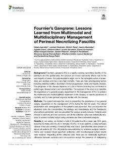
Fig. 3. What can be learnt from computer simulations in heart failure. Multi-scale electromechanical models of heart failure (HF) help to understand arrhythmogenic mechanisms. At the cellular level, electrophysiological remodeling can be modeled by alterations in ion channels, in β-adrenergic system, and in T-tubules (image taken from [148]) and is responsible for significant changes in action potential and Ca2+ transient waveforms, which might lead to EADs and DADs generation. At the tissue and organ level, structural remodeling (intercellular uncoupling and fibrosis) lead to discontinuous and decreased conduction velocity and electrophysiological heterogeneities. Triggered events and heterogeneous substrate set the stage for ventricular arrhythmias in the failing heart. Incorporating the mechanical response (electro-mechanical model reproduced with permission from [132]) in a HF model will improve the understanding of the mechanical dysfunction of the failing heart and the design of cardiac resynchronization therapy.
Please cite this article as: J.F. Gomez, et al., Lessons learned from multi-scale modeling of the failing heart, J Mol Cell Cardiol (2015), http:// dx.doi.org/10.1016/j.yjmcc.2015.10.016
J.F. Gomez et al. / Journal of Molecular and Cellular Cardiology xxx (2015) xxx–xxx
computational models could guide and improve CRT, as in Constantino et al., who suggested that an adequate pacing location would be the site of longest electromechanical delay [132]. 6. Concluding remarks and future challenges This article reviews the main computational studies on HF from cell to organ. These studies illustrate how modeling and computer simulations may reveal the mechanisms of arrhythmogenic processes and therapies in the setting of HF. In this last section, the main advances, as well as limitations and future challenges of the computational approach of HF are presented. In addition to the generic limitations of cardiac models [147], several improvements in the pathological models of HF should be highlighted to guide research towards future challenges. As summarized in Fig. 3, simulations at the cellular level have contributed to elucidating the role of ion channel remodeling in AP and Ca2 + handling HF-induced changes. However, the effects of changes in the T-tubule system [148] and β-adrenergic pathways in the failing cell need further analysis. Simulations of AP prolongation and altered Ca2 + transient have also furthered our understanding of the mechanisms of triggered activity: EADs and DADs in failing cells. 1D, 2D, and 3D models of the failing tissue have made it possible to simulate the effects of structural remodeling in the failing heart. The increase of cellular uncoupling and fibrosis in heart failure may have a significant role in setting an arrhythmogenic substrate, leading to cardiac arrhythmias. Although fibroblast interaction with cardiomyocytes has been addressed in several multi-scale computational studies in normal physiological conditions, the literature is scarce in the setting of HF. In addition, only the work of Chilton and coworkers [90] provides experimental data to inform an electrical model of ventricular fibroblasts. Further experiments are needed to improve these models, such as the work of Chatelier et al. [149], who reported the expression of voltage gated sodium channels in human atrial fibroblasts. Recently a great effort has been made to improve electromechanical models of the heart, which identify the link between electrical activity and especially Ca2+ transient and cellular and organ contraction (see Fig. 3). In terms of HF, electromechanical simulation studies are scarce and further studies are needed. These computational models can help to evaluate mechanical heart dysfunction and improve CRT. With regard to drug therapy in HF, apart from several studies focusing on sodium channel blockers, there is a lack of computational work. The analysis of the effects of β-blockers in the failing heart through computer simulations would help to understand why these drugs are effective in these patients. Recently, safety pharmacology assessments have highlighted the use of computer simulations in combination with in vitro experiments on human induced pluripotent stem cell-derived cardiomyocytes (iPSC-CMs) for drug safety screening. β-blockers have been proved to improve the function of iPSC-DC from dilated cardiomyopathy [150], and because this technology offers the possibility to obtain disease-specific cell lines, this could also be tested for other HF etiologies. Furthermore, mathematical models of hiPSC-CMs have been recently formulated [151], allowing in silico studies for drug safety and supporting the evaluation of this new in vitro technology [152]. It is important to mention that the efforts recently made in the development of patient-specific models, based on high-resolution images, will be of great help if they are extended to different stages of HF and different etiologies. Finally, a crucial challenge is the integration of computer pathological models and simulations into clinical practice. This integration of computer models in the clinical routine requires the development of automatic tools to personalize models and validate them not only in the laboratory setting but also in a clinical environment, in which clinicians have to deal with patients with different characteristics. Research effort needs to be made to build the necessary user-friendly interfaces for clinicians and provide the clinically relevant answers to the specific questions of the physicians.
11
Acknowledgements This work was partially supported by (i) the “VI Plan Nacional de Investigación Científica, Desarrollo e Innovación Tecnológica” from the Ministerio de Economía y Competitividad of Spain and the European Commission (European Regional Development Funds ERDF-FEDER) (grant number TIN2012-37546-C03-01), and by (ii) Programa Prometeo de la Conselleria d’Educació Formació I Ocupació, Generalitat Valenciana (grant number PROMETEO/2012/030).
References [1] R. Coronel, J.R. De Groot, J.J. Van Lieshout, Defining heart failure, Cardiovasc. Res. 50 (3) (2001) 419–422. [2] R. Coronel, R. Wilders, A.O. Verkerk, R.F. Wiegerinck, D. Benoist, O. Bernus, Electrophysiological changes in heart failure and their implications for arrhythmogenesis, Biochim. Biophys. Acta Mol. basis Dis. 1832 (12) (2013) 2432–2441. [3] G.F. Tomaselli, E. Marbán, Electrophysiological remodeling in hypertrophy and heart failure, Cardiovasc. Res. 42 (2) (May-1999) 270–283. [4] V.A. Maltsev, N. Silverman, H.N. Sabbah, A.I. Undrovinas, Chronic heart failure slows late sodium current in human and canine ventricular myocytes: implications for repolarization variability, Eur. J. Heart Fail. 9 (3) (Mar. 2007) 219–227. [5] C. Valdivia, W. Chu, J. Pu, Increased late sodium current in myocytes from a canine heart failure model and from failing human heart, J. Mol. Cell. Cardiol. 38 (3) (Mar. 2005) 475–483. [6] S. Despa, M.A. Islam, C.R. Weber, S.M. Pogwizd, D.M. Bers, Intracellular Na+ concentration is elevated in heart failure but Na/K pump function is unchanged, Circulation 105 (21) (May 2002) 2543–2548. [7] S. Despa, D.M. Bers, Na+ transport in the normal and failing heart — remember the balance, J. Mol. Cell. Cardiol. 61 (Aug. 2013) 2–10. [8] G.R. Li, J. Feng, L. Yue, M. Carrier, Transmural heterogeneity of action potentials and Ito1 in myocytes isolated from the human right ventricle, Am. J. Physiol. 275 (2 Pt 2) (Aug. 1998) H369–H377. [9] G.-R. Li, C.-P. Lau, A. Ducharme, J.-C. Tardif, S. Nattel, Transmural action potential and ionic current remodeling in ventricles of failing canine hearts, Am. J. Physiol. Heart Circ. Physiol. 283 (3) (2002) H1031–H1041. [10] G.-R. Li, C.-P. Lau, T.-K. Leung, S. Nattel, Ionic current abnormalities associated with prolonged action potentials in cardiomyocytes from diseased human right ventricles, Heart Rhythm. 1 (4) (2004) 460–468. [11] E. Wagner, M.A. Lauterbach, T. Kohl, V. Westphal, G.S.B. Williams, J.H. Steinbrecher, J.-H. Streich, B. Korff, H.-T.M. Tuan, B. Hagen, S. Luther, G. Hasenfuss, U. Parlitz, M.S. Jafri, S.W. Hell, W.J. Lederer, S.E. Lehnart, Stimulated emission depletion live-cell super-resolution imaging shows proliferative remodeling of T-tubule membrane structures after myocardial infarction, Circ. Res. 111 (4) (Aug. 2012) 402–414. [12] A.R. Lyon, K.T. MacLeod, Y. Zhang, E. Garcia, G.K. Kanda, M.J. Lab, Y.E. Korchev, S.E. Harding, J. Gorelik, Loss of T-tubules and other changes to surface topography in ventricular myocytes from failing human and rat heart, Proc. Natl. Acad. Sci. U. S. A. 106 (16) (2009) 6854–6859. [13] F.G. Akar, R.D. Nass, S. Hahn, E. Cingolani, M. Shah, G.G. Hesketh, D. DiSilvestre, R.S. Tunin, D.A. Kass, G.F. Tomaselli, Dynamic changes in conduction velocity and gap junction properties during development of pacing-induced heart failure, Am. J. Physiol. Heart Circ. Physiol. 293 (2) (Aug. 2007) H1223–H1230. [14] F.G. Akar, R.C. Wu, G.J. Juang, Y. Tian, M. Burysek, D. Disilvestre, W. Xiong, A.A. Armoundas, G.F. Tomaselli, Molecular mechanisms underlying K+ current downregulation in canine tachycardia-induced heart failure, Am. J. Physiol. Heart Circ. Physiol. 288 (6) (Jun. 2005) H2887–H2896. [15] E. Dupont, T. Matsushita, R.A. Kaba, C. Vozzi, S.R. Coppen, N. Khan, R. Kaprielian, M.H. Yacoub, N.J. Severs, Altered connexin expression in human congestive heart failure, J. Mol. Cell. Cardiol. 33 (2) (Feb. 2001) 359–371. [16] R.F. Wiegerinck, T.A.B. van Veen, C.N. Belterman, C.A. Schumacher, M. Noorman, J.M.T. de Bakker, R. Coronel, Transmural dispersion of refractoriness and conduction velocity is associated with heterogeneously reduced connexin43 in a rabbit model of heart failure, Heart Rhythm. 5 (8) (2008) 1178–1185. [17] P. Kohl, Heterogeneous cell coupling in the heart: an electrophysiological role for fibroblasts, Circ. Res. 93 (5) (Sep. 2003) 381–383. [18] G. Gaudesius, M. Miragoli, S.P. Thomas, S. Rohr, Coupling of cardiac electrical activity over extended distances by fibroblasts of cardiac origin, Circ. Res. 93 (5) (Sep. 2003) 421–428. [19] M. Miragoli, G. Gaudesius, S. Rohr, Electrotonic modulation of cardiac impulse conduction by myofibroblasts, Circ. Res. 98 (6) (Mar. 2006) 801–810. [20] M. Miragoli, N. Salvarani, S. Rohr, Myofibroblasts induce ectopic activity in cardiac tissue, Circ. Res. 101 (8) (Oct. 2007) 755–758. [21] L. Priebe, D.J. Beuckelmann, Simulation study of cellular electric properties in heart failure, Circ. Res. 82 (11) (1998) 1206–1223. [22] C. Weber, V. Piacentino, S. Houser, D. Bers, Dynamic regulation of sodium/calcium exchange function in human heart failure, Circulation 108 (18) (Nov. 2003) 2224–2229. [23] D.M. Bers, S. Despa, J. Bossuyt, Regulation of Ca2+ and Na+ in normal and failing cardiac myocytes, Ann. N. Y. Acad. Sci. 1080 (2006) 165–177. [24] R.L. Winslow, J. Rice, S. Jafri, E. Marbán, B. O'Rourke, Mechanisms of altered excitation-contraction coupling in canine tachycardia-induced heart failure, II: model studies, Circ. Res. 84 (5) (Mar. 1999) 571–586.
Please cite this article as: J.F. Gomez, et al., Lessons learned from multi-scale modeling of the failing heart, J Mol Cell Cardiol (2015), http:// dx.doi.org/10.1016/j.yjmcc.2015.10.016
12
J.F. Gomez et al. / Journal of Molecular and Cellular Cardiology xxx (2015) xxx–xxx
[25] J.L. Puglisi, D.M. Bers, LabHEART: an interactive computer model of rabbit ventricular myocyte ion channels and Ca transport, Am. J. Physiol. Cell Physiol. 281 (6) (2001) C2049–C2060. [26] T.R. Shannon, F. Wang, D.M. Bers, Regulation of cardiac sarcoplasmic reticulum Ca release by luminal [Ca] and altered gating assessed with a mathematical model, Biophys. J. 89 (6) (2005) 4096–4110. [27] Y.L. Hashambhoy, J.L. Greenstein, R.L. Winslow, Role of CaMKII in RyR leak, EC coupling and action potential duration: a computational model, J. Mol. Cell. Cardiol. 49 (4) (Oct. 2010) 617–624. [28] T. O'Hara, Y. Rudy, Arrhythmia formation in subclinical (‘silent’) long QT syndrome requires multiple insults: quantitative mechanistic study using the KCNQ1 mutation Q357R as example, Heart Rhythm. 9 (2) (2012) 275–282. [29] E. Grandi, J. Puglisi, S. Wagner, L. Maier, Simulation of Ca-calmodulin-dependent protein kinase II on rabbit ventricular myocyte ion currents and action potentials, Biophys. J. 93 (11) (Dec. 2007) 3835–3847. [30] L. Lu, L. Xia, X. Zhu, Simulation of arrhythmogenic effect of rogue RyRs in failing heart by using a coupled model, Comput. Math. Methods Med. 2012 (2012) 1–10. [31] J.D. Moreno, P.C. Yang, J.R. Bankston, E. Grandi, D.M. Bers, R.S. Kass, C.E. Clancy, Ranolazine for congenital and acquired late iNa-linked arrhythmias: in silico pharmacological screening, Circ. Res. 113 (7) (Sep. 2013) e50–e61. [32] S. Zlochiver, Subthreshold parameters of cardiac tissue in a bi-layer computer model of heart failure, Cardiovasc. Eng. 10 (4) (2010) 190–200. [33] K.H.W.J. ten Tusscher, D. Noble, P.J. Noble, A.V. Panfilov, A model for human ventricular tissue, Am. J. Physiol. Heart Circ. Physiol. 286 (4) (Apr. 2004) H1573–H1589. [34] Y. Zhang, L. Xia, M. Tang, Q. Sun, S. Zhang, Simulation study of transmural cellular electrical properties in failed human heart, Chin. J. Biomed. Eng. 27 (4) (2008) 526–532. [35] B. Trenor, K. Cardona, J.F. Gomez, S. Rajamani, J.M. Ferrero, L. Belardinelli, J. Saiz, Simulation and mechanistic investigation of the arrhythmogenic role of the late sodium current in human heart failure, PLoS One 7 (3) (2012), e32659. [36] C.M. Ambrosi, K.A. Yamada, J.M. Nerbonne, I.R. Efimov, Gender differences in electrophysiological gene expression in failing and non-failing human hearts, PLoS One 8 (1) (Jan. 2013), e54635. [37] J. Walmsley, J.F. Rodriguez, G.R. Mirams, K. Burrage, I.R. Efimov, B. Rodriguez, mRNA expression levels in failing human hearts predict cellular electrophysiological remodeling: a population-based simulation study, PLoS One 8 (2) (Jan. 2013), e56359. [38] R.L. Winslow, D.F. Scollan, A. Holmes, C.K. Yung, J. Zhang, M.S. Jafri, Electrophysiological modeling of cardiac ventricular function: from cell to organ, Annu. Rev. Biomed. Eng. 2 (1) (2000) 119–155. [39] N. Morita, A.A. Sovari, Y. Xie, M.C. Fishbein, W.J. Mandel, A. Garfinkel, S.-F. Lin, P.-S. Chen, L.-H. Xie, F. Chen, Z. Qu, J.N. Weiss, H.S. Karagueuzian, Increased susceptibility of aged hearts to ventricular fibrillation during oxidative stress, Am. J. Physiol. Heart Circ. Physiol. 297 (5) (2009) H1594–H1605. [40] S.M. Narayan, J.D. Bayer, G. Lalani, N.A. Trayanova, Action potential dynamics explain arrhythmic vulnerability in human heart failure: a clinical and modeling study implicating abnormal calcium handling, J. Am. Coll. Cardiol. 52 (22) (Nov. 2008) 1782–1792. [41] J.D. Moreno, Z.I. Zhu, P.-C. Yang, J.R. Bankston, M.-T. Jeng, C. Kang, L. Wang, J.D. Bayer, D.J. Christini, N.A. Trayanova, C.M. Ripplinger, R.S. Kass, C.E. Clancy, A computational model to predict the effects of class I anti-arrhythmic drugs on ventricular rhythms, Sci. Transl. Med. 3 (98) (Aug. 2011), 98ra83. [42] C.E. Clancy, Y. Chen-Izu, D.M. Bers, L. Belardinelli, P.A. Boyden, L. Csernoch, S. Despa, B. Fermini, L.C. Hool, L. Izu, R.S. Kass, W.J. Lederer, W.E. Louch, C. Maack, A. Matiazzi, Z. Qu, S. Rajamani, C.M. Rippinger, O.M. Sejersted, B. O'Rourke, J.N. Weiss, A. Varró, A. Zaza, Deranged sodium to sudden death, J. Physiol. 593 (6) (2015) 1331–1345. [43] C. Antzelevitch, V. Nesterenko, J.C. Shryock, S. Rajamani, Y. Song, L. Belardinelli, The role of late I Na in development of cardiac arrhythmias, Handbook of Experimental Pharmacology, 221 2014, pp. 137–168. [44] G.F. Tomaselli, D.P. Zipes, What causes sudden death in heart failure? Circ. Res. 95 (8) (Oct. 2004) 754–763. [45] A. Zaza, L. Belardinelli, J.C. Shryock, Pathophysiology and pharmacology of the cardiac ‘late sodium current, Pharmacol. Ther. 119 (3) (Sep. 2008) 326–339. [46] A.I. Undrovinas, L. Belardinelli, N.A. Undrovinas, H.N. Sabbah, Ranolazine improves abnormal repolarization and contraction in left ventricular myocytes of dogs with heart failure by inhibiting late sodium current, J. Cardiovasc. Electrophysiol. 17 (Suppl. 1) (May 2006) S169–S177. [47] I. Biktasheva, R. Simitev, R. Suckley, V. Biktashev, Asymptotic properties of mathematical models of excitability, Philos. Trans. R. Soc. A Math. Phys. Eng. Sci. 364 (1842) (2006) 1283–1298. [48] C.E. Clancy, Y. Rudy, Na+ channel mutation that causes both Brugada and long-QT syndrome phenotypes: a simulation study of mechanism, Circulation 105 (10) (Feb. 2002) 1208–1213. [49] P. Milberg, C. Pott, M. Fink, G. Frommeyer, T. Matsuda, A. Baba, N. Osada, G. Breithardt, D. Noble, L. Eckardt, Inhibition of the Na+/Ca2+ exchanger suppresses torsades de pointes in an intact heart model of long QT syndrome-2 and long QT syndrome-3, Heart Rhythm. 5 (10) (2008) 1444–1452. [50] T.J. Hund, Y. Rudy, Rate dependence and regulation of action potential and calcium transient in a canine cardiac ventricular cell model, Circulation 110 (20) (Nov. 2004) 3168–3174. [51] E. Grandi, F.S. Pasqualini, D.M. Bers, A novel computational model of the human ventricular action potential and Ca transient, J. Mol. Cell. Cardiol. 48 (1) (2010) 112–121. [52] T. O'Hara, L. Virág, A. Varró, Y. Rudy, Simulation of the undiseased human cardiac ventricular action potential: model formulation and experimental validation, PLoS Comput. Biol. 7 (5) (May 2011), e1002061.
[53] J.F. Gomez, K. Cardona, L. Romero, J.M. Ferrero, B. Trenor, Electrophysiological and structural remodeling in heart failure modulate arrhythmogenesis. 1D simulation study, PLoS One 9 (9) (2014), e106602. [54] J.D. Moreno, P.-C. Yang, J.R. Bankston, E. Grandi, D.M. Bers, R.S. Kass, C.E. Clancy, Ranolazine for congenital and acquired late INa-linked arrhythmias: in silico pharmacological screening, Circ. Res. 113 (7) (2013) e50–e61. [55] E. Grandi, J.L. Puglisi, S. Wagner, L.S. Maier, S. Severi, D.M. Bers, Simulation of Ca-calmodulin-dependent protein kinase II on rabbit ventricular myocyte ion currents and action potentials, Biophys. J. 93 (11) (2007) 3835–3847. [56] S. Morotti, A.G. Edwards, A.D. McCulloch, D.M. Bers, E. Grandi, A novel computational model of mouse myocyte electrophysiology to assess the synergy between Na+ loading and CaMKII, J. Physiol. 592 (Pt 6) (Mar. 2014) 1181–1197. [57] E. Grandi, A.W. Herren, CaMKII-dependent regulation of cardiac Na+ homeostasis, Front. Pharmacol. 5 (March, 2014) 1–10. [58] N. Jost, L. Virág, P. Comtois, B. Ordög, V. Szuts, G. Seprényi, M. Bitay, Z. Kohajda, I. Koncz, N. Nagy, T. Szél, J. Magyar, M. Kovács, L.G. Puskás, C. Lengyel, E. Wettwer, U. Ravens, P.P. Nánási, J.G. Papp, A. Varró, S. Nattel, Ionic mechanisms limiting cardiac repolarization reserve in humans compared to dogs, J. Physiol. 591 (Pt 17) (Sep. 2013) 4189–4206. [59] L. Li, W.E. Louch, S.A. Niederer, J.M. Aronsen, G. Christensen, O.M. Sejersted, N.P. Smith, Sodium accumulation in SERCA knockout-induced heart failure, Biophys. J. 102 (9) (2012) 2039–2048. [60] J.Q. He, M.W. Conklin, J.D. Foell, M.R. Wolff, R.A. Haworth, R. Coronado, T.J. Kamp, Reduction in density of transverse tubules and L-type Ca2+ channels in canine tachycardia-induced heart failure, Cardiovasc. Res. 49 (2) (2001) 298–307. [61] W. Louch, Reduced synchrony of Ca2+ release with loss of T-tubules — a comparison to Ca2+ release in human failing cardiomyocytes, Cardiovasc. Res. 62 (1) (2004) 63–73. [62] V.O. Nikolaev, A. Moshkov, A.R. Lyon, M. Miragoli, P. Novak, H. Paur, M.J. Lohse, Y.E. Korchev, S.E. Harding, J. Gorelik, Beta2-adrenergic receptor redistribution in heart failure changes cAMP compartmentation, Science 327 (5973) (2010) 1653–1657. [63] M. Schäfer, K. Frischkopf, G. Taimor, H.M. Piper, K.D. Schlüter, Hypertrophic effect of selective beta(1)-adrenoceptor stimulation on ventricular cardiomyocytes from adult rat, Am. J. Physiol. Cell Physiol. 279 (2) (2000) C495–C503. [64] J. Desantiago, X. Ai, M. Islam, G. Acuna, M.T. Ziolo, D.M. Bers, S.M. Pogwizd, Arrhythmogenic effects of β2-adrenergic stimulation in the failing heart are attributable to enhanced sarcoplasmic reticulum Ca load, Circ. Res. 102 (11) (2008) 1389–1397. [65] R.B. Clark, A. Tremblay, P. Melnyk, B.G. Allen, W.R. Giles, C. Fiset, T-tubule localization of the inward-rectifier K(+) channel in mouse ventricular myocytes: a role in K(+) accumulation, J. Physiol. 537 (Pt 3) (2001) 979–992. [66] L.-F. Cheng, F. Wang, A.N. Lopatin, Metabolic stress in isolated mouse ventricular myocytes leads to remodeling of t tubules, AJP Heart Circ. Physiol. 301 (2011) H1984–H1995. [67] N. Gaur, Y. Rudy, Multiscale modeling of calcium cycling in cardiac ventricular myocyte: macroscopic consequences of microscopic dyadic function, Biophys. J. 100 (12) (2011) 2904–2912. [68] W.E. Louch, J. Hake, G.F. Jølle, H.K. Mørk, I. Sjaastad, G.T. Lines, O.M. Sejersted, Control of Ca2+ release by action potential configuration in normal and failing murine cardiomyocytes, Biophys. J. 99 (5) (2010) 1377–1386. [69] M. Nivala, Z. Song, J.N. Weiss, Z. Qu, T-tubule disruption promotes calcium alternans in failing ventricular myocytes: mechanistic insights from computational modeling, J. Mol. Cell. Cardiol. 79 (2015) 32–41. [70] P.T. Wright, V.O. Nikolaev, T. O'Hara, I. Diakonov, A. Bhargava, S. Tokar, S. Schobesberger, A.I. Shevchuk, M.B. Sikkel, R. Wilkinson, N.A. Trayanova, A.R. Lyon, S.E. Harding, J. Gorelik, Caveolin-3 regulates compartmentation of cardiomyocyte beta2-adrenergic receptor-mediated cAMP signaling, J. Mol. Cell. Cardiol. 67 (2014) 38–48. [71] R.F. Wiegerinck, A.O. Verkerk, C.N. Belterman, T.A.B. Van Veen, A. Baartscheer, T. Opthof, R. Wilders, J.M.T. De Bakker, R. Coronel, Larger cell size in rabbits with heart failure increases myocardial conduction velocity and QRS duration, Circulation 113 (6) (2006) 806–813. [72] S.B. Wachter, E.M. Gilbert, Beta-adrenergic receptors, from their discovery and characterization through their manipulation to beneficial clinical application, Cardiology 122 (2) (2012) 104–112. [73] J. Heijman, P.G.A. Volders, R.L. Westra, Y. Rudy, Local control of β-adrenergic stimulation: effects on ventricular myocyte electrophysiology and Ca(2+)-transient— data supplement, J. Mol. Cell. Cardiol. 50 (5) (2011) 863–871. [74] J.J. Saucerman, L.L. Brunton, A.P. Michailova, A.D. McCulloch, Modeling betaadrenergic control of cardiac myocyte contractility in silico, J. Biol. Chem. 278 (48) (Nov. 2003) 47997–48003. [75] J.J. Saucerman, S.N. Healy, M.E. Belik, J.L. Puglisi, A.D. McCulloch, Proarrhythmic consequences of a KCNQ1 AKAP-binding domain mutation: computational models of whole cells and heterogeneous tissue, Circ. Res. 95 (12) (Dec. 2004) 1216–1224. [76] A.R. Soltis, J.J. Saucerman, Synergy between CaMKII substrates and β-adrenergic signaling in regulation of cardiac myocyte Ca(2+) handling, Biophys. J. 99 (7) (Oct. 2010) 2038–2047. [77] Y. Xie, E. Grandi, J.L. Puglisi, D. Sato, D.M. Bers, β-adrenergic stimulation activates early afterdepolarizations transiently via kinetic mismatch of PKA targets, J. Mol. Cell. Cardiol. 58 (2013) 153–161. [78] Y. Xie, E. Grandi, D.M. Bers, D. Sato, How does adrenergic signalling affect the transitions from ventricular tachycardia to ventricular fibrillation? Europace 16 (3) (2014) 452–457. [79] D. Lang, K. Holzem, C. Kang, M. Xiao, H.J. Hwang, G.A. Ewald, K.A. Yamada, I.R. Efimov, Arrhythmogenic remodeling of 2 versus 1 adrenergic signaling in the human failing heart, Circ. Arrhythmia Electrophysiol. (2015).
Please cite this article as: J.F. Gomez, et al., Lessons learned from multi-scale modeling of the failing heart, J Mol Cell Cardiol (2015), http:// dx.doi.org/10.1016/j.yjmcc.2015.10.016
J.F. Gomez et al. / Journal of Molecular and Cellular Cardiology xxx (2015) xxx–xxx [80] O.R. de Peuter, F. Lussana, R.J.G. Peters, H.R. Büller, P.W. Kamphuisen, A systematic review of selective and non-selective beta blockers for prevention of vascular events in patients with acute coronary syndrome or heart failure, Neth. J. Med. 67 (9) (2009) 284–294. [81] M.H. Ruwald, A.-C.H. Ruwald, C. Jons, J. Alexis, S. McNitt, W. Zareba, A.J. Moss, Effect of metoprolol versus carvedilol on outcomes in MADIT-CRT (multicenter automatic defibrillator implantation trial with cardiac resynchronization therapy), J. Am. Coll. Cardiol. 61 (14) (2013) 1518–1526. [82] V.E. Bondarenko, A compartmentalized mathematical model of the β1-adrenergic signaling system in mouse ventricular myocytes, PLoS One 9 (2) (2014), e89113. [83] M. Tanaka, H. Fujiwara, T. Onodera, D.J. Wu, M. Matsuda, Y. Hamashima, C. Kawai, Pathogenetic role of myocardial fiber disarray in the progression of cardiac fibrosis in normal hearts, hypertensive hearts and hearts with hypertrophic cardiomyopathy, Jpn. Circ. J. 51 (6) (1987) 624–630. [84] T. Kawara, R. Derksen, J.R. de Groot, R. Coronel, S. Tasseron, A.C. Linnenbank, R.N. Hauer, H. Kirkels, M.J. Janse, J.M. de Bakker, Activation delay after premature stimulation in chronically diseased human myocardium relates to the architecture of interstitial fibrosis, Circulation 104 (25) (2001) 3069–3075. [85] P. Camelliti, T.K. Borg, P. Kohl, Structural and functional characterisation of cardiac fibroblasts, Cardiovasc. Res. 65 (1) (Jan. 2005) 40–51. [86] P. Kohl, A.G. Kamkin, I.S. Kiseleva, D. Noble, Mechanosensitive fibroblasts in the sino-atrial node region of rat heart: interaction with cardiomyocytes and possible role, Exp. Physiol. 79 (6) (1994) 943–956. [87] P. Kohl, P. Camelliti, F.L. Burton, G.L. Smith, Electrical coupling of fibroblasts and myocytes: relevance for cardiac propagation, J. Electrocardiol. 38 (4) (2005) 45–50. [88] P. Camelliti, C.R. Green, I. LeGrice, P. Kohl, Fibroblast network in rabbit sinoatrial node: structural and functional identification of homogeneous and heterogeneous cell coupling, Circ. Res. 94 (6) (Apr. 2004) 828–835. [89] P. Kohl, R.G. Gourdie, Fibroblast–myocyte electrotonic coupling: does it occur in native cardiac tissue? J. Mol. Cell. Cardiol. 70 (2014) 37–46. [90] L. Chilton, S. Ohya, D. Freed, E. George, V. Drobic, Y. Shibukawa, K.A. Maccannell, Y. Imaizumi, R.B. Clark, I.M.C. Dixon, W.R. Giles, K + currents regulate the resting membrane potential, proliferation, and contractile responses in ventricular fibroblasts and myofibroblasts, Am. J. Physiol. Heart Circ. Physiol. 288 (6) (Jun. 2005) H2931–H2939. [91] K. Andrew MacCannell, H. Bazzazi, L. Chilton, Y. Shibukawa, R.B. Clark, W.R. Giles, A mathematical model of electrotonic interactions between ventricular myocytes and fibroblasts, Biophys. J. 92 (11) (2007) 4121–4132. [92] F.B. Sachse, A.P. Moreno, J.A. Abildskov, Electrophysiological modeling of fibroblasts and their interaction with myocytes, Ann. Biomed. Eng. 36 (1) (2008) 41–56. [93] M.M. Maleckar, J.L. Greenstein, W.R. Giles, N.A. Trayanova, Electrotonic coupling between human atrial myocytes and fibroblasts alters myocyte excitability and repolarization, Biophys. J. 97 (8) (2009) 2179–2190. [94] I. Turner, C.L.-H. Huang, R.C. Saumarez, Numerical simulation of paced electrogram fractionation: relating clinical observations to changes in fibrosis and action potential duration, J. Cardiovasc. Electrophysiol. 16 (2) (2005) 151–161. [95] Y. Xie, A. Garfinkel, P. Camelliti, P. Kohl, J. Weiss, Z. Qu, Effects of fibroblast-myocyte coupling on cardiac conduction and vulnerability to reentry: a computational study, Heart Rhythm. 6 (11) (Nov. 2009) 1641–1649. [96] V.S. Petrov, G.V. Osipov, J. Kurths, Fibroblasts alter spiral wave stability, Chaos An Interdiscip. J. Nonlinear Sci. 20 (4) (2010), 045103. [97] Z.J. Engelman, M.L. Trew, B.H. Smaill, Structural heterogeneity alone is a sufficient substrate for dynamic instability and altered restitution, Circ. Arrhythmia Electrophysiol. 3 (2) (2010) 195–203. [98] T.P. Nguyen, Z. Qu, J.N. Weiss, Cardiac fibrosis and arrhythmogenesis: the road to repair is paved with perils, J. Mol. Cell. Cardiol. 70 (2014) 83–91. [99] A. Greisas, S. Zlochiver, Modulation of spiral-wave dynamics and spontaneous activity in a fibroblast/myocyte heterocellular tissue — a computational study, IEEE Trans. Biomed. Eng. 59 (5) (2012) 1398–1407. [100] R.V. Oren, C.E. Clancy, Determinants of heterogeneity, excitation and conduction in the sinoatrial node: a model study, PLoS Comput. Biol. 6 (12) (2010), e1001041. [101] J.F. Gomez, K. Cardona, L. Martinez, J. Saiz, B. Trenor, Electrophysiological and structural remodeling in heart failure modulate arrhythmogenesis. 2D simulation study, PLoS One 9 (7) (2014), e103273. [102] A.R. Nayak, T.K. Shajahan, A.V. Panfilov, R. Pandit, Spiral-wave dynamics in a mathematical model of human ventricular tissue with myocytes and fibroblasts, PLoS One 8 (9) (2013), e72950. [103] R. Majumder, A.R. Nayak, R. Pandit, Nonequilibrium arrhythmic states and transitions in a mathematical model for diffuse fibrosis in human cardiac tissue, PLoS One 7 (10) (2012), e45040. [104] M. Aguilar, X.Y. Qi, H. Huang, P. Comtois, S. Nattel, Fibroblast electrical remodeling in heart failure and potential effects on atrial fibrillation, Biophys. J. 107 (10) (2014) 2444–2455. [105] K.S. McDowell, H.J. Arevalo, M.M. Maleckar, N.A. Trayanova, Susceptibility to arrhythmia in the infarcted heart depends on myofibroblast density, Biophys. J. 101 (6) (Sep. 2011) 1307–1315. [106] H.J. Jongsma, R. Wilders, Gap junctions in cardiovascular disease, Circ. Res. 86 (2000) 1193–1197. [107] N.S. Peters, C.R. Green, P.A. Poole-Wilson, N.J. Severs, Reduced content of connexin43 gap junctions in ventricular myocardium from hypertrophied and ischemic human hearts, Circulation 88 (3) (1993) 864–875. [108] A.V. Glukhov, V.V. Fedorov, P.W. Kalish, V.K. Ravikumar, Q. Lou, D. Janks, R.B. Schuessler, N. Moazami, I.R. Efimov, Conduction remodeling in human end-stage nonischemic left ventricular cardiomyopathy, Circulation 125 (15) (Apr. 2012) 1835–1847.
13
[109] K. Gima, Y. Rudy, Ionic current basis of electrocardiographic waveforms: a model study, Circ. Res. 90 (8) (2002) 889–896. [110] J.F. Gomez, K. Cardona, L. Romero, J. Saiz, L. Belardinelli, S. Rajamani, B. Trenor, U.P. De València, G. Science, Increase in late sodium current and cellular uncoupling exacerbates transmural dispersion of repolarization in heart failure, Comput. Cardiol. 39 (2012) 233–236. [111] V.A. Maltsev, A. Undrovinas, Late sodium current in failing heart: friend or foe? Prog. Biophys. Mol. Biol. 96 (1–3) (2008) 421–451. [112] A. Baartscheer, C. Schumacher, C. Belterman, R. Coronel, J. Fiolet, SR calcium handling and calcium after-transients in a rabbit model of heart failure, Cardiovasc. Res. 58 (1) (Apr. 2003) 99–108. [113] Y. Xie, D. Sato, A. Garfinkel, Z. Qu, J.N. Weiss, So little source, so much sink: requirements for afterdepolarizations to propagate in tissue, Biophys. J. 99 (5) (2010) 1408–1415. [114] F.G. Akar, D.S. Rosenbaum, Transmural electrophysiological heterogeneities underlying arrhythmogenesis in heart failure, Circ. Res. 93 (7) (Oct. 2003) 638–645. [115] Q. Lou, D.L. Janks, K. Holzem, D. Lang, B. Onal, C.M. Ambrosi, V.V. Fedorov, I.-W. Wang, I.R. Efimov, Right ventricular arrhythmogenesis in failing human heart: the role of conduction and repolarization remodeling, AJP: Heart Circ. Physiol. 303 (12) (Dec-2012) H1426–H1434. [116] R. Coronel, F.J.G. Wilms-Schopman, T. Opthof, M.J. Janse, Dispersion of repolarization and arrhythmogenesis, Heart Rhythm. 6 (4) (2009) 537–543. [117] A.V. Glukhov, V.V. Fedorov, Q. Lou, V.K. Ravikumar, P.W. Kalish, R.B. Schuessler, N. Moazami, I.R. Efimov, Transmural dispersion of repolarization in failing and nonfailing human ventricle, Circ. Res. 106 (5) (Mar. 2010) 981–991. [118] Q. Lou, V.V. Fedorov, A.V. Glukhov, N. Moazami, V.G. Fast, I.R. Efimov, Transmural heterogeneity and remodeling of ventricular excitation–contraction coupling in human heart failure, Circulation 123 (17) (May 2011) 1881–1890. [119] M.M. Elshrif, P. Shi, E.M. Cherry, Electrophysiological properties under heart failure conditions in a human ventricular cell: a modeling study, Conf. Proc. … Annu. Int. Conf. IEEE Eng. Med. Biol. Soc. IEEE Eng. Med. Biol. Soc. Annu. Conf., vol. 2014, no. 1 2014, pp. 4324–4329. [120] P.N. Jordan, D.J. Christini, Action potential morphology influences intracellular calcium handling stability and the occurrence of alternans, Biophys. J. 90 (2) (2006) 672–680. [121] P.N. Jordan, D.J. Christini, Characterizing the contribution of voltage- and calciumdependent coupling to action potential stability: implications for repolarization alternans, Am. J. Physiol. Heart Circ. Physiol. 293 (4) (2007) H2109–H2118. [122] S.A. Gaeta, T. Krogh-Madsen, D.J. Christini, Feedback-control induced pattern formation in cardiac myocytes: a mathematical modeling study, J. Theor. Biol. 266 (3) (2010) 408–418. [123] A. Garfinkel, Y.H. Kim, O. Voroshilovsky, Z. Qu, J.R. Kil, M.H. Lee, H.S. Karagueuzian, J.N. Weiss, P.S. Chen, Preventing ventricular fibrillation by flattening cardiac restitution, Proc. Natl. Acad. Sci. U. S. A. 97 (11) (2000) 6061–6066. [124] V. Jacquemet, C.S. Henriquez, Genesis of complex fractionated atrial electrograms in zones of slow conduction: a computer model of microfibrosis, Heart Rhythm. 6 (6) (Jun. 2009) 803–810. [125] V. Jacquemet, C.S. Henriquez, Modulation of conduction velocity by nonmyocytes in the low coupling regime, IEEE Trans. Biomed. Eng. 56 (3) (2009) 893–896. [126] R.A. Gray, A.M. Pertsov, J. Jalife, Spatial and temporal organization during cardiac fibrillation, Nature 392 (May, 1998) 75–78. [127] K. Umapathy, K. Nair, S. Masse, S. Krishnan, J. Rogers, M.P. Nash, K. Nanthakumar, Phase mapping of cardiac fibrillation, Circ. Arrhythm. Electrophysiol. 3 (1) (Feb. 2010) 105–114. [128] Q. Lou, C.M. Ripplinger, P.V. Bayly, I.R. Efimov, Quantitative panoramic imaging of epicardial electrical activity, Ann. Biomed. Eng. 36 (10) (Oct. 2008) 1649–1658. [129] S. Zlochiver, V. Muñoz, K.L. Vikstrom, S.M. Taffet, O. Berenfeld, J. Jalife, Electrotonic myofibroblast-to-myocyte coupling increases propensity to reentrant arrhythmias in two-dimensional cardiac monolayers, Biophys. J. 95 (9) (2008) 4469–4480. [130] K. Tanaka, S. Zlochiver, K.L. Vikstrom, M. Yamazaki, J. Moreno, M. Klos, A.V. Zaitsev, R. Vaidyanathan, D.S. Auerbach, S. Landas, G. Guiraudon, J. Jalife, O. Berenfeld, J. Kalifa, Spatial distribution of fibrosis governs fibrillation wave dynamics in the posterior left atrium during heart failure, Circ. Res. 101 (8) (Oct. 2007) 839–847. [131] K.R. Chi, News & analysis revolution dawning in cardiotoxicity testing, Nat. Rev. Drug Discov. 12 (August, 2013) 565–567. [132] J. Constantino, Y. Hu, N.A. Trayanova, A computational approach to understanding the cardiac electromechanical activation sequence in the normal and failing heart, with translation to the clinical practice of CRT, Prog. Biophys. Mol. Biol. 110 (2–3) (2012) 372–379. [133] L. Galeotti, P.M. van Dam, Z. Loring, D. Chan, D.G. Strauss, Evaluating strict and conventional left bundle branch block criteria using electrocardiographic simulations, Europace 15 (12) (Dec. 2013) 1816–1821. [134] M. Potse, D. Krause, L. Bacharova, R. Krause, F.W. Prinzen, A. Auricchio, Similarities and differences between electrocardiogram signs of left bundle-branch block and left-ventricular uncoupling, Europace 14 (Suppl. 5) (Nov. 2012) v33–v39. [135] T.P. Usyk, A.D. McCulloch, Electromechanical model of cardiac resynchronization in the dilated failing heart with left bundle branch block, J. Electrocardiol. 36 (2003) 57–61. [136] C. Leclercq, O. Faris, R. Tunin, J. Johnson, R. Kato, F. Evans, J. Spinelli, H. Halperin, E. McVeigh, D.A. Kass, Systolic improvement and mechanical resynchronization does not require electrical synchrony in the dilated failing heart with left bundle-branch block, Circulation 106 (14) (2002) 1760–1763. [137] P. Sogaard, H. Egeblad, A.K. Pedersen, W.Y. Kim, B. Kristensen, P.S. Hansen, P.T. Mortensen, Sequential versus simultaneous biventricular resynchronization for severe heart failure: evaluation by tissue Doppler imaging, Circulation 106 (16) (2002) 2078–2084.
Please cite this article as: J.F. Gomez, et al., Lessons learned from multi-scale modeling of the failing heart, J Mol Cell Cardiol (2015), http:// dx.doi.org/10.1016/j.yjmcc.2015.10.016
14
J.F. Gomez et al. / Journal of Molecular and Cellular Cardiology xxx (2015) xxx–xxx
[138] T. Aiba, A.S. Barth, G.G. Hesketh, Y.L. Hashambhoy, K. Chakir, R.S. Tunin, J.L. Greenstein, R.L. Winslow, D.A. Kass, G.F. Tomaselli, Cardiac resynchronization therapy improves altered Na channel gating in canine model of dyssynchronous heart failure, Circ. Arrhythm. Electrophysiol. 6 (3) (Jun. 2013) 546–554. [139] B. Rodríguez, J.C. Eason, N. Trayanova, Differences between left and right ventricular anatomy determine the types of reentrant circuits induced by an external electric shock. A rabbit heart simulation study, Prog. Biophys. Mol. Biol. 90 (1–3) (2006) 399–413. [140] D. Romero, R. Sebastian, B.H. Bijnens, V. Zimmerman, P.M. Boyle, E.J. Vigmond, A.F. Frangi, Effects of the Purkinje system and cardiac geometry on biventricular pacing: a model study, Ann. Biomed. Eng. 38 (4) (2010) 1388–1398. [141] M. Sermesant, R. Chabiniok, P. Chinchapatnam, T. Mansi, F. Billet, P. Moireau, J.M. Peyrat, K. Wong, J. Relan, K. Rhode, M. Ginks, P. Lambiase, H. Delingette, M. Sorine, C.A. Rinaldi, D. Chapelle, R. Razavi, N. Ayache, Patient-specific electromechanical models of the heart for the prediction of pacing acute effects in CRT: a preliminary clinical validation, Med. Image Anal. 16 (1) (2012) 201–215. [142] J. Aguado-Sierra, A. Krishnamurthy, C. Villongco, J. Chuang, E. Howard, M.J. Gonzales, J. Omens, D.E. Krummen, S. Narayan, R.C.P. Kerckhoffs, A.D. McCulloch, Patient-specific modeling of dyssynchronous heart failure: a case study, Prog. Biophys. Mol. Biol. 107 (1) (2011) 147–155. [143] N.H.L. Kuijpers, E. Hermeling, P.H.M. Bovendeerd, T. Delhaas, F.W. Prinzen, Modeling cardiac electromechanics and mechanoelectrical coupling in dyssynchronous and failing hearts: insight from adaptive computer models, J. Cardiovasc. Transl. Res. 5 (2) (Apr. 2012) 159–169. [144] R. Huang, Y. Zhang, L. Xia, Cellular level electromechanical modeling and simulation of heart failure, Conf. Proc. … Annu. Int. Conf. IEEE Eng. Med. Biol. Soc. IEEE Eng. Med. Biol. Soc. Annu. Conf., 7 2005, pp. 7301–7304. [145] A. Krishnamurthy, C.T. Villongco, J. Chuang, L.R. Frank, V. Nigam, E. Belezzuoli, P. Stark, D.E. Krummen, S. Narayan, J.H. Omens, A.D. McCulloch, R.C. Kerckhoffs, Patient-specific models of cardiac biomechanics. J. Comput. Phys. 244 (2013) 4–21.
[146] V. Gurev, J. Constantino, J.J. Rice, N.A. Trayanova, Distribution of electromechanical delay in the heart: insights from a three-dimensional electromechanical model, Biophys. J. 99 (3) (2010) 745–754. [147] G. Plank, L. Zhou, J.L. Greenstein, S. Cortassa, R.L. Winslow, B. O'Rourke, N.A. Trayanova, From mitochondrial ion channels to arrhythmias in the heart: computational techniques to bridge the spatio-temporal scales, Philos. Trans. A Math. Phys. Eng. Sci. 366 (1879) (Sep. 2008) 3381–3409. [148] M. Miragoli, A. Moshkov, P. Novak, A. Shevchuk, V.O. Nikolaev, I. El-Hamamsy, C.M.F. Potter, P. Wright, S.H.S.A. Kadir, A.R. Lyon, J.A. Mitchell, A.H. Chester, D. Klenerman, M.J. Lab, Y.E. Korchev, S.E. Harding, J. Gorelik, Scanning ion conductance microscopy: a convergent high-resolution technology for multi-parametric analysis of living cardiovascular cells, J. R. Soc. Interface 8 (60) (2011) 913–925. [149] A. Chatelier, A. Mercier, B. Tremblier, O. Thériault, M. Moubarak, N. Benamer, P. Corbi, P. Bois, M. Chahine, J.F. Faivre, A distinct de novo expression of Nav1.5 sodium channels in human atrial fibroblasts differentiated into myofibroblasts, J. Physiol. 590 (2012) 4307–4319. [150] N. Sun, M. Yazawa, J. Liu, L. Han, V. Sanchez-Freire, O.J. Abilez, E.G. Navarrete, S. Hu, L. Wang, A. Lee, A. Pavlovic, S. Lin, R. Chen, R.J. Hajjar, M.P. Snyder, R.E. Dolmetsch, M.J. Butte, E.A. Ashley, M.T. Longaker, R.C. Robbins, J.C. Wu, Patient-specific induced pluripotent stem cells as a model for familial dilated cardiomyopathy, Sci. Transl. Med. 4 (130) (2012), 130ra47. [151] M. Paci, J. Hyttinen, K. Aalto-Setälä, S. Severi, Computational models of ventricularand atrial-like human induced pluripotent stem cell derived cardiomyocytes, Ann. Biomed. Eng. 41 (11) (2013) 2334–2348. [152] M. Paci, J. Hyttinen, B. Rodriguez, S. Severi, Human induced pluripotent stem cellderived versus adult cardiomyocytes: an in silico electrophysiological study on ionic current block effects, Br. J. Pharmacol. (Aug. 2015).
Please cite this article as: J.F. Gomez, et al., Lessons learned from multi-scale modeling of the failing heart, J Mol Cell Cardiol (2015), http:// dx.doi.org/10.1016/j.yjmcc.2015.10.016