Lessons we Learned from High-Throughput and Top-Down Systems Biology Analyses about Glioma Stem Cells
1
Andreas Mock, 2,3Sara Chiblak, 1Christel Herold-Mende
1
Division of Experimental Neurosurgery, Department of Neurosurgery, University of Heidelberg, INF 400, 69120 Heidelberg, Germany 2
Molecular and Translational Radiation Oncology, National Center for Tumor Diseases (NCT), German Cancer Research Center (DKFZ), Heidelberg, Germany 3
Heidelberg Institute for Radiation Oncology (HIRO), Heidelberg Ion Therapy Center (HIT), Department of Radiation Oncology, University of Heidelberg Medical School, Heidelberg, Germany
Accepted version of the peer-reviewed article The published manuscript is available at http://benthamscience.com/journals/current-pharmaceutical-design/volume/20/issue/1/page/66/
Short Running Title:
Omics and systems biology of GSCs
Journal Category:
Review
Address of Correspondence:
Prof. Dr. rer. nat. Christel Herold-Mende Experimentelle Neurochirurgie Neurochirurgische Universitätsklinik INF 400 69120 Heidelberg, Germany Tel.: 49 6221 566405 Fax.: 49 6221 5633979 Email:
[email protected]
- 1 -
ABSTRACT A growing body of evidence suggests that glioma stem cells (GSCs) account for tumor initiation, therapy resistance, and the subsequent regrowth of gliomas. Thus, continuous efforts have been undertaken to further characterize this subpopulation of less differentiated tumor cells. Although we are able to enrich GSCs, we still lack a comprehensive understanding of GSC phenotypes and behavior. The advent of high-throughput technologies raised hope that incorporation of these newly developed platforms would help to tackle such questions. Since then a couple of comparative genome, transcriptome- and proteome-wide studies on GSCs have been conducted giving new insights in GSC biology. However, lessons had to be learned in designing high-throughput experiments and some of the resulting conclusions fell short of expectations because they were performed on only a few GSC lines or at one molecular level instead of an integrative poly-omics approach. Despite these shortcomings, our knowledge of GSC biology has markedly expanded due to a number of survivalassociated biomarkers as well as glioma-relevant signaling pathways and therapeutic targets being identified. In this article we review recent findings obtained by comparative high-throughput analyses of GSCs. We further summarize fundamental concepts of systems biology as well as its applications for glioma stem cell research.
KEY WORDS: Stem cells, glioblastoma, systems biology, profiling, top-down, high-throughput, omics, integrative analysis.
- 2 -
1. INTRODUCTION The cancer stem cell (CSC) model suggests that tumors are driven by an immature subpopulation of tumor cells. According to this concept, CSCs are hierarchically organized and are endowed with an increased tumorigenicity [1]. The first successful identification of CSC was described for acute myeloid leukemia [2,3]. Soon after, their existence was proposed in a number of solid malignancies including tumors of the lung, breast, colon, head and neck and the brain [4-9]. The CD133 stem cell antigen has been successfully used to enrich tumor cells with increased tumorigenicity in gliomas as well as other types of cancer [6,8-11]. However, there has been a controversial discussion surrounding the existence of glioma stem cells (GSC) mostly due to the fact that CD133-positive cells do not constitute the only tumorigenic subpopulation in gliomas [12,13]. Furthermore, we do not have proper markers to define the different levels of the hierarchy below GSCs. Currently, the CSC debate seems to be resolved based on very recent publications identifying small subsets of tumor cells that arise de novo during tumor formation in intact organs such as skin, intestine and brain and behave like cancer stem cells [14-16]. However, in gliomas the analysis of CSCs is complicated due to the occurrence of different GSC phenotypes within the same tumors and a marked influence of the tumor microenvironment on stem cell properties [13,14,17]. This emphasizes the need to learn more about GSC phenotypes and GSC biology. For this purpose, systems biology seems to be one of the most attractive approaches because it integrates data from “-omics” technologies at different molecular levels such as DNA, mRNA, miRNA and protein. Although the majority of analyses up to now have only used one of these approaches, notably microarray expression profiling, it is evident that a more comprehensive understanding that can be translated into clinical applications will not be achieved by concentrating on single genes. Therefore, we will review current concepts and experimental designs of systems biology and delineate how these are applied to study GSC biology, hierarchy, heterogeneity, therapy resistance, biomarkers, and the putative cell of origin. Since analyses of tumor tissues as a source of CSC might be compromised by stromal cell components and heterogeneous tumor cell populations, we will strictly concentrate on high-throughput studies performed on isolated GSCs as the experimental input.
- 3 -
2. CONCEPTS OF SYSTEMS BIOLOGY Molecular cell biology of the 20th century was dominated by reductionist
approaches
aimed at
generating information about individual cellular components, their biochemical compositions or their biological functions [18]. However, the advent of high-throughput technologies substantially increased the amount of information obtained by a single experiment. Today, for a growing number of organisms whole-genome data is available [19-23] paving the way for systems
biology. This more
holistic approach constitutes a principal change of paradigm in biology. Its ultimate goal is to develop a system-level understanding of biological processes [24]. But how can we look at cells as systems? High-throughput technologies are only the basis of a more complete understanding as they provide us with a comprehensive list of cellular components and some state variables. The pure generation of e.g. transcriptome data is therefore not per
se
systems biology, but it is often deemed as such. What turns a
transcriptome analysis into a systems biology approach is the integrative analysis of interactions that relies on bioinformatics and methods for systems analysis. The central role is therefore assigned to mathematics in this paradigm change. In the end, these mathematical approaches might even produce results that could not have been generated before, a property sometimes referred to as emergence [18]. Since systems biology is a highly diverse field, it is convenient to discriminate between bottom-up and top-down approaches as a means of distinguishing the methodologies (Fig. 1). In the following chapter, we will delineate these two main approaches to systems biology with a special emphasis on glioma stem cells.
- 4 -
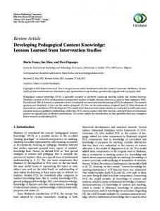
Fig. (1). Approaches to Systems Biology: Bottom-up approaches start with the generation of computational network models of defined subcellular systems, which are confirmed by experimental data. Starting point of topdown systems biology are high-throughput approaches of whole cell material followed by bioinformatical analyses.
3. APPROACHES TO SYSTEMS BIOLOGY a) Top-down Approach Top-down approaches to systems biology start with the generation of high-throughput data using microarrays or other new measurement technologies and are often referred to as discovery science. The term relates to the fact that high-throughput analyses enumerate the elements of a system irrespective of any hypotheses on how the system functions [25]. Nevertheless, top-down approaches and hypothesis-driven research are rather complementary, the latter providing the testing ground for systems biology. However, data from single-omics approaches should always be viewed with some caution because of the occurrence of false-negative and false-positive results. Moreover, these single level analyses are not sufficient to understand if an expression change is correlated with an altered gene function. To increase the reliability of gene function annotation, multiple independent datasets need to be integrated [26].
- 5 -
b) Bottom-up Approach Bottom-up approaches to systems biology are mechanistic in nature and have an engineering flavor. They start with network models of cell parts, which are largely built on existing biological knowledge [27]. Therefore, these approaches are particularly suited when most of the interactions in a network are already known, enabling us to find the last few missing pieces to the puzzle. The main workflow of bottom-up approaches is (1) to build precise simulation models with known interactions, (2) to analyze the dynamic properties of a system by changing parameters in silico and (3) to confirm the generated simulation results with experimental data [27]. However, currently we are far from modeling a whole eukaryotic cell, even less the complex biology of glioma stem cells in a way that bottom-up approaches will lead to novel insights. Therefore, most research conducted so far on glioma stem cells, employs a top-down systems biology approach.
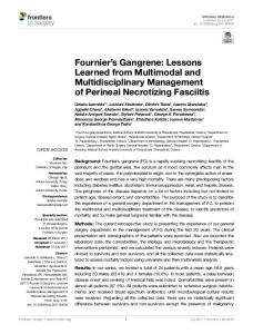
Fig. (2). Experimental workflow of top-down systems biology approaches in GSC research: The illustration shows the outline of commonly in GSC research applied top-down systems biology approaches. GSCs were mainly compared with NSCs, non-stem glioma cells, brain tumor or normal brain tissues. Depending on the biological questions high-throughput analyses differed regarding analytes and technical platforms used. A key role in top-down systems biology approaches is assigned to subsequent bioinformatics providing various outputs. PDB ID: 9ICD (Hurley et al. 1991 [28]).
- 6 -
4. EXPERIMENTAL DESIGN As previously stated, top-down systems biology analyses follow a dedicated workflow where the generation of high-throughput data comprises the starting point. (Fig. 2) delineates the current framework of top-down systems biology analysis in GSC research. For reasons of clarity and comprehensibility, only the most commonly conducted analyses are subsumed.
4.1. Experimental input. In order to shed light on GSC biology, mostly comparative analysis with other cell lines were performed. Here, GSCs were compared with neural stem cells (NSCs) [29-31], non-stem like glioma cells including differentiated GSCs [32,33], brain tumor tissues or normal brain tissue [31]. Particularly those genes which not only are differentially but also seem to be exclusively expressed by GSCs are of special interest for therapeutic developments [29,32,34]. Moreover, in some experiments, the heterogeneity among GSCs themselves was assessed [12,35,36].
4.2. High-throughput analyses. High-throughput analyses were conducted on different levels of biology as well as on various technical platforms (Fig. 2). Common high-throughput analytes were: DNA [37], mRNA [12,35], miRNA [29,31,33] and proteins [38]. Occasionally, additional “-omics” approaches such as glycoproteomics [39], phosphoproteomics [40, 41], and kinomics [42] were performed.
4.3. Experimental output. Most experiments resulted in the identification of differentially expressed genes, proteins etc. In some studies, a few of the top genes were further functionally validated in order to strengthen the obtained results, while others continued with in-depth descriptive in silico analysis of gene functions. The obtained findings were matched to gene ontologies [36,43], signal transduction networks [44] or protein-protein-interaction networks [43]. Open-access databases such as KEGG, the Gene Ontology, STRING or REACTOME have the advantage of being freely available for every researcher, whereas commercial databases such as Metacore or Ingenuity Pathway Analysis seem to offer considerably more disease-relevant and especially cancer-relevant pathways.
- 7 -
5. BIOLOGICAL QUESTIONS 5.1. Stem Cell Biology To study the characteristic features and the functional role of glioma stem cells by high-throughput analyses three central questions were addressed 1.) What are the similarities and differences between GSCs and their normal counterparts? 2.) How do GSCs differ from non-GSCs? 3.) Which changes occur when GSCs are treated? Accordingly, GSCs were compared on different molecular levels to NSCs, non-GSCs and GSCs treated in different ways. GSCs versus NSCs: Comparison of purified GSCs with NSCs has been performed in multiple studies to elaborate on the occurrence of stem cell-associated characteristics in GSCs and on the other hand to elucidate properties specific to tumor-initiating cells. Although the miRNA profile of glioma tissues was shown to be reminiscent of neural precursor cells [31], in a combined microarray and deep sequencing analysis on 3 GSC and 3 NSC lines, Lang et al. could identify a set of 10 differentially expressed miRNAs. In 3 independent GSC lines as well as in glioma tissues, 5 of them could be validated including upregulation of miR-10a, miR-10b, and miR-140 and downregulation of miR-124 and miR-874. Furthermore, downstream targets of miR-124 (the oncogenes NRAS and PIM3) and miR10a/b (the tumor suppressor gene CSMD1) were also validated at the mRNA level [45]. Another study described miR-138 to be GSC-specific as compared to NSC and allows the identification of bona fide tumor-initiating cells [29]. Moreover, Engstrom and coworkers identified a set of 29 genes that could clearly distinguish GSCs from NSCs using mRNA expression profiling [44]. On the protein level, Morfouace et al. applying a two-dimensional DIGE and MS/MS approach identified 44 differentially expressed proteins between rat GSCs and NSCs with glycolysis and pyruvate metabolism among the most significantly enriched pathways in GSC [30]. Subsequent in-vitro analyses confirmed GSCs to be completely dependent on glucose, which they metabolized through a non-oxidative pathway, whereas NSCs were capable of using alternative substrates. In a therapeutic setting the authors made use of this observation by treating GSC with dichloroacetate (DCA), a compound capable of shifting metabolism from anaerobic glycolysis to glucose oxidation via an inhibition of pyruvate dehydrogenase kinase. Accordingly, DCA had no effect on rat NSC but forced rat GSC into oxidative phosphorylation. Interestingly, this did not lead to apoptosis in these cancer cells. However,
- 8 -
when applied in combination with radiotherapy or etoposide, BAX-induced apoptosis was observed in vitro accompanied by a reduced proliferation in vivo suggesting DCA as a promising drug for clinical studies. GSCs versus non-GSCs: Discriminating non- or less-tumorigenic tumor cells from GSCs within a given tumor mass still poses a major challenge. As a source of non-GSCs several studies used retinoic acid (RA) or serum-differentiated GSCs. One of the most comprehensive analyses was performed by Ernst and colleagues [46]. They investigated the molecular mechanism underlying RA-induced differentiation at the mRNA, miRNA and DNA methylation level in 5 GSC lines. Besides the identification of 99 up- and 6 (>2-fold) downregulated genes, inhibition of TGFß/BMP, Wnt/ß-Cat and IGF pathways were observed upon differentiation. This could be linked to a decreased expression of the miR-17-92 cluster. Subsequent miR-17-92 inhibition experiments resulted in increased apoptosis and decreased proliferation and revealed connective tissue growth factor (CTGF) as one of the corresponding mRNA downstream targets. In accordance with these findings, DNA methylation analysis revealed CTGF demethylation upon differentiation. This integrative study identified CTGFmediated differentiation to be regulated on multiple levels suggesting an important role of CTGF in stem cell differentiation. Katsushima and colleagues also elaborated on differential miRNA signatures in 2 GSC lines as compared to their differentiated non-GSC counterpart [33]. Using RNA expression arrays they identified 13 up- and 34 downregulated miRNAs. By focusing on miRNAs, which have relevant targets closely associated with neural cell differentiation, a profound relationship was found between miR-1275 and Claudin11. MiR-1275 inhibited Claudin11 expression in GSC lines while antimiR-1275 treatment led to significant growth suppression suggesting a contribution of microRNA1275 to Claudin11 protein suppression in human GSCs. In another profiling approach performed by Yan and colleagues, differential expression was analyzed in freshly isolated FACS-sorted CD133positive and CD133-negative glioblastoma cells derived from the same patients [35]. In this analysis 89 up- and 125 downregulated genes constituted a CD133-related gene expression signature which resembled human embryonic stem cells, distinguished glioblastoma from low grade gliomas and identified an aggressive glioblastoma subtype preferentially seen in younger patients with excessive mutations. The authors interpreted their findings as molecular and genetic support for the stem-like
- 9 -
nature of CD133-positive glioma cells. In one of the few proteomics approaches Lu and colleagues focused on GSC-specific proteins by using a non-gel shotgun multidimensional liquid chromatography protein separation technique [47]. Comparison of one GSC line with and without RA treatment resulted in the identification of 175 differentially expressed proteins. Among those proteins, CNTFR showed a 3.7x higher expression in untreated GSCs and therefore was suggested as a marker for glioma-initiating cells. Furthermore, functional experiments revealed induction of differentiation upon treatment with CNTF indicating the relevance of the CNTFR pathway for differentiation of GSC. To identify differentiation-regulated glycoproteins He and coworkers analyzed HSR-GBM1 cells before and after differentiation using a multilectin affinity chromatography and quantitative glycoproteomics approach [34]. Three lectins including concanavalin A (Con A), wheat germ agglutinin (WGA), and peanut agglutinin (PNA) were used to capture glycoproteins, followed by LC-MS/MS analysis. A total of 73 and 79 high-confidence glycoproteins were identified in undifferentiated and differentiated cells, respectively. Label-free quantitation resulted in the discovery of 18 differentially expressed glycoproteins in GSC. Among the top candidates, upregulation was observed for PTPRZ1, PLOD1, P4HA1 and TN-C, while EGFR, Cathepsin D, and PSAP were downregulated in undifferentiated GSC. These glycoprotein markers might help to characterize the differentiation status of GSCs which may be useful in stem-cell targeted therapy of glioblastoma. In a similar study performed by the same group, HSR-GBM1 were compared to U373 cells, a serum-cultivated conventional cell line [41]. Here, GSCspecific expression of PTPRZ1 and TN-C could be confirmed. In addition, increased expression of the glycoproteins MSCP, PODXL, and CD90 in GSC was reported. Further analysis of these novel markers in other GSC lines as well as their functional validation should prove the suitability of these proteins in order to exclude a bias caused by the use of one single GSC line. GSCs Versus GSCs Treated with Signaling Molecule Inhibitors: Analyzing changes in the GSC phenotype upon treatment with inhibitors targeting relevant pathways for GSC maintenance might help to improve our understanding of GSC biology and the drug- induced molecular effects. Following this line of reasoning, He and coworkers studied the response of GSC11 glioblastoma stem cells to STAT3 phosphorylation inhibition by WP1193 [41]. In a descriptive glycotranscriptomics approach concentrating on changes in the mRNA expression of 359 glycogenes, increase of B3GAT2 transcripts
- 10 -
and decrease of some glycosphingolipid transcripts including UGCGL2, a glycosylceramide synthase was assessed. This analysis was complemented by determining multiple changes in the glycolipid and phospholipid profile. In parallel, Nilsson and coworkers assessed the effect of WP1193 on GSC11 glioblastoma stem cells in a comprehensive quantitative phosphoproteomic approach by employing phosphoprotein and -peptide enrichment followed by tandem MS [40]. This analysis generated a large and complex data set which needs to be validated in future studies on more GSC lines to ultimately decide the relevance of these findings for glioma stem cell biology. Based on the well-known importance of the NOTCH pathway for self-renewal of GSCs [48], Dai et al treated one GSC line with a gamma-secretase inhibitor (GSI) and performed a dose-dependent quantitative MS-based proteomic analysis [38]. Besides some potent anti-GSC effects such as reduced proliferation, increased differentiation, elevated apoptotic activity, and a phenotypic shift towards non-tumorigenic cells, a multilevel filtering strategy was developed to detect the most promising candidates. Thereby, an attenuated NFR2-mediated oxidative stress response and a downregulation of glycolysis were identified as consequences of GSI treatment. 5.2 GSC Hierarchy / Heterogeneity GSC hierarchy: A crucial criticism regarding the brain tumor stem cell hypothesis is the postulated hierarchical organization. Unlike leukemic stem cells, there are no reliable markers or marker combinations available for defining a distinct hierarchy of tumor cells within gliomas. However, just recently the cancer stem cell concept could be corroborated by Luis Parada’s group. In a genetically engineered mouse glioma model they demonstrated that regrowth in temozolomide-treated tumors was caused by a relatively quiescent subset of glioma stem cells which gave rise to a transient populations of highly proliferative cells [49]. Moreover, Heidi Phillip’s group provided evidence for a hierarchy of self-renewing tumor-initiating cells in GBM [14]. By extensive expression profiling of cultivated subclones from one PTEN-deficient GSC, they identified a linear hierarchy with decreasing selfrenewal capacities among different clones. Both studies would be an appropriate framework for deciphering the hierarchy underlying glioblastoma cells through further integrated high-throughput analyses.
- 11 -
GSC heterogeneity. There is accumulating evidence for the existence of different GSC phenotypes. For instance, Günther and coworkers employed an unsupervised analysis of the transcriptome of nine GSCs resulting in two distinct clusters. Cluster-1 was characterized by a stem-like phenotype, a high tumorigenicity and CD133 expression whereas cluster-2 displayed a restricted stem-like phenotype and low CD133 expression [36]. A similar subtyping was found by Lottaz et al, when comparing the transcriptome of 17 GSCs. Moreover, consensus clustering of the top 500 variable genes revealed a “proneural” signature for type I GSC and mesenchymal transcription profile for type II GSCs. In concordance with the study by Günther and coworkers, type I GSCs were CD133 positive and grew as neurospheres whereas type II GSCs displayed (semi-) adherent growth and lacked CD133 expression. Finally, they established a 24-gene signature distinguishing the two GSC types [12]. Of note, a marked influence of the tumor microenvironment on the GSC phenotype cannot be excluded. It has been shown that in vivo GSC plasticity is not only promoted by intrinsic signaling, but also essentially influenced by multiple interactions with stromal cells, immune cells, adjacent endothelial cells and the extracellular matrix in hypoxic or acidic tumor areas, which is often referred to as the GSC niche [recently reviewed in [50]]. 5.3. Therapeutic Targets The assumption that growth of primary and recurrent gliomas is caused by immature GSCs consequently implies the search for therapeutic targets in GSCs, which under the most optimal circumstances would spare normal stem cells. With the help of large scale screens this issue has been addressed in several ways: 1.) as a result of a comparative screen, 2.) by knockdown of a large number of molecules in parallel under the same conditions, or 3.) by “-omics” analyses after manipulation of single genes in GSCs. Comparative high-throughput screens: As mentioned earlier, comparison of GSCs with NSCs or nonGSCs by high-throughput technologies identified a couple of GSC-enriched if not GSC-specific molecules which could serve as targets. For some of them therapeutic suitability has already been investigated. For instance, sequence-specific functional inhibition of miR-138, which has been described before to be GSC-specific as compared to NSC has been shown to prevent tumor formation
- 12 -
in vitro and in vivo [29]. With miR-145, another miRNA was selected for the opposite behaviour. In this case CD133-positive and corresponding CD133-negative cells isolated from 5 glioblastoma tissues were studied in a combined miRNA/mRNA microarray analysis [51]. Noteworthy, miR-145 downstream targets include genes which are important for self- renewal such as Sox2 and Oct4. In vitro application of miR145 with cationic polyurethane-short branch PEI as a vehicle inhibited cancer stem-like properties and reduced chemo- and radioresistance of GSC. In addition, in vivo delivery of PU-PEI-miR145 alone was able to significantly suppress tumorigenesis and stemness as well as synergistically improved survival rate in combination with radio- and temozolomide-based chemotherapy and thereby presenting a promising new treatment option. Furthermore, Speranza and colleagues confirmed low expression of miR145 in GSCs and glioblastoma tissues as compared to low-grade glioma by microarray analysis and identified NEDD9 as a novel target of miR-145, which increases the invasiveness of glioblastomas [52]. However, before future use of miRNAs as therapeutic targets some major problems need to be solved including i) unknown further downstream targets and thus unwanted side effects when targeting specific miRs as well as ii) finding a suitable drug vector for targeted delivery to the tumor cells. High-throughput silencing approaches: Knockdown experiments have been widely used to identify therapeutic targets in GSC. Only few of them were performed in high-throughput formats. In one of these exceptional studies, Wurdak and colleagues targeted ~500 human kinases in altogether 5 GSC lines by RNAi. With respect to 50 kinases, silencing resulted in GSC differentiation and could be validated in 5 independent GSC lines. The strongest differentiation effect with the highest reproducibility in GSCs was found after knockdown of TRRAP. Silencing of TRRAP decreased selfrenewal and proliferation of GSC and increased their sensitivity to apoptosis while negatively affecting cell cycle progression. This not only implies a critical role for TRRAP in maintaining a tumorigenic stem-like state but mandates its further validation as a therapeutic target. The complete kinome and also the complete phosphatome were addressed in a comprehensive RNAi screen performed by Goidts and coworkers to identify kinases and phosphatases essential for survival of GSCs [53]. GSCs were transduced with lentiviral vector-based shRNA silencing 682 kinases and 180 phosphatases. Subsequently, the most promising candidates were validated in 3 independent GSC
- 13 -
lines. Altogether, 46 candidates including a number of genes involved in metabolism, especially glycolysis such as PDK1, PKM2 and PFKFB4 significantly influenced GSC survival. Functional studies confirmed an essential role of PFKFB4 for stem cell maintenance. Corroborating these findings, high PFKFB4 expression in tumor tissues was related to a shorter survival of the corresponding glioblastoma patients. These observations underline the importance of the glycolytic pathway in the maintenance of GSCs and imply tumor metabolism as a promising therapeutic target in glioblastoma. However, due to the well-known tumor heterogeneity and the occurrence of clonal evolution upon targeted therapy, blocking of a single pathway is doomed to fail. Comparative “-omics” of genetically manipulated cells: Al- though an increasing number of studies use high-throughput analyses to characterize cells after loss- or gain-of function experiments, data on GSCs are still rare. In a thorough study, Kamal and colleagues analyzed the functional role of the repressor element 1 silencing transcription factor (REST) which is a transcriptional repressor of many neuronal differentiation genes [54]. Loss- and gain-of-function experiments on GSC indicated REST to maintain self-renewal and resulted in histopathologically distinct tumors displaying an increased invasiveness, growth and impaired apoptotic properties. Genome-wide mRNA expression analysis of REST-overexpressing and REST-silenced cells identified 301 genes that were more than 1.5 fold overexpressed. Ingenuity® pathway analysis attributed these genes to pathways involved in cellular movement, cellular growth and invasion proposing REST as another promising target molecule which deserves further investigation.
6) IDENTIFICATION OF BIOMARKERS Following the assumption that GSCs account for an increased therapy resistance and ultimately tumor regrowth [55], identifying GSC-associated genes is likely to yield prognostic biomarkers in glioblastoma patients. Most frequently, comparative high-throughput studies in GSCs were based on mRNA microarrays. When comparing 20 GSCs with NSCs and normal brain tissues a set of differentially expressed genes (AJAP1, EMP3, PDPN) could be verified to be prognostic on the protein level with the help of tissue microarrays [37]. A very recent study conducted high-throughput
- 14 -
sequencing of transcript tags (Tag-seq) on 3 adherent GSC and 2 NSC lines. The resulting differentially expressed genes were analyzed regarding enriched pathways and validated on a larger panel of GSC and NSC lines with quantitative PCR, leading to a set of 29 genes distinguishing GSCs from NSCs. Both, this GSC expression signature and individual up- (DDIT3, HOXD10, PDE1C and PLS3) and downregulated (PTEN and TUSC3) genes showed prognostic value in a variety of public data sets. In the very same study, the influence of copy number alterations on the GSC transcriptome was assessed conducting comparative genome hybridization (CGH). Integrating the CGH and transcriptome data revealed a distinct correlation [44]. Noteworthy, the open access to a growing number of public databases does not only allow validation of genes of interest but is used more and more to expand hypothesis-driven comparative analysis. For instance Ng and coworkers derived a gene signature distinguishing oligodendroglial from glioblastoma GSCs by combining their own small data set (n=6) with publically available data from two other studies (n=14). They obtained an oligodendroglial GSC signature which was significantly associated with patient survival (REMBRANDT: p value = 1.93E-05; Gravendeel p value = 0.0082) [56]. miRNA profiling of GSCs constitutes another emerging research field. Alterations in the expression of miRNAs may play a crucial role in cancer initiation, progression and metastasis [57]. Here, an impaired miRNA expression could also be identified to markedly contribute to brain tumor pathogenesis [58,59]. A recent comparative miRNA profiling of GSCs and NSCs revealed strikingly similar profiles with the exception of miR-138, which was significantly upregulated in GSCs. Consequent functional experiments assigned miR-138 a vital role in promoting glioma growth. Furthermore, loss-of-function gene expression analysis depicted its regulatory network. Finally, the prognostic value of miR-138 was assessed using TCGA miRNA data (n=197; p < 0.05) [29]. High-throughput analyses on the protein level leading to disease-relevant biomarkers are very rare. In one of the few studies, the proteome of untreated GSCs was compared to one CNTF-treated and thereby differentiated GSC line. Using a new multidimensional capillary isoelectric focusing nanoreversed-phase liquid chromatography platform with tandem MS Xu and co-workers could identify neuronatin (NNAT) to be exclusively expressed in GSCs. In a validation study sample of 59 GBM
- 15 -
patients, NNAT constituted an independent risk factor (p = 0.006) for decreased patient survival [32].
7. CELL OF ORIGIN Soon after the discovery of GSCs, it was proposed that these cells might originate from NSCs for several reasons: similar to NSCs, GSCs are not only cultivated in the same serum-free media as neurospheres, but are able to self-renew, and to express astrocytic, neuronal, as well as oligodendroglial lineage markers upon differentiation [8]. Although data from experimental mouse models support this notion [49,60] very recent data from Inder Verma’s and Heidi Phillips group indicate that gliomas can not only arise from NSC, but also from lineage-committed neural precursor cells, astrocytes, and even mature neurons [61,62]. What complicates the search for the cell of origin even more are recent profiling data suggesting several subtypes of glioblastoma which might be maintained by different types of GSCs and might therefore be derived from different cells of origin [63]. When addressing this question by performing high-throughput analyses on GSCs, miRNA profiling of 2 GSC lines revealed a miRNA profile reminiscent of neural precursor cells [31]. In a more comprehensive approach 9 GSC lines were profiled and yielded 2 distinct clusters with differing growth behavior and stem cell marker expression indicating different types of founder cells [36]. These findings could be substantiated by Lottaz and colleagues who compared transcriptional profiles of 17 GSC lines with several cultures of fetal NSC (fNSC), adult NSC (aNSC), astrocytes, and neurons [12]. Again GSC profiles could be assigned to two clusters. While GSC in cluster one displayed a proneural signature and resembled fNSC, GSC lines in cluster two exhibited a mesenchymal signature and expression profiles similar to aNSC. Moreover, specifically in the latter components of the TGFß/BMP signaling pathway and ECM-related molecules were upregulated. Interestingly, no differences in stemness pathways were observed between these two groups of GSC. From these data the authors conclude that glioblastoma may derive from different cells that have preserved or acquired properties of either fNSC or aNSC but lost the corresponding differentiation potential. Since these analyses were only able to recapitulate the proneural and the mesenchymal glioblastoma subtypes future work is not only needed to confirm these initial findings but also to learn more about the putative cell of origin of the other glioblastoma subtypes.
- 16 -
CONCLUSION Due to recent developments in molecular biology, researchers have been able to address scientific questions in a growing number of large scale formats. This does not only enable us to obtain a comprehensive view on a single molecular level, but also allows for the integration of data sets obtained from RNA, DNA, protein and metabolite analyses. Moreover, increasing numbers of public available data sets like TCGA, Rembrandt and others are powerful tools to perform in silico validation and to generate new hypotheses. It is apparent that such systems biology approaches are multidisciplinary research efforts, demanding both specialized research facilities and the cooperative work from experts in different disciplines. Although data integration of different molecular levels is still at the very beginning, high-throughput analyses of GSCs improved our understanding of the complex cellular processes involved in tumor biology in several aspects. Assessment of differences in expression profiles when comparing GSCs to their normal or less tumorigenic counterparts or different types of tissues led to the identification of tumor-relevant pathways, new survival-associated biomarkers, novel therapeutic targets and shed light on the intratumoral organization and the putative cell of origin. However, since our current GSC data do not mirror all glioblastoma subtypes and because most of the analyses were performed in only a few GSC lines, future work is needed to enlarge the number of GSC-associated data sets to compensate for these limitations. Also, most of the available “-omics” analyses were performed by mRNA and miRNA expression profiling and even less frequently by proteomics. In contrast, there is almost no knowledge about the contribution of DNA methylation, metabolism and fluxomics to GSC maintenance and behavior. Hopefully, improvement of large scale technologies and the expanding field of systems biology will help to fill these gaps in order to improve our therapeutic strategies against one of the deadliest human cancers.
CONFLICT OF INTEREST The authors confirm that this article content has no conflicts of interest.
- 17 -
ACKNOWLEDGEMENTS We thank Thomas M. Kaffenberger, Christoph Geisenberger, Dr. Christine Jungk, Steffen Dettling for critically reading the manuscript and Prof. Dr. Wolfgang Wiechert for inspiring introductions to the field of systems biology.
REFERENCES 1.
Visvader JE, Lindeman GJ. Cancer stem cells: current status and evolving complexities. Cell Stem Cell. 2012 Jun 14;10(6):717–28.
2.
Lapidot TT, Sirard CC, Vormoor JJ, Murdoch BB, Hoang TT, Caceres-Cortes JJ, et al. A cell initiating human acute myeloid leukaemia after transplantation into SCID mice. Nature. 1994 Feb 16;367(6464):645–8.
3.
Bonnet DD, Dick JEJ. Human acute myeloid leukemia is organized as a hierarchy that originates from a primitive hematopoietic cell. Nat Med. 1997 Jun 30;3(7):730–7.
4.
Eramo AA, Lotti FF, Sette GG, Pilozzi EE, Biffoni MM, Di Virgilio AA, et al. Identification and expansion of the tumorigenic lung cancer stem cell population. Cell Death Differ. 2008 Feb 29;15(3):504–14.
5.
Al-Hajj M, Wicha MS, Benito-Hernandez A, Morrison SJ, Clarke MF. Prospective identification of tumorigenic breast cancer cells. Proc. Natl. Acad. Sci. U.S.A. 2003 Apr 1;100(7):3983–8.
6.
Ricci-Vitiani L, Lombardi DG, Pilozzi E, Biffoni M, Todaro M, Peschle C, et al. Identification and expansion of human colon-cancer-initiating cells. Nature. 2007 Jan 4;445(7123):111–5.
7.
Prince ME, Sivanandan R, Kaczorowski A, Wolf GT, Kaplan MJ, Dalerba P, et al. Identification of a subpopulation of cells with cancer stem cell properties in head and neck squamous cell carcinoma. Proc. Natl. Acad. Sci. U.S.A. 2007 Jan 16;104(3):973–8.
8.
Singh SK, Clarke ID, Terasaki M, Bonn VE, Hawkins C, Squire J, et al. Identification of a cancer stem cell in human brain tumors. Cancer Res. 2003 Sep 15;63(18):5821–8.
9.
Singh SK, Hawkins C, Clarke ID, Squire JA, Bayani J, Hide T, et al. Identification of human brain tumour initiating cells. Nature. 2004 Nov 18;432(7015):396–401.
10.
Collins ATA, Berry PAP, Hyde CC, Stower MJM, Maitland NJN. Prospective identification of tumorigenic prostate cancer stem cells. Cancer Res. 2005 Nov 30;65(23):10946–51.
11.
O'Brien CA, Pollett A, Gallinger S, Dick JE. A human colon cancer cell capable of initiating tumour growth in immunodeficient mice. Nature. 2007 Jan 3;445(7123):106–10.
12.
Lottaz C, Beier D, Meyer K, Kumar P, Hermann A, Schwarz J, et al. Transcriptional Profiles of CD133+ and CD133- Glioblastoma-Derived Cancer Stem Cell Lines Suggest Different Cells of Origin. Cancer Res. 2010 Feb 8;70(5):2030–40.
13.
Campos B, Herold-Mende CC. Insight into the complex regulation of CD133 in glioma. Int J Cancer. 2011 Jan 31;128(3):501–10.
14.
Chen R, Nishimura MC, Bumbaca SM, Kharbanda S, Forrest WF, Kasman IM, et al. A Hierarchy of Self-Renewing Tumor-Initiating Cell Types in Glioblastoma. Cancer Cell. 2010 Apr 12;17(4):14–4.
- 18 -
15.
Schepers AGA, Snippert HJH, Stange DED, van den Born MM, van Es JHJ, van de Wetering MM, et al. Lineage tracing reveals Lgr5+ stem cell activity in mouse intestinal adenomas. Science. 2012 Aug 9;337(6095):730–5.
16.
Driessens GG, Beck BB, Caauwe AA, Simons BDB, Blanpain CC. Defining the mode of tumour growth by clonal analysis. Nature. 2012 Aug 22;488(7412):527–30.
17.
Campos BB, Zeng LL, Daotrong PHP, Eckstein VV, Unterberg AA, Mairbäurl HH, et al. Expression and regulation of AC133 and CD133 in glioblastoma. Glia. 2011 Nov 30;59(12):1974–86.
18.
Palsson BO. Systems Biology. Cambridge University Press; 2006.
19.
Blattner FRF, Plunkett GG, Bloch CAC, Perna NTN, Burland VV, Riley MM, et al. The complete genome sequence of Escherichia coli K-12. Science. 1997 Sep 4;277(5331):1453–62.
20.
Goffeau A, Aert R, Agostini-Carbone ML, Ahmed A. The yeast genome directory. Nature. 1997.
21.
C. elegans Sequencing Consortium. Genome sequence of the nematode C. elegans: a platform for investigating biology. Science. 1998 Dec 11;282(5396):2012–8.
22.
Adams MD, Celniker SE, Holt RA, Evans CA, Gocayne JD, Amanatides PG, et al. The genome sequence of Drosophila melanogaster. Science. 2000 Mar 23;287(5461):2185–95.
23.
Lander ESE, Linton LML, Birren BB, Nusbaum CC, Zody MCM, Baldwin JJ, et al. Initial sequencing and analysis of the human genome. Nature. 2001 Feb 14;409(6822):860–921.
24.
Kitano H. Perspectives on systems biology. NGCO. 2000 Aug 31;18(3):199–216.
25.
Aebersold R, Hood LE, Watts JD. Equipping scientists for the new biology. Nat. Biotechnol. 2000 Apr;18(4):359.
26.
Ge H, Walhout AJM, Vidal M. Integrating “omic” information: a bridge between genomics and systems biology. Trends Genet. 2003 Oct;19(10):551–60.
27.
Kitano H. Foundations of systems biology. 2001.
28.
Hurley JHJ, Dean AMA, Koshland DED, Stroud RMR. Catalytic mechanism of NADP(+)-dependent isocitrate dehydrogenase: implications from the structures of magnesium-isocitrate and NADP+ complexes. Biochemistry. 1991 Sep 2;30(35):8671–8.
29.
Chan XHDX, Nama SS, Gopal FF, Rizk PP, Ramasamy SS, Sundaram GG, et al. Targeting Glioma Stem Cells by Functional Inhibition of a Prosurvival OncomiR-138 in Malignant Gliomas. Cell Rep. 2012 Sep 26;2(3):591–602.
30.
Morfouace MM, Lalier LL, Bahut MM, Bonnamain VV, Naveilhan PP, Guette CC, et al. Comparison of spheroids formed by rat glioma stem cells and neural stem cells reveals differences in glucose metabolism and promising therapeutic applications. J Biol Chem. 2012 Sep 27;287(40):33664–74.
31.
Lavon I, Zrihan D, Granit A, Einstein O, Fainstein N, Cohen MA, et al. Gliomas display a microRNA expression profile reminiscent of neural precursor cells. Neuro Oncol. 2010 Apr 30;12(5):422–33.
32.
Xu DS, Yang C, Proescholdt M, Bründl E, Brawanski A. Neuronatin in a Subset of Glioblastoma Multiforme Tumor Progenitor Cells Is Associated with Increased Cell Proliferation and Shorter Patient Survival. PLoS One. 2012.
33.
Katsushima KK, Shinjo KK, Natsume AA, Ohka FF, Fujii MM, Osada HH, et al. Contribution of microRNA-1275 to Claudin11 protein suppression via a polycomb-mediated silencing mechanism in human glioma stem-like cells. J Biol Chem. 2012 Aug 9;287(33):27396–406.
34.
He J, Liu Y, Zhu TS, Xie X, Costello MA, Talsma CE, et al. Glycoproteomic analysis of glioblastoma stem cell differentiation. J Proteome Res. 2011 Jan 7;10(1):330–8.
- 19 -
35.
Yan X, Ma L, Yi D, Yoon J, Diercks A. A CD133-related gene expression signature identifies an aggressive glioblastoma subtype with excessive mutations. 2011.
36.
Günther HS, Schmidt NO, Phillips HS, Kemming D, Kharbanda S, Soriano R, et al. Glioblastomaderived stem cell-enriched cultures form distinct subgroups according to molecular and phenotypic criteria. Oncogene. 2008 Apr 30;27(20):2897–909.
37.
Ernst A, Hofmann S, Ahmadi R, Becker N, Korshunov A, Engel F, et al. Genomic and expression profiling of glioblastoma stem cell-like spheroid cultures identifies novel tumor-relevant genes associated with survival. Clin Cancer Res. 2009 Oct 31;15(21):6541–50.
38.
Dai L, He J, Liu Y, Byun J, Vivekanandan A, Pennathur S, et al. Dose-dependent proteomic analysis of glioblastoma cancer stem cells upon treatment with γ-secretase inhibitor. Proteomics. 2011 Nov 30;11(23):4529–40.
39.
He JJ, Liu YY, Xie XX, Zhu TT, Soules MM, DiMeco FF, et al. Identification of cell surface glycoprotein markers for glioblastoma-derived stem-like cells using a lectin microarray and LC-MS/MS approach. Audio, Transactions of the IRE Professional Group on. 2010 May 6;9(5):2565–72.
40.
Nilsson CL, Dillon R, Devakumar A, Shi SD-H, Greig M, Rogers JC, et al. Quantitative Phosphoproteomic Analysis of the STAT3/IL-6/HIF1α Signaling Network: An Initial Study in GSC11 Glioblastoma Stem Cells. J Proteome Res. 2010 Jan 3;9(1):430–43.
41.
He H, Nilsson CL, Emmett MR, Marshall AG, Kroes RA, Moskal JR, et al. Glycomic and Transcriptomic Response of GSC11 Glioblastoma Stem Cells to STAT3 Phosphorylation Inhibition and Serum-Induced Differentiation. J Proteome Res. 2010 May 6;9(5):2098–108.
42.
Wurdak H, Zhu S, Romero A, Lorger M, Watson J, Chiang C-Y, et al. An RNAi Screen Identifies TRRAP as a Regulator of Brain Tumor-Initiating Cell Differentiation. Cell Stem Cell. 2010 Jan 7;6(1):11–1.
43.
Garcia JL, Perez-Caro M, Gomez-Moreta JA. Molecular analysis of ex-vivo CD133+ GBM cells revealed a common invasive and angiogenic profile but different proliferative signatures among high grade …. BMC …. 2010.
44.
Engstrom PG, Tommei D, Stricker SH, Ender C, Pollard SM, Bertone P. Digital transcriptome profiling of normal and glioblastoma-derived neural stem cells identifies genes associated with patient survival. Genome Med. 2012 Oct 8;4(10):76–6.
45.
Lang M-F, Yang S, Zhao C, Sun G, Murai K, Wu X, et al. Genome-wide profiling identified a set of miRNAs that are differentially expressed in glioblastoma stem cells and normal neural stem cells. PLoS One. 2012;7(4):e36248–8.
46.
Ernst A, Campos B, Meier J, Devens F, Liesenberg F, Wolter M, et al. De-repression of CTGF via the miR-17-92 cluster upon differentiation of human glioblastoma spheroid cultures. Oncogene. 2010 Mar 21;29(23):3411–22.
47.
Lu J, Ksendzovsky A, Yang C, Mehta GU, Yong RL, Weil RJ, et al. CNTF receptor subunit α as a marker for glioma tumor-initiating cells and tumor grade. J. Neurosurg. 2012 Dec;117(6):1022–31.
48.
Fan X, Khaki L, Zhu TS, Soules ME, Talsma CE, Gul N, et al. NOTCH pathway blockade depletes CD133-positive glioblastoma cells and inhibits growth of tumor neurospheres and xenografts. Stem Cells. 2010 Jan;28(1):5–16.
49.
Chen J, Li Y, Yu T-S, McKay RM, Burns DK, Kernie SG, et al. A restricted cell population propagates glioblastoma growth after chemotherapy. Nature. 2012 Aug 23;488(7412):522–6.
50.
Charles NA, Holland EC, Gilbertson R, Glass R, Kettenmann H. The brain tumor microenvironment. Glia. 2012 Mar;60(3):502–14.
- 20 -
51.
Yang Y-P, Chien Y, Chiou G-Y, Cherng J-Y, Wang M-L, Lo W-L, et al. Inhibition of cancer stem celllike properties and reduced chemoradioresistance of glioblastoma using microRNA145 with cationic polyurethane-short branch PEI. Biomaterials. 2012 Jan 31;33(5):15–5.
52.
Speranza MCM, Frattini VV, Pisati FF, Kapetis DD, Porrati PP, Eoli MM, et al. NEDD9, a novel target of miR-145, increases the invasiveness of glioblastoma. Oncotarget. 2012 Jun 30;3(7):723–34.
53.
Goidts V, Bageritz J, Puccio L, Nakata S, Zapatka M, Barbus S, et al. RNAi screening in glioma stemlike cells identifies PFKFB4 as a key molecule important for cancer cell survival. Oncogene. 2012 Jul 5;31(27):3235–43.
54.
Ballas N, Grunseich C, Lu DD, Speh JC, Mandel G. REST and its corepressors mediate plasticity of neuronal gene chromatin throughout neurogenesis. Cell. 2005.
55.
Bao S, Wu Q, McLendon RE, Hao Y, Shi Q, Hjelmeland AB, et al. Glioma stem cells promote radioresistance by preferential activation of the DNA damage response. Nature. 2006 Dec 6;444(7120):756–60.
56.
Ng FS-LF, Toh TBT, Ting EH-LE, Koh GR-HG, Sandanaraj EE, Phong MM, et al. Progenitor-like traits contribute to patient survival and prognosis in oligodendroglial tumors. Clin Cancer Res. 2012 Jul 31;18(15):4122–35.
57.
Calin GAG, Croce CMC. MicroRNA signatures in human cancers. Nat Rev Cancer. 2006 Oct 31;6(11):857–66.
58.
Bottoni A, Piccin D, Tagliati F, Luchin A, Zatelli MC, Uberti ECD. miR-15a and miR-16-1 downregulation in pituitary adenomas. J Cell Physiol. 2005 Jun 30;204(1):280–5.
59.
Chan JAJ, Krichevsky AMA, Kosik KSK. MicroRNA-21 is an antiapoptotic factor in human glioblastoma cells. Cancer Res. 2005 Jul 14;65(14):6029–33.
60.
Alcantara Llaguno S, Chen J, Kwon C-H, Jackson EL, Li Y, Burns DK, et al. Malignant astrocytomas originate from neural stem/progenitor cells in a somatic tumor suppressor mouse model. Cancer Cell. 2009 Jan 6;15(1):45–56.
61.
Friedmann-Morvinski D, Bushong EA, Ke E, Soda Y, Marumoto T, Singer O, et al. Dedifferentiation of neurons and astrocytes by oncogenes can induce gliomas in mice. Science. 2012 Nov 23;338(6110):1080–4.
62.
Lai AA, Kharbanda SS, Pope WBW, Tran AA, Solis OEO, Peale FF, et al. Evidence for sequenced molecular evolution of IDH1 mutant glioblastoma from a distinct cell of origin. J Clin Oncol. 2011 Nov 30;29(34):4482–90.
63.
Phillips HS, Kharbanda S, Chen R, Forrest WF, Soriano RH, Wu TD, et al. Molecular subclasses of high-grade glioma predict prognosis, delineate a pattern of disease progression, and resemble stages in neurogenesis. Cancer Cell. 2006 Mar;9(3):157–73.
- 21 -