Eps15-homology (EH) domains are protein-interaction domains of about 100 amino acids found in a large variety of species ranging from nematodes to mam-.
Journal of Biomolecular NMR, 14: 97–98, 1999. KLUWER/ESCOM © 1999 Kluwer Academic Publishers. Printed in the Netherlands.
97
Letter to the Editor: Sequence-specific 1 H, 13C and 15N assignment and secondary structure of the apo EH2 domain of mouse Eps15 Alfonso Carotenutoa,∗ , Marco Tessaria , Brian Whiteheada , Jan M.A. Aelena , Paul M.P. van Bergen en Henegouwenb & Geerten W. Vuistera,∗∗ a Nijmegen
SON Research Center, Department of Biophysical Chemistry, University of Nijmegen, Toernooiveld, 6525 ED Nijmegen, The Netherlands b Department of Molecular Cell Biology, Institute of Biomembranes, University of Utrecht, Padualaan 8, 3584 CH Utrecht, The Netherlands Received 4 January 1999; Accepted 26 February 1999
Key words: Ca2+ -binding protein, EH domain, NMR assignments, secondary structure Biological context
Methods and results
Eps15-homology (EH) domains are protein-interaction domains of about 100 amino acids found in a large variety of species ranging from nematodes to mammals (Wong et al., 1995). EH domains are frequently present in multiple copies and were shown to bind NPF or F/WF containing peptides (Paoluzi et al., 1998). EH-domain containing proteins are thought to function in the sorting and routing steps of important biological processes such as neuronal development, synaptic transmission, and endocytosis (Salcini et al., 1997). Eps15 is involved in the endocytosis of the activated EGF-receptor (Benmerah et al., 1995) and contains three EH domains. Previous studies on the apo EH1 domain of mouse (mEH1) (Whitehead et al., 1998; Whitehead et al., submitted) and the Ca2+ complexed form of the human EH2 domain (hEH2) (de Beer et al., 1998) showed that both polypeptides contained two EF-hand motifs connected by a short antiparallel β-sheet. In addition, mEH1 contained an additional N- and C-terminal helix and a long structured loop. Here, we present near complete 1 H, 13 C, and 15 N assignments and the secondary structure for the apo EH2 domain of mouse Eps15 (mEH2).
Escherichia coli K10 cells, transformed with the pGex-2T expression vector (Pharmacia, Uppsala, Sweden) containing the EH2 domain from mouse Eps15 (residues 119–221, (Fazioli et al., 1993) and two additional N-terminal residues resulting from the cloning procedure) were grown at 30 ◦ C. Purification of the EH2, which was expressed as a GSTfusion protein, was performed as described previously (Whitehead et al., 1998). The yield was 5 mg L−1 in minimal media. Protein samples were dialysed against 40 mM EDTA in Tris buffer (20 mM, pH 7.1), followed by dialysis against pure H2 O, lyophilisation, and dissolving in 350 µl acetate-buffered saline solution (95/5% H2 O/D2 O, 50 mM CD3 COONa, 100 mM NaCl, pH 5.0, 2.5 mM deuterated DDT, 5 mM NaN3 ) to a final concentration of ∼1 mM. Using this protocol, approximately 90% of the protein was in the apo state. Water flip-back, pulsed-field gradient, and sensitivity-enhanced versions of the 3D HNCO, 3D HNCA, 3D C(CO)NH and 3D 15 N-separated NOESY-HSQC and HMQC-NOESY-HSQC experiments were used for assignments of the backbone resonances. NMR data were acquired at 15 ◦ C on Bruker DRX600, Varian Unity Inova 500 and Varian Unity Inova 750 spectrometers. The program NMRPipe (Delaglio et al., 1995) was used for transformation of all data. The 2D 1 H/15 N HSQC spectrum of EH2 allowed for the production of a list of 1 H/15 N shift pairs for analysis of the 3D spectra in the program XEASY (Bartels et al., 1995). Using the commonly
∗ On leave from: Facolta di Farmicia, Universita di Salerno, Piazza Vittorio Emanuele 20, Penta di Fisciano (Salerno), Italy. ∗∗ To whom correspondence should be addressed.
98 120
125
130
135
140
145
150
155
160
165
170
175
180
185
190
195
200
205
210
215
220
S E L PWA VK S E DKAKY D A I F D S L S P V DG F L S GDKVK P V L L N S K L P V E I L GR VWE L S D I DHDGK L D R D E F A V AMF L V Y C A L E K E P V PMS L P P A L V P P S KRK TWV V S
dαN dNN dαN(i,i+3) dαN(i,i+4) dαβ(i,i+3) 1 0 -1 4 13 0 ∆ Cα(ppm) -4 4 13 0 ∆ Cβ(ppm) -4 ∆Hα(ppm)
doubling α1
β1
α2
α3
β2
α4
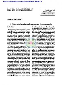
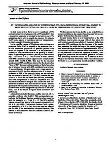
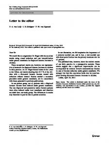
Figure 1. Summary of the sequential and medium-range NOEs, and deviations from random coil chemical shifts for 1 Hα , 13 Cα , and 13 Cβ of the apo EH2 domain of mouse Eps15. Secondary structural elements as occur in the Ca2+ -complexed form of hEH2 (de Beer et al., 1998) are also indicated (solid). Helix 2 appears to be extended by two residues in apo mEH2 (dashed). Stars denote residues subject to multiple (2–4) conformations in slow chemical exchange (see also text).
followed protocol, sequential assignments were obtained by connecting the shift pairs by means of the HNCA, CBCA(CO)NH and 15 N-separated NOESYHSQC data and mapping unique connected fragments onto the primary sequence by means of the Cα and Cβ chemical shift information in conjunction with the 1 H spin-system topologies. Assignments of the non-aromatic side chains were obtained using 3D HC(C)H- and (H)CCH-TOCSY and C(CO)NH experiments. Aromatic resonances were assigned using 13 C-HSQC, HCCH-TOCSY, and 15 N-filtered 2D NOESY and TOCSY spectra. Figure 1 shows a summary of the sequential and medium-range NOEs, and deviations from random coil chemical shifts for 1 Hα , 13 Cα , and 13 Cβ . Analysis of these results indicates the presence of four α-helices (residues 126–138, 148–156, 162–172, 182– 197) which are also indicated. The helices largely coincide with those observed in the Ca2+ -complexed hEH2 (de Beer et al., 1998), albeit that helix 1 appears to be two residues longer. As in hEH2, a small antiparallel β-sheet is present in apo mEH2, comprising residues 145–147 and 179–181. In all, the data indicate that, upon binding Ca2+ , the secondary structure remains conserved. In the course of 1 to 4 weeks, several residues (indicated by stars) experience a severe broadening or even multiple (2–4) conformations in slow chemical exchange. Most likely, this results from a non-specific oligomerization, as the process is accompanied by an overall increase in 15 N T2 . The effect can be partially reversed by addition of DTT, implicating disulfide formation by the solvent-exposed Cys194 residue.
Extent of assignments and data deposition Backbone and side-chain 1 H, 15 N, and 13 C backbone resonances were assigned except for resonances of backbone nitrogens of prolines, carbonyl resonances of residues preceding Pro, and side-chain resonances of Arg and Lys beyond Cδ and Cε , respectively. The assignments for the apo EH2 domain from mouse Eps15 at pH 5.0 and 15 ◦ C have been deposited in the BioMagResBank (accession number 4288). Acknowledgements This research was supported by the Netherlands Foundation for Chemical Research (CW) with financial assistance from the Netherlands Organisation for Scientific Research (NWO). References Bartels, C., Xia, T.H., Billeter, M., Guntert, P. and Wüthrich, K. (1995) J. Biomol. NMR, 6, 1–10. Benmerah, A., Gagnon, J., Begue, B., Megarbane, B., DautryVarsat, A. and Cerf-Bensussan, N. (1995) J Cell Biol., 131, 1831–1838. de Beer, T., Carter, R.E., Lobel-Rice, K.E., Sorkin, A. and Overduin, M. (1998) Science, 281, 1357–1360. Delaglio, F., Grzesiek, S., Vuister, G.W., Zhu, G., Pfeifer, J. and Bax, A. (1995) J. Biomol. NMR, 6, 277–293. Fazioli, F., Minichiello, L., Matoskova, B., Wong, W.T. and Di Fiore, P.P. (1993) Mol. Cell Biol., 13, 5814–5828. Paoluzi, S., Castagnoli, L., Lauro, I., Salcini, A.E., Coda, L., Fre’, S., Confalonieri, S., Pelicci, P.G., Paolo, D.F. and Cesareni, G. (1998) EMBO J., 17, 6541–6550. Salcini, A.E., Confalonieri, S., Doria, M., Santolini, E., Tassi, E., Minenkova, O., Cesareni, G., Pelicci, P.G. and Di Fiore, P.P. (1997) Genes Dev., 11, 2239–2249. Whitehead, B., Tessari, M., Versteeg, H.H., van Delft, S., van Bergen en Henegouwen, P.M.P. and Vuister, G.W. (1998) J. Biomol. NMR, 12, 465–466. Wong, W.T., Schumacher, C., Salcini, A.E., Romano, A., Castagnino, P., Pelicci, P.G. and Di Fiore, P. (1995) Proc. Natl. Acad. Sci. USA, 92, 9530–9534.