Leukemia
Molgó, et al., J Leuk 2015, 3:4 http://dx.doi.org/10.4172/2329-6917.1000197
Case Report
Open access
Radiotherapy in Cutaneous B-Cell Lymphomas Treatment: Report of Three Cases Montserrat Molgó1, Josefina Rodríguez2, Camila Arriagada2*, Eugenio Vines3, Félix Fich1, Katherine Droppelmann1 and Sergio González4 1Department 2Resident
of Dermatology, School of Medicine, Pontificia Universidad Católica de Chile, Chile
of Dermatology, School of Medicine, Pontificia Universidad Católica de Chile, Chile
3Department
of Hematology, School of Medicine, Pontificia Universidad Católica de Chile, Chile
4Department
of Anatomic Pathology, School of Medicine, Pontificia Universidad Católica de Chile, Chile
*Corresponding
author: Camila Arriagada, Resident of Dermatology, School of Medicine, Pontificia Universidad Católica de Chile, Chile, Tel: 562-23548622; E-mail:
[email protected] Received date: October 28, 2015; Accepted date: December 07 2015; Published date: December 12, 2015 Copyright: © 2015 Montserrat M, et al. This is an open-access article distributed under the terms of the Creative Commons Attribution License, which permits unrestricted use, distribution, and reproduction in any medium, provided the original author and source are credited.
Abstract Introduction: Primary cutaneous lymphomas (PCL) are a heterogeneous group of extra nodal non-Hodgkin lymphomas defined as malignant tumor derived from B, T or natural killer cells. Primary cutaneous follicle centre lymphoma (PCFCL), represents the most common type of primary cutaneous B- Cell Lymphomas. Case Report:We present the case of three female patients with facial PCFCL, all of them with excellent respond to radiotherapy. Discussion: Multiple treatments for PCFCL have been described, including systemic or intralesional Rituximab, surgery or radiotherapy. We present these 3 cases of PCFLC treated with radiotherapy in our service, because of the excellent response, confirming radiotherapy as a therapeutic alternative.
Keywords: Cutaneous B-cell lymphomas; Primary cutaneous follicle centre lymphoma; Radiotherapy
Introduction Primary cutaneous lymphomas (PCL) are a heterogeneous group of extra nodal non-Hodgkin lymphomas defined as malignant tumor derived from B, T or natural killer cells. PLC present manifestations only in the skin without involving another locations at the moment of diagnosis [1]. PCL represent the second most common extranodal lymphoma location after primary gastrointestinal lymphoma [2]. Approximately 25% of PCL are type B- Cell Lymphomas. According to the latest classification these are divided into 3 groups (WHO EORTC): Primary cutaneous follicle centre lymphoma (PCFCL), primary cutaneous marginal zone lymphoma (PCMZL) and primary cutaneous diffuse large B- cell lymphoma, leg type (PCDLBCL, LT) [3,4]. We present the case of three female patients attending our service.
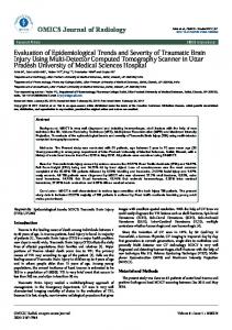
Case Report 1. 37- year-old female patient, with no medical history, presents with a 5 years evolution indurated erythematous tumor on her left cheek. Biopsy reports a cutaneous follicle centre lymphoma. Systemic involvement study was negative. 3D Radiotherapy was indicated (40 Gy in 20 fractions, performed on a Cobalt-60 treatment device), with complete resolution of the tumor (Figure 1).
J Leuk ISSN:2329-6917 JLU, an open access journal
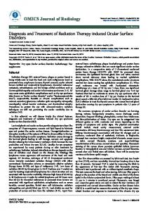
Figure 1: a) Indurated erythematous tumor on the left cheek. b) After Radiotherapy 2. 45- year-old female patient, with no medical history, presents with an infiltrated erythematous tumor on her left cheek, she refers a 18 years history of slow growing, asymptomatic. Biopsy reports a cutaneous follicle centre lymphoma. Systemic involvement study was negative. Radiotherapy (40 Gy in 20 fractions, with appositional
Volume 3 • Issue 4 • 1000197
Citation:
Montserrat Molgó, Josefina Rodríguez, Camila Arriagada, Eugenio Vines, Félix Fich, et al. (2015) Radiotherapy in Cutaneous B-Cell Lymphomas Treatment: Report of Three Cases. J Leuk 3: 197. doi:10.4172/2329-6917.1000197
Page 2 of 3 electron field technique) associated to intralesional corticoids was indicated, with complete resolution of the tumor (Figure 2).
Figure 2: a) Infiltrated erythematous tumor on the left cheek. b) After Radiotherapy associated to intralesional corticoids treatment. 3. 65- year-old female patient, with no medical history, presents with erythematous infiltrated papules on cheeks and nose. First biopsy reports granulomatous rosacea, after no response to multiple treatments a second biopsy was indicated reporting a cutaneous follicle centre lymphoma. Systemic involvement study was negative. Radiotherapy was also indicated (30 Gy in 15 fractions, with a single electron field technique), with complete resolution of the tumor (Figure 3). We interpreted as an agminate form.
The available therapeutic modalities are diverse and include surgical excision, radiotherapy, corticosteroids, antibiotics, monoclonal antibodies and interferon, as well as monotherapy or polychemotherapy schemas. Treatment should be tailored according to the risk and selected based on the type of lymphoma and on the clinical stage, as well as on the characteristics of the patient and the number, type, extent, location and distribution of the skin lesions [7]. In general, treatment may vary depending on the presentation of disease, with patients presenting with a solitary lesion or lesions limited to a single anatomic site being approached differently from patients presenting with generalized or treatment-refractory disease or evidence of extracutaneous spread. Data supporting these forms of treatment are limited to small retrospective or case studies, and consensus opinions of a group of experts [6]. Radiotherapy for PCL should be highly individualized, depending on tumor histology and location. In general, in patients undergoing radiation therapy, cumulative dose per field ranges from 15 Gy to 45 Gy, generally fractionated into doses of 2 Gy to 2.5 Gy per irradiation field per week, and usually including a margin of 1 cm to 5 cm of normal skin around the affected skin. Most of the patients have a complete response (CR), while recurrence occurs in about half of cases [8]. Adverse effects regarding the use of radiation is dose- dependent, acute toxicity usually presents with erythema and dry desquamation, and late toxicity includes skin fibrosis, telangiectasia, temporary anhidrosis and irreversible alopecia [9,10]. In the case of our patients the principal side effects where erythema and dry desquamation, well managed with topical treatment. We present these 3 cases of PCFLC treated with radiotherapy in our service, because of the excellent response, supporting radiotherapy as a therapeutic alternative. Our experience is in concordance to the findings reported in the literature. It is important to determine an appropriate treatment to achieve a better quality of life for patients.
Figure 3: a) Erythematous infiltrated papules on cheeks and nose b) After Radiotherapy.
Discussion The most common type of primary cutaneous B- Cell Lymphomas is the PCFCL, which represents 60% of all cases [5]. The median age of patients is 51 years, with a slight male predominance [5]. PCFCL typically presents as plaques, papules or erythematous solitary tumors, painless, firm, usually located on the head and trunk and rarely present in lower limbs [2]. Normally skin lesions presents for several years, with resistance to multiple treatments before diagnostic confirmation is made. Diagnostic confirmation is made by histological study, immunohistochemistry and clonal rearrangement [2]. After the diagnosis is confirmed, staging procedures in addition to history and physical examination should be performed to evaluate for extracutaneous disease and anticipate treatment [6]. This type of cutaneous lymphoma has a good prognosis, with a 5-year survival greater than 95%, but a high risk of recurrence [5].
J Leuk ISSN:2329-6917 JLU, an open access journal
Our experience over 25 years in a university medical institution is different from international reports. We have 30 patients with primary cutaneous B- Cell Lymphomas, from which 63,3% present PCMZL, 20% PCFCL and 16,7% PCDLBCL, LT. Also, we have one case associated with chronic Helicobacter pylori infection and other case with positive Borrelia serology.
References 1. 2. 3. 4.
5.
6.
Kim MJ, Hong ME, Maeng CH, Jung HA, Hong JY, et al. (2015) Clinical features and treatment outcomes of primary cutaneous B-cell lymphoma: a single-center analysis in South Korea. Int J Hematol 101: 273-278. Vermeer MH, Willemze R (2014) Recent advances in primary cutaneous B-cell lymphomas. Curr Opin Oncol 26: 230-236. Wilcox RA (2015) Cutaneous B- cell lymphomas: 2015 uptodate on diagnosis, risk- stratification, and management. Am J Hematol 90: 73-76. Specht L, Dabaja B, Illidge T, Wilson LD, Hoppe RT (2015) Modern radiation therapy for primary cutaneous lymphomas: field and dose guidelines from the International Lymphoma Radiation Oncology Group. Int J Radiat Oncol Biol Phys 92: 32-39. Senff NJ, Hoefnagel JJ, Jansen PM, Vermeer MH, van Baarlen J, et al. (2007) Reclassification of 300 primary cutaneous B-cell lymphomas according to the new WHO-EORTC classification for cutaneous lymphomas: comparison with previous classifications and identification of prognostic markers. J Clin Oncol 25 :1581-1587. Pinter-Brown LC (2015) Diagnosis and Management of Cutaneous B-cell Lymphoma. Dermatol Clin 33: 835-840.
Volume 3 • Issue 4 • 1000197
Citation:
Montserrat Molgó, Josefina Rodríguez, Camila Arriagada, Eugenio Vines, Félix Fich, et al. (2015) Radiotherapy in Cutaneous B-Cell Lymphomas Treatment: Report of Three Cases. J Leuk 3: 197. doi:10.4172/2329-6917.1000197
Page 3 of 3 7.
8. 9.
Peñate Y, Hernández-Machín B, Pérez-Méndez LI, Santiago F, Rosales B, et al. (2012) Intralesional rituximab in the treatment of indolent primary cutaneous B-cell lymphomas: an epidemiological observational multicentre study. The Spanish Working Group on Cutaneous Lymphoma. Br J Dermatol 167: 174-179. Lima M (2015) Cutaneous primary B-cell lymphomas: from diagnosis to treatment. An Bras Dermatol 90: 687-706. Hoppe RT, Harrison C, Tavallaee M, Bashey S, Sundram U, et al. (2015) Low-dose total skin electron beam therapy as an effective modality to
J Leuk ISSN:2329-6917 JLU, an open access journal
10.
reduce disease burden in patients with mycosis fungoides: results of a pooled analysis from 3 phase-II clinical trials. J Am Acad Dermatol 72: 286-292. Lowry L, Smith P, Qian W, Falk S, Benstead K, et al. (2011) Reduced dose radiotherapy for local control in non-Hodgkin lymphoma: a randomised phase III trial. Radiother Oncol 100: 86-92.
Volume 3 • Issue 4 • 1000197