Letters to the Editor
Mohammad Kazem Fallahzadeh, Mohammad Reza Namazi Endogenous Opiates
IL-2, IFNIL-4, IL-5
Macrophage Function
Antibody Production
UV Light
CRH, ACTH, Cortisol,
NO
Department of Dermatology, Shiraz University of Medical Sciences, Shiraz, Iran Address for correspondence: correspondence: Dr. Mohammad Reza Namazi, Dermatology Department, Faghihi Hospital, Shiraz, Iran. E-mail:
[email protected]
Neutral Endopeptidase
TH1 suppression
DOI: 10.4103/0378-6323.57735 - PMID: 19915254
REFERENCES Immunosuppression
1. 2.
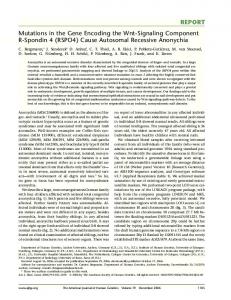
Figure 1: Mechanisms of immunosuppression induced by ultraviolet-released endogenous opioids
Morphine inhibits the expression of antigenic markers for T-helper cells and also the respiratory burst of these cells.[6] This drug suppresses antibody production in response to the T cell-dependent antigens.[6] It also leads to elevated plasma levels of corticotropinreleasing hormone (CRH), adrenocorticotropic hormone (ACTH) and glucocorticoids.[6] Therefore, through suppression of the immune system, morphine increases the susceptibility to various types of opportunistic infections.[6] A functional relationship between endogenous opiates and the immune system is based on the demonstration of special opiate receptors (µ3) on immune cells, which enables these compounds to directly inhibit the immune activities.[6] Under stressful conditions, endogenous morphine helps other immunosuppressive compounds such as ACTH and IL-10 to lower the hyperstimulation of stimulatory molecules such as IL-1 and TNF-α.[6] Morphine stimulation by µ3 leads to nitric oxide (NO) release.[6] Basal unstimulated NO is released in the body to oppose the pro-inflammatory state and to downregulate immuoncytes.[6] Morphine may enhance this inhibitory state by enhancing the normal basal actions of NO.[6] In short, given the immunomodulatory effects of opioids, we suggest that a part of UV-induced immunosuppression occurs through the release of endogenous opiates [Figure 1]. Therefore, we conclude that topical opioid antagonists could serve as a novel class of protective agents against UV-induced skin cancers and their addition to the popular cancer preventive agents could provide a better protective effect.
3. 4. 5.
6. 7.
Schwarz T. Mechanisms of UV-induced immunosuppression. Keio J Med 2005;54:165-71. Feldman SR, Liguori A, Kucenic M, Rapp SR, Fleischer AB Jr, Lang W, et al. Ultraviolet exposure is a reinforcing stimulus in frequent indoor tanners. J Am Acad Dermatol 2004;51:45-51. Warthan MM, Uchida T, Wagner RF Jr. UV light tanning as a type of substance-related disorder. Arch Dermatol 2005;141:963-6. Levins PC, Carr DB, Fisher JE, Momtaz K, Parrish JA. Plasma beta-endorphin and beta-lipoprotein response to ultraviolet radiation. Lancet 1983;2:166. Kaur M, Liguori A, Lang W, Rapp SR, Fleischer AB Jr, Feldman SR. Induction of withdrawal-like symptoms in a small randomized, controlled trial of opioid blockade in frequent tanners. J Am Acad Dermatol 2006;54:709-11. Stefano GB, Kream R. Endogenous opiates, opioids, and immune function: evolutionary brokerage of defensive behaviors. Semin Cancer Biol 2008;18:190-8. Roy S, Wang J, Gupta S, Charboneau R, Loh HH, Barke RA. Chronic morphine treatment differentiates T helper cells to Th2 effector cells by modulating transcription factors GATA 3 and T-bet. J Neuroimmunol 2004;147:78-81.
Linear scleroderma with partial anonychia Sir, Linear scleroderma, a variant of morphea, is characterized by band-like induration of the skin, often with pigmentary changes, and it frequently occurs in children in the first or second decade.[1] The limbs are most commonly affected followed by the face. Various studies have shown different rates of frequency of different morphologic variants of morphea. A study of 239 patients with morphea, of which 126 were children, showed that 22 (17.5%) of the children had linear scleroderma.[2] Thus, although linear scleroderma is not a rare disease, yet, so far, no case of this condition with anonychia has been reported. We herein report a case of linear scleroderma with partial anonychia.
Indian J Dermatol Venereol Leprol | November-December 2009 | Vol 75 | Issue 6
623
Letters to the Editor
A 22-year-old Indian girl presented with hyperpigmented linear macular and thickened lesions extending from the ulnar side of the middle of the right forearm up to the ring and little fingers for the past 10 years. The lesion first appeared on the middle part of the ulnar side of the right forearm and gradually progressed to involve the dorsal aspect of the little and ring fingers within 5 years. This included involvement of the nail of the ring finger, which led to partial loss of nail plate. The disease process has remained localized and static for the last 5 years. There was no history suggestive of Raynaud’s phenomenon and there were no systemic complaints. Examination showed a browncolored linear band-like lesion with irregular and sharp borders. The lesion was atrophic in the proximal part and demonstrated induration and binding down in the distal part [Figure 1]. Hair was absent on the affected parts. The lesion was encroaching on the nail apparatus of the ring finger where partial anonychia with a small remnant of nail plate and loss of cuticle were present [Figure 2]. There was ulnar deviation of the right ring finger and mild radial deviation of the little finger. Systemic examination was normal. Biopsy was taken from the proximal part of the lesion on the hand. Histopathology showed a sparse superficial and deep perivascular and periappendageal lymphohistiocytic infiltrate with occasional plasma cells [Figure 3]. There was marked thickening of collagen bundles in the reticular and papillary dermis. The thickened bundles were generally parallel to the epidermis and were closely packed to give a hyalinized appearance. These findings were consistent with the diagnosis of scleroderma. As the lesion of scleroderma visibly involved the nail apparatus and biopsy from the lesion showed typical features of scleroderma [Figure 3], we did not take nail matrix biopsy. Patient’s urinalysis (routine and microscopy) was normal. Her rheumatoid factor was negative and antinuclear antibody (ANA) test revealed equivocal result. Some patients with linear scleroderma may be at some risk for developing systemic collagen-vascular diseases.[3] The present patient is being followedup closely due to her equivocal ANA result. Among different variants of morphea, there is only one report of anonychia in a patient with pansclerotic morphea of childhood.[4] The same patient appears to have been reported again as part of a case series[5] because several details (age, weight, laboratory investigations, treatment and outcome) of these two cases match. No
624
Figure 1: Clinical photograph showing linear scleroderma with partial anonychia
Figure 2: Close-up showing partial anonychia of the right ring finger
Figure 3: Histopathology showing sparse perivascular and periappendageal lymphohistiocytic infiltrate and marked thickening of collagen bundles giving a hyalinized appearance. (H and E, x100)
Indian J Dermatol Venereol Leprol | November-December 2009 | Vol 75 | Issue 6
Letters to the Editor
patient with linear scleroderma has been reported so far with anonychia. Basically, the loss of nail plate results from the involvement of the nail apparatus due to the sclerotic process. This change will be irreversible. Anonychia is generally described in textbooks as a congenital anomaly; acquired causes are usually trauma and lichen planus.[6] The present case shows, for the first time, that linear scleroderma can cause anonychia thus documenting a so far unreported cause of acquired anonychia.
Sanjay Singh, Surendra Kumar Department of Dermatology, Institute of Medical Sciences, Banaras Hindu University, Varanasi - 221 005, India Address for correspondence: correspondence: Dr. Sanjay Singh, C-9, New Medical Enclave, Banaras Hindu University, Varanasi - 221 005, India. E-mail:
[email protected] DOI: 10.4103/0378-6323.57736 - PMID: 19915255
REFERENCES 1.
2.
3. 4. 5.
6.
Falanga V, Killoran CE. Morphea. In: Wolff K, Goldsmith LA, Katz SI, Gilchrest BA, Paller AS, Leffell DJ, editors. Fitzpatrick’s Dermatology in General Medicine. 7th ed. New York: McGrawHill; 2008. p. 543-6. Marzano AV, Menni S, Parodi A, Borghi A, Fuligni A, Fabbri P, et al. Localized scleroderma in adults and children. Clinical and laboratory investigations on 239 cases. Eur J Dermatol 2003;13:171-6. Woo TY, Rasmussen JE. Juvenile linear scleroderma associated with serological abnormalities. Arch Dermatol 1985;121:14035. Wollina U, Buslau M, Weyers W. Squamous cell carcinoma in pansclerotic morphea of childhood. Pediatr Dermatol 2002;19:151-4. Wollina U, Buslau M, Heinig B, Petrov I, Unger E, Kyriopoulou E, et al. Disabling pansclerotic morphea of childhood poses a high risk of chronic ulceration of the skin and squamous cell carcinoma. Int J Low Extrem Wounds 2007;6:291-8. de Barkers DAR, Baran R, Dawber RPR. Disorders of nails. In: Burns T, Breathnach S, Cox N, Griffiths C, editors. Rook’s Textbook of Dermatology. 7th ed. Oxford: Blackwell; 2004. p. 62.1-62.
Tzanck smear Þnding of Dorfman-Chanarin syndrome Sir, Dorfman-Chanarin syndrome (DCS), also referred as neutral lipid storage disease with ichthyosis, is an autosomal recessive inherited disorder characterized by lipid vacuoles in peripheral leukocytes and several tissues. Genetic studies have identified a causative mutation in hydrolase CGI-58 gene on human
chromosome 3p21. Because of these mutations, triacylglycerols accumulate in cytosolic droplets in multiple organs.[1] Tzanck smear findings of DCS were not reported previously. A 21-year-old man was admitted in the dermatology department with generalized ichthyosis present since birth. He had no family history of a similar condition. Dermatologic examination revealed generalized scaly skin lesions in face, abdomen, arms, and legs [Figure 1]. There was marked involvement of flexures. The individual scales over the trunk were white, fine, translucent, and semiadherent while those on the face were grey brown scale-crust. Bullous lesions, erosions, and keratosis pilaris were absent. The palms, soles, teeth, and nails were normal. Ophthalmological examination revealed early corticonuclear cataract, hyperopia, and ectropion. Audiological examination revealed bilateral sensory neural hearing loss. Abdominal and neurologic examinations were normal. Laboratory parameters revealed increased levels of aspartate aminotransaminase (75 U/L; normal range: 0–40), alanine aminotransaminase (81 U/L; normal range: 0–40), and creatine phosphokinase (785 U/L; normal range: 25–175). The complete blood count, fasting blood sugar, renal function tests, thyroid function tests, lipid levels, albumin, and bilirubin were within normal range. Hepatitis markers were negative. Giemsa stained peripheral blood smear of patient showed lipid vacuoles in neutrophils consistent with Jordan’s anomaly [Figure 2a]. Chest X-ray, electrocardiogram, electromyelography, cranial computerized tomography were normal. Abdominal ultrasonography revealed increased echotexture suggestive of grade II–III steatohepatitis. Liver biopsy was planned, but the patient did not allow. After cleaning with alcohol, the skin of the face was grasped between the thumb and forefinger of the left hand. A superficial incision was made with a blade (no. 15) and the tissue was scraped through the incision. The cellular materials were then spread as a thin layer onto two microscopic slides and the air-dried specimens were stained by Giemsa. Tzanck smear examination showed vacuoles in the cytoplasm of keratinocytes [Figure 2b]. Histopathologic examination of skin biopsy confirmed lipid vacuoles in keratinocytes [Figure 3]. Based on the clinical, laboratory, and histological
Indian J Dermatol Venereol Leprol | November-December 2009 | Vol 75 | Issue 6
625