Review
TRENDS in Microbiology
Vol.12 No.4 April 2004
Linking sequence to phenotype in Phytophthora – plant interactions Edgar Huitema1, Jorunn I.B. Bos1, Miaoying Tian1, Joe Win1, Mark E. Waugh2 and Sophien Kamoun1 1
Department of Plant Pathology, The Ohio State University, Ohio Agricultural Research and Development Center, Wooster, OH 44691, USA 2 National Center for Genome Resources, 2935 Rodeo Park Drive East, Santa Fe, NM 87505, USA
Oomycetes, such as Phytophthora spp., establish pathogenic interactions with a diversity of plants, but the molecular mechanisms underlying these diseases remain poorly characterized. However, research on Phytophthora pathosystems has accelerated significantly with ongoing advances in microbial and plant genomics and the resulting resources. A variety of functional analyses are being used to associate gene sequences with key processes that regulate interactions between these important pathogens and their hosts. Data from such analyses are starting to shed light on the relationship between pathogen molecules that manipulate host cell structure and function and the innate defense response in plants. Oomycetes, such as members of the genus Phytophthora, establish close interactions with a diversity of plants resulting in important diseases in crop, ornamental and native plants [1]. Despite superficial morphological similarities with fungi, oomycetes form a distinct group of eukaryotic organisms that are more closely related to brown algae and diatoms [2]. Until recently, Phytophthora spp. have been chronically understudied at the molecular level. With the continuing advances in genomics and the resulting resources, research on Phytophthora has accelerated significantly and is facing a new era [3,4]. Genome sequencing projects are under way for the potato and tomato late blight pathogen Phytophthora infestans, the soybean root rot pathogen Phytophthora sojae, and the sudden oak death pathogen Phytophthora ramorum. Numerous expressed sequence tags (ESTs) are also increasingly available for Phytophthora spp. and other oomycetes. In parallel, genome sequence resources have been accumulating for several economically important host plants of Phytophthora, such as tomato, potato and soybean, as well as experimental hosts, such as Arabidopsis thaliana and Nicotiana spp. The goal in this postgenomics era is to link sequences to phenotypes in a rapid and efficient manner. To meet this challenge, the Phytophthora and plant research communities have embarked on a diversity of functional analyses to associate gene sequences with key processes that regulate interactions between these pathogens and their hosts. Corresponding author: Sophien Kamoun (
[email protected]).
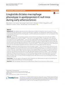
Molecular crosstalk between Phytophthora and plants involves a multitude of signal exchanges. The pathogen produces effectors; these are molecules that manipulate host cell structure and function by facilitating infection (virulence factors) or triggering defense responses (avirulence factors or specific elicitors). They interact directly or indirectly with components of the defense response pathways of plants, which can be resistance proteins or various other plant molecules generally termed virulence targets (Figure 1). Functional genetic analyses of Phytophthora – plant interactions involve identification and characterization of these various molecules and the processes that they control. With the availability of genome sequences, novel functional genomics strategies to link sequences to phenotypes in a robust and efficient manner have become
Phytophthora
Phytophthora
Suppress defense, enhance susceptibility, disease symptoms Susceptible plant
Effector
HR Resistant plant
Virulence target
Resistance protein TRENDS in Microbiology
Figure 1. A simplified view of molecular interactions between Phytophthora and plants. Phytophthora secretes effector proteins (green half circles) that interact with plant molecules known as virulence targets (purple crescents). These virulence targets are thought to be components of the plant defense response that are being inactivated by pathogen effectors. In susceptible plants, the interaction between effectors and virulence targets results in molecular events that facilitate colonization, such as suppression of defense responses, enhanced disease susceptibility, and elicitation of disease symptoms. In resistant plants, plant resistance (R) proteins recognize the effector– virulence target complex resulting in the activation of the hypersensitive response (HR). The objective of molecular studies of Phytophthora–plant interactions is to identify and functionally characterize these various molecular players. The symbol o-o depicts protein– protein interactions that are thought to be crucial for the outcome of the infection.
www.sciencedirect.com 0966-842X/$ - see front matter q 2004 Elsevier Ltd. All rights reserved. doi:10.1016/j.tim.2004.02.008
Review
194
TRENDS in Microbiology
available (see Figures 2 – 4 for an illustration of some of the methodologies incorporated into these strategies). In this review we discuss recent advances in classical and genome-scale functional genetic analyses of Phytophthora pathosystems (the biological system that comprises pathogen and host) and provide an outlook on how functional genomics can impact these analyses. Pathogen effector genes Penetration and colonization of host tissue Phytophthora spp. produce motile spores, or zoospores, that reach leaf or root surfaces. They then encyst, germinate and penetrate plant tissue [5– 7]. Germinating cysts produce germ tubes, which swell to form appressoria or appressorium-like structures that facilitate adhesion and penetration of plant surfaces. In root-infecting species, penetration can occur between cells without the aid of an appressorium [5]. Phytophthora spp. are thought to have an arsenal of genes that facilitate or contribute to these early infection events. The Car genes of P. infestans (GenBank accession numbers AF061186 and AF061185) are upregulated in germinating cysts and appressoria
Vol.12 No.4 April 2004
shortly before penetration of the plant tissue [8]. They encode extracellular mucin-like proteins that have been suggested to facilitate adhesion [8]. More recently, the CBEL gene (cellulose-binding and elicitor, GenBank accession number X97205), which encodes a cellulosebinding protein of Phytophthora parasitica, was shown to be essential in adhesion to cellulosic substrates [9]. However, although P. parasitica strains silenced for the CBEL gene were impaired in their ability to attach to cellophane membranes, they remained able to infect tobacco plants [9]. Several genes with significant similarity to degradative enzymes, such as cutinases, proteases, endo- and exoglucanases, and chitinases, have been identified in EST libraries and are thought to facilitate infection by breaking down physical barriers in the plant [10–12]. A handful of Phytophthora genes that encode degradative enzymes have been characterized in detail, including phospholipases [13], (a) CPP
LB 35S
Replicase
8
25
CPP
Candidate gene
CP nos
RB
12 (a)
LB
RB 35S
(b)
Candidate gene
nos
(b)
(c)
(d)
(e)
(f)
(c)
(g) (d)
TRENDS in Microbiology
TRENDS in Microbiology
Figure 2. Agroinfiltration: transient gene expression in plants using Agrobacterium tumefaciens-mediated transformation. (a) Strategy for agroinfiltration. The gene of interest (candidate gene) is cloned in a plant gene expression cassette in a T-DNA binary vector, and the recombinant plasmid is transformed into A. tumefaciens. The A. tumefaciens strain carrying the binary plasmid is cultured, washed and incubated in a solution that contains acetosyringone to induce the bacterial vir genes. The bacterial solution is then infiltrated with a syringe into leaf panels of a mature plant, such as Nicotiana benthamiana. Within 48 hours, most of the plant cells within the infiltrated area are transiently transformed with the T-DNA and express the candidate gene. (b) Symptoms observed on leaves of N. benthamiana following infiltration with A. tumefaciens strain containing a binary vector expressing the hypersensitive response (HR)-inducing gene inf1 of Phytophthora infestans. Inoculated leaves were photographed five days after inoculation with A. tumefaciens containing the binary vector p35S-INF1 (bottom left and top right sides of the leaf), and the negative control pGUSi (top left and bottom right). UV autofluoresence observed in N. benthamiana leaf panels corresponding to (c) the negative control and (d) the p35S-INF1 infiltration areas. Cell-death-associated fluorescence is observed in panel (d), whereas background red fluorescence is caused by chloroplasts. See Refs. [65,66] for more information. www.sciencedirect.com
Figure 3. Agroinfection: transient gene expression in plants using Agrobacterium tumefaciens carrying a binary potato virus X (PVX) vector. (a) Strategy for agroinfection. The gene of interest (candidate gene) is cloned into a binary PVX vector and the recombinant plasmid is transformed into Agrobacterium tumefaciens. The A. tumefaciens strain carrying the binary PVX plasmid is inoculated by wounding plant leaves with a toothpick. T-DNA will be transferred to a few cells surrounding the wounded area resulting in expression of PVX, systemic spreading of the virus in the plant, and expression of the candidate gene in plant tissue. (b– e) Symptoms observed in Nicotiana benthamiana following inoculation with A. tumefaciens carrying a binary PVX vector expressing the hypersensitive response (HR)-inducing gene inf1 of Phytophthora infestans. Leaves were photographed eight days after inoculation with A. tumefaciens containing (b,d) a binary PVX vector and (c,e) a PVX:INF1 construct. The arrows indicate the sites of inoculation. Note the HR lesion induced by the INF1 protein around the wounded area (c,e). Wild-type PVX induces no symptoms at the inoculation site and a mild yellowing, known as mosaic symptoms, in the upper leaves (b). (f,g) Large scale agroinfection assays on tobacco leaves. (f) Inoculation of a tobacco leaf with a 96-needle colony replicator dipped in a microtiter plate containing A. tumefaciens strains carrying recombinant PVX plasmids. (g) Symptoms observed on a tobacco leaf ten days after inoculation. The necrotic lesions correspond to sites inoculated with PVX:INF1. For clarity, only 40 of the 96 inoculations are shown. See Refs. [27,30,67] for more information.
Review
TRENDS in Microbiology
(a) LB
RB
35S Rep 134 K
Rep 194 K
LB 35S Coat protein
(b)
MP
16 K 35-T
RB
(c)
Gene X
nos
(d)
(e)
(f)
(g)
TRENDS in Microbiology
Figure 4. Virus-induced gene silencing (VIGS). (a) Strategy for VIGS using Agrobacterium tumefaciens binary vectors carrying Tobacco Rattle Virus (TRV) RNA1 and RNA 2 genomes. The plant gene of interest (gene X) is cloned into a binary TRV RNA 2 vector and the recombinant plasmid is transformed into A. tumefaciens. The A. tumefaciens strain carrying the recombinant binary TRV RNA 2 plasmid and a strain carrying the RNA 1 genome are cultured, washed in inducing buffer and mixed 1:1 before infiltration with a syringe into leaf panels. TRV is allowed to infect the plant resulting in systemic gene silencing. At approximately three weeks after infiltration, a secondary challenge is performed using pathogen inoculation, elicitor treatment or expression of a heterologous gene by agroinfiltration or agroinfection. The effect of VIGS on the challenge treatment is then monitored. (b,c) Symptoms observed in Nicotiana benthamiana silenced for the ubiquitin ligase-associated gene SGT1 following inoculation with A. tumefaciens carrying a binary PVX vector expressing the hypersensitive response (HR)-inducing gene inf1 of Phytophthora infestans. N. benthamiana plants were inoculated with A. tumefaciens carrying a (b) TRV:SGT1 construct or the (c) TRV vector, and then challenged after three weeks with A. tumefaciens carrying a binary PVX vector (top of b and c), and a PVX:INF1 plasmid (bottom of b and c). Leaves were photographed eight days after the secondary PVX challenge. Note the absence of necrotic lesion in the SGT1-silenced leaf (b) suggesting that SGT1 is required for INF1-mediated HR [63]. (d– g) UV autofluoresence observed in N. benthamiana leaf panels corresponding to the (d,f) TRV:SGT1-silenced plant and (e,g) TRV control plants. Silenced plants were challenged with (d,e) PVX or (f,g) PVX:INF1. Celldeath-associated fluorescence is observed in panel (g), whereas background red fluorescence is caused by chloroplasts. See Refs. [57,68,69] for more information.
a b-glucosidase/xylosidase [14], exo-1,3-b-glucanases [12], an endo-1,3-b-glucanase [12] and endopolygalacturonases (endoPGs) [15,16]. The endoPG family is remarkable in many respects. In Phytophthora cinnamomi, endoPGs form a major family that contains at least 19 members [15]. Birth-and-death evolution, reticulate evolution and diversifying selection were all detected in this gene family and might have contributed to the evolution of this structurally diverse and complex class of enzymes [15]. Phylogenetic analyses have indicated that Phytophthora endoPGs, exo1,3-b-glucanases and an endo-1,3-b-glucanase are more similar to fungal genes than to their plant and bacterial counterparts [12,15,16]. These observations are in sharp www.sciencedirect.com
Vol.12 No.4 April 2004
195
contrast to phylogenies that have been constructed from ribosomal sequences or compiled protein sequences from mitochondrial and housekeeping chromosomal genes, which consistently group oomycetes with brown algae and diatoms [2,17,18]. The apparent discrepancies between these phylogenies could reflect a convergent evolution in the arsenal of hydrolytic enzymes between oomycetes and fungi, two distantly related groups of filamentous pathogenic microbes that have similar life strategies and hosts [15,16,19]. These observations suggest that there are common mechanisms of infection among filamentous microbes. In the future, comparative genomics analyses between plant-pathogenic oomycetes and fungi will help define a common set of virulence genes. Suppression of host defense responses Following infection, plants exhibit complex defense responses through the production of pathogen-inducible antimicrobial enzymes, for example, glucanases and proteases that degrade microbial cell walls and proteins. Phytophthora can suppress these defense responses by producing inhibitory molecules that target host enzymes [4,20 –22]. In P. infestans, water-soluble glucans have been reported to suppress host defenses in a plant cultivarspecific manner [21,22]. Recently, genes encoding secreted proteins that inhibit soybean endo-b-1,3 glucanase have been cloned from P. sojae [23]. These proteins, termed glucanase inhibitor proteins (GIPs, GenBank accession numbers AF406607– AF406609), share significant structural similarity to the trypsin class of serine proteases, but contain mutated catalytic residues and are proteolytically non-functional as a consequence [23]. GIPs are thought to function as counterdefensive molecules that inhibit the degradation of b-1,3/1,6 glucans in the pathogen cell wall and/or the release of defense-eliciting oligosaccharides by host endo-b-1,3 glucanases [23]. Data mining of P. infestans ESTs revealed an additional class of secreted inhibitory proteins that contain domains typical of Kazal serine protease inhibitors. One of these proteins, EPI1 from P. infestans, was found to inhibit and physically interact with tomato proteases suggesting a novel type of defensecounterdefense mechanism between plants and Phytophthora (M. Tian and S. Kamoun, abstract 322, XXII Fungal Genetics Conference, Asilomar, CA, 2003, http://www.fgsc.net/asil2003/asil2003abs.htm). Induction of defense responses and disease-like symptoms Several Phytophthora effector molecules are known to induce a variety of cellular defense responses in plants [24 –31]. Some of these effectors induce defense responses in both susceptible and resistant plants and are referred to as general elicitors. Other effectors induce defense responses specifically in resistant plants and are known as specific elicitors. However, in many cases the contribution of elicitors to the infection process remains ambiguous owing to the lack of direct evidence obtained from knockout or overexpression Phytophthora mutants. General elicitors have been compared to pathogen-associated molecular patterns (PAMPs) of animal pathogens, which are surface-derived molecules that induce the
196
Review
TRENDS in Microbiology
expression of defense response genes and the production of antimicrobial compounds in host cells [32,33]. Nonetheless, it cannot be ruled out that some elicitors might function as toxins that facilitate colonization of host tissue during the late phase of Phytophthora infections, when host tissue collapses and turns necrotic [4,27]. Originally, Phytophthora elicitors were identified and purified using biochemical methods and were subsequently cloned using reverse genetics and hybridization methods. This classical strategy led to the identification of genes encoding INF1 elicitin from P. infestans (GenBank accession number U50844) [34], cryptogein from Phytophthora cryptogea (GenBank accession number Z34459) [35], PsojNIP and GPE1 from P. sojae (GenBank accession numbers AF320326 and U10471) [27,29], NPP1 and CBEL from P. parasitica (GenBank accession numbers AF352031 and X97205) [24,31], and PcF from Phytophthora cactorum (GenBank accession number AF354650) [26]. More recently, elicitors from Phytophthora have been identified using functional genomic strategies [27,28,30]. These approaches typically involve two steps: (i) data mining for genes that fulfill particular criteria, and (ii) functional genetic analyses to identify the desired genes among the selected candidates. Examples of computational tools that were developed to mine sequence datasets include PexFinder (with Pex standing for Phytophthora extracellular protein), an algorithm based on SignalP v2.0 [36] and designed to identify putative secreted or membrane-associated proteins from ESTs [27,30]. Other data mining tools are embedded into EST analysis pipelines, such as (I) the XGI system of the National Center for Genome Resources (NCGR, http://www.ncgr.org) [37,38], which performs automated BLAST searches [39] against a variety of target databases, (ii) BLIMP searches of the BLOCKS þ protein motif database [40], (iii) PexFinder analyses [30], and (iv) eight separate analyses as part of InterProScan searches [41] against the InterPro database [42] [see the Phytophthora Functional Genomics Database (PFGD, www.pfgd.org) as an example]. The XGI system also performs automated assignment of gene ontology annotations based on high-scoring homologies with Swiss-Prot data as well as curated gene ontology (http://www. geneontology.org/) annotations available through InterPro. Rapid functional assays for expressing Phytophthora genes in planta are well established and are ideal for discovering genes with elicitor function [43] (Figures 2 and 3). Using an Agrobacterium tumefaciens binary vector carrying the potato virus X (PVX) genome, Torto et al. [30] screened 63 candidate Pex cDNAs for elicitor activity in planta and recovered two novel necrosis-inducing cDNAs, crn1 and crn2 (GenBank accession numbers AF424675 and AF424677). These cDNAs encode extracellular proteins that belong to a large and complex protein family in Phytophthora. The crn genes are expressed in P. infestans during colonization of the host plant tomato, and it is crn2 that induces the defense response genes. Qutob et al. [27] also used the PVX vector to express 16 Pex cDNAs from P. sojae in Nicotiana benthamiana. One of these cDNAs encodes PsojNIP, a 26 kDa protein that is similar in sequence to necrosis-inducing proteins from various www.sciencedirect.com
Vol.12 No.4 April 2004
eukaryotic and prokaryotic species. PsojNIP induces necrosis and cell death in tobacco and also the host plant soybean. Interestingly, Fellbrich et al. [24] independently identified an ortholog of the PsojNIP gene by purification of a necrosis-inducing protein, NPP1, from culture filtrates of P. parasitica and by cloning using reverse genetics. These two studies, which were published side-by-side, offer a comparison between classical and functional genomics approaches to elicitor discovery. Bos et al. [44] described another functional genomics strategy that combines data mining with intraspecific comparative genomics and functional analyses to identify novel avirulence genes from Phytophthora. This approach provides a rapid and efficient alternative to classical positional cloning strategies for isolating avirulence genes that match known disease resistance genes (R genes) and has the potential to uncover ‘orphan’ avirulence genes for which corresponding R genes have not been previously identified [44]. Plant defense genes Disease resistance (R) genes Several R genes that target Phytophthora spp. have been genetically defined. Cloning of these R genes has been achieved in a few cases. Eleven late blight R genes were introgressed into potato from the Mexican wild species Solanum demissum using classical breeding. One of these genes, R1 (GenBank accession number AF447489), was cloned using a combination of positional cloning and the candidate gene approach [45]. The R1 gene is predicted to encode a polypeptide of 1293 amino acids that belongs to the CC-NBS-LRR (coiled-coil motif, nucleotide-binding site and leucine-rich repeat domain) class of plant R genes [46,47]. Another late blight R gene, RB (GenBank accession number AAP45164), was recently cloned from the wild diploid potato species Solanum bulbocastanum using a combination of map-based cloning and long-range polymerase chain reaction [48]. RB is predicted to encode a protein of 970 amino acids that also belongs to the CC-NBS-LRR class [48]. As with several other R genes, both R1 and RB belong to complex loci that carry several R gene analogs (RGAs) of the CC-NBS-LRR class [45,48]. In contrast to R1, which is only effective against races of P. infestans that carry Avr1 [45], RB is effective against all tested races of the pathogen and holds great promise to help achieve sustainable management of late blight [48,49]. In fact, somatic hybrids between potato and the parental S. bulbocastanum clone PT29, as well as several backcrossed progenies, exhibited broad-spectrum and persistent resistance in a variety of field trials over several years [48,49]. Whether this phenotype is solely due to RB, or whether additional resistance genes are involved remains to be determined. The P. infestans Avr (avirulence) genes that trigger R1and RB-mediated resistance responses are unknown. Sequence analyses suggest that the R1 and RB proteins are localized inside the plant cell [45,48]. How these proteins perceive molecular signals from the avirulent pathogen is one of the trying research questions in Phytophthora – plant interactions. The most prevalent theory maintains that Phytophthora and other oomycete
Review
TRENDS in Microbiology
and fungal pathogens deliver effector molecules into plant cells through specialized structures, such as haustoria, even though the exact mechanism of how these molecules would cross the plant plasma membrane remains unknown [50,51]. Cloning and functional analysis of Avr– R gene pairs in Phytophthora pathosystems will provide insight into this issue. Defense signaling in Arabidopsis The model plant Arabidopsis thaliana is emerging as an experimental system for the study of resistance to Phytophthora. Arabidopsis exhibits both host resistance (against Phytophthora spp. that can cause disease on this plant) and non-host resistance (against Phytophthora spp. that cannot cause disease on Arabidopsis). Two Phytophthora spp. have been reported that infect Arabidopsis and provide pathosystems that are more amenable to genetic analysis than the agronomically important Phytophthora diseases. Cabbage isolates of Phytophthora brassicae (previously known as Phytophthora porri) [52,53] and several isolates of Phytophthora cinnamomi [54] can infect and extensively colonize Arabidopsis. Both species exhibit marked variation in the responses that they induce in different Arabidopsis ecotypes. Most Arabidopsis defense mutants did not show any alteration in their resistance to P. brassicae. However, pad2, a mutant with reduced production of the phytoalexin camalexin, was heavily colonized resulting in a hypersusceptibility phenotype [52]. To date, the molecular identity of pad2 has not been reported. Other Phytophthora spp., such as P. infestans and P. sojae, cannot colonize and cause disease on Arabidopsis, resulting in non-host interactions [55,56]. Cytological and molecular analyses indicate that non-host resistance of Arabidopsis to these species is associated with the hypersensitive response (HR) and other active defense responses [55,56]. Considering the impressive genetic and functional genomic resources that are available, Arabidopsis offers good prospects for dissecting the complex interactions between non-host plants and Phytophthora. For example, pen2, an Arabidopsis mutant that is deficient in a cell wall glycosyl hydrolase, was recently shown to allow enhanced penetration and HR in response to P. infestans (P. Schulze-Lefert, abstract 19, European Plant Science Organization Conference, Brunnen, Switzerland, 2002, http://www.epsoweb.org/ catalog/Conf2002.htm). Defense signaling in Nicotiana benthamiana Research on genetic dissection of defense response pathways in plants is greatly benefiting from the emergence of Nicotiana benthamiana as an alternative and complementary model system to Arabidopsis. N. benthamiana allows rapid and high-throughput analysis of gene function using virus-induced gene silencing (VIGS) [57] (Figure 4). Using VIGS, a loss-of-function phenotype can be generated for a given candidate gene within a one-month timescale. VIGS can also be used in forward genetic screens. For example, Lu et al. [58] identified 79 N. benthamiana cDNAs that are required for HR against the bacterial pathogen www.sciencedirect.com
Vol.12 No.4 April 2004
197
Pseudomonas syringae by screening 4992 cDNAs that were randomly picked from a normalized library. VIGS is also facilitating the genetic dissection of the HR of N. benthamiana to P. infestans and the elicitin INF1. Yoshioka et al. [59] showed that two respiratory burst oxidase homologs of N. benthamiana, known as NbrbohA and NbrbohB (GenBank accession numbers BAC56864 and BAC56865), are required for H2O2 accumulation and resistance to P. infestans. VIGS of the Nbrboh genes also led to a reduction of HR cell death that was observed as a result of INF1, suggesting that the oxidative burst is required for full development of the HR. Kanzaki et al. [60] reported that two cytosolic molecular chaperone proteins of N. benthamiana, known as NbHSP90c-1 and NbHSP70c-1 (GenBank accession numbers AB105429 and AB105430), are required for INF1-mediated HR and non-host resistance to the bacterial pathogen Pseudomonas cichorii. Cytosolic HSP90 proteins were also recently implicated in a variety of R gene-mediated responses to bacterial, fungal and viral pathogens [58,61]. NbHSP90c-1 interacts with NbSIPK (GenBank accession number AB098730), a mitogen-activated protein (MAP) kinase that is activated during INF1-mediated HR [62]. However, VIGS of NbSIPK and another MAPK kinase (NbWIPK, GenBank accession number AB098729) did not result in a loss of HR to INF1, suggesting that activation of these MAPK kinase cascades is not required for response to INF1 [62]. Peart et al. [63] also used VIGS to show that the response of N. benthamiana to INF1 was dependent on the ubiquitin ligase-associated protein NbSGT1 (GenBank accession numbers AF516180 and AF516181), which is also required for non-host resistance to bacterial plant pathogens. Takahashi et al. [61] reported that cytosolic HSP90 interacts with SGT1 and another resistance signaling protein, RAR1 (GenBank accession number AY438026), suggesting that these proteins might function with HSP90 as co-chaperones that regulate the activity and stability of substrate proteins essential for disease resistance signaling. Databases for Phytophthora– plant genomics A crucial component of functional genomics is the dissemination of large datasets to the research community through public databases, which is a more effective alternative to traditional publications. Ideally, a functional genomics database should be iterative and interactive, allowing users to develop hypotheses, query the database with specific questions, collect information, test hypotheses, and finally revisit the database to refine their queries or deposit new information. In recent years, several genomics databases have emerged for Phytophthora spp. and their host plants, mainly through projects funded by the NSF Plant Genome Research Program (Table 1). The PFGD is a publicly accessible resource that interrelates functional and sequence data. It builds upon cDNA and genome sequences derived from other databases, including the former Phytophthora Genome Consortium (PGC) database and other public P. infestans sequence data. Sequences are screened, clustered, analyzed and annotated using NCGR’s XGI system [37,38]. PFGD also includes functional
198
Review
TRENDS in Microbiology
Vol.12 No.4 April 2004
Table 1. Major public databases and genomic resources for Phytophthora spp. and their host plantsa
a
Database
url
Organism(s)
Key features
Phytophthora Functional Genomics Database (PFGD)
http://www.pfgd.org/
Phytophthora infestans, Phytophthora sojae
Processed, assembled and annotated ESTs, functional assays and expression data
Consortium for the Genomics of Microbial Eukaryotes (COGEME)
http://www.cogeme.man.ac.uk/
P. infestans, P. sojae and various fungal species
Assembled and annotated ESTs
Phytophthora sojae genome sequencing project at DOE Joint Genome Institute (JGI)
http://genome.jgi-psf.org/physo00/physo00.home.html
P. sojae
Whole genome shotgun sequence of P. sojae
Solanaceae Genomics Network (SGN)
http://www.sgn.cornell.edu/
Lycopersicon esculentum, Lycopersicon pimpinellifolium, Lycopersicon hirsutum, Lycopersicon pennellii, Solanum tuberosum, Solanum melongena
Comparative genetic maps, marker sequences, ESTs, phylogenetic information, a tomato –Arabidopsis synteny map, and a mutant phenotype database named ’The Genes That Make Tomatoes’
Solanaceae Genomics Database (SolGD)
http://www.solgd.org/
L. esculentum, S. tuberosum, Nicotiana tabacum, Nicotiana benthamiana, Nicotiana otophora
Annotated unigene sequences
SolGenes
http://grain.jouy.inra.fr/cgi-bin/webace/ webace?db ¼ solgenes
Lycopersicon, Solanum, and Capsicum spp.
Genetic maps and marker information
NSF Potato Genome Project (UC Berkeley)
http://www.potatogenome.org
Mainly Solanum spp.
Genome sequences and resources in the Solanaceae R Gene Sequences Database (SOLAR)
NSF Potato Functional Genomics (TIGR)
http://www.tigr.org/tdb/potato/
S. tuberosum
Annotated and assembled ESTs
Solanaceae Gene Expression Database (SGED)
http://www.tigr.org/tdb/potato/SGED_index2.shtml
S. tuberosum
DNA microarray gene expression profiling data
C.M. Rick Tomato Genetics Resource Center (TGRC)
http://tgrc.ucdavis.edu/
Lycopersicon spp.
Seed bank of tomato germplasm
Tomato TIGR Gene Index
http://www.tigr.org/tdb/tgi/lgi/
L. esculentum
Tomato Expression Database (TED)
http://ted.bti.cornell.edu/
L. esculentum
Annotated and assembled ESTs DNA microarray gene expression profiling data
Transcriptome Analysis of BY-2 (TAB)
http://mrg.psc.riken.go.jp/strc/
N. tabacum
ESTs from tobacco BY-2 cell culture
Legume Information System (LIS)
http://www.comparative-legumes.org/
Medicago truncatula and various leguminae
Annotated EST unigenes and genome sequences
SoyBase
http://www.soybase.org/
Glycine max and various leguminae
Genetic maps and marker information
The plant list focuses on two major botanical families, the solanacaeae and the leguminae, which are hosts to two major Phytophthora pathogens, P. infestans and P. sojae.
annotation of P. infestans candidate effector genes. To enable an integrated analysis to be performed using solanaceous sequences and those of P. infestans, the Solanaceae Genomics Database (SolGD) has been developed as a sister database for PFGD. Currently, SolGD contains unigenes derived from ESTs of solanaceous www.sciencedirect.com
plants, including tomato, potato, N. benthamiana, Nicotiana tabacum and Nicotiana otophora. Interlinkage between PFGD and SolGD facilitates analysis of the ‘interaction transcriptome’, which consists of host and pathogen genes whose expression might be regulated during infection [64].
Review
TRENDS in Microbiology
Other resources include the Solanaceae Genomics Network (SGN), which provides genomic information about solanaceous species, including comparative maps, marker sequences, ESTs and phylogenetic information. In addition, comprehensive resources on tomato and potato ESTs are available on The Institute for Genome Research (TIGR) Tomato Gene Index and Potato Gene Index, respectively. In addition, the Solanaceae Gene Expression Database (SGED) stores gene expression data that are obtained from the TIGR expression profiling service of 10 000-clone potato cDNA microarrays, and the Tomato Expression Database (TED) contains basic information about tomato ESTs and cDNA microarray data. Finally, the Solanaceae R Gene Sequences Database (SOLAR) archives genomic resources centered on R genes from various solanaceous plants, such as wild potato, tomato and pepper. SOLAR includes physical and genetic maps, genomic sequences of R gene loci and candidate late blight R genes, and evolutionary models and bioinformatic tools for R gene analysis. The web addresses of the databases described here are listed in Table 1. Future perspectives Functional genomics of Phytophthora – plant interactions is a promising area of research. There is already a respectable set of data mining and functional genomics tools that have been incorporated into expanding databases. In addition, the field is now embracing comparative genomics as a result of the exponential increases in sequence data for oomycetes and their hosts. Nevertheless, this field is in its infancy and requires additional technical developments in bioinformatics and functional analysis. In summary, listed here are some of the key themes that will immediately impact the development of functional genomics of Phytophthora pathosystems: † Completion of genome sequencing of Phytophthora and host species. † Identification of candidate effector and defense genes using bioinformatics and wet laboratory approaches. † Proteomics approaches to molecular interactions at the infection interface. † High-throughput functional analyses of Phytophthora effector genes. † High-throughput functional analyses of plant defense genes using Arabidopsis genetics and VIGS. † Development of databases that incorporate functional and sequence data. These and other advances in genome-scale functional analyses of Phytophthora pathosystems will lead to a greater understanding of the basic molecular mechanisms underlying the interactions between these economically important pathogens and their plant hosts. Ultimately, this will allow the identification of novel genetic targets for efficient pathogen management and improvement of crops. Acknowledgements We are grateful to colleagues in the Phytophthora field and previous members of our laboratory for useful discussions and access to unpublished information. We thank Saskia Hogenhout and anonymous reviewers for suggestions on the manuscript. Research in our laboratory is www.sciencedirect.com
Vol.12 No.4 April 2004
199
supported by NSF Plant Genome Research Program grant DBI-0211659, Syngenta Biotechnology, and State and Federal Funds appropriated to the Ohio Agricultural Research and Development Center, the Ohio State University. We dedicate this paper to Don Nuss for his pioneering support of Phytophthora genomics.
References 1 Erwin, D.C. and Ribeiro, O.K. (1996) Phytophthora Diseases Worldwide, APS Press 2 Baldauf, S.L. et al. (2000) A kingdom-level phylogeny of eukaryotes based on combined protein data. Science 290, 972 – 977 3 Birch, P.R.J. and Whisson, S. (2001) Phytophthora infestans enters the genomics era. Mol. Plant Pathol. 2, 257 – 263 4 Kamoun, S. (2003) Molecular genetics of pathogenic oomycetes. Eukaryot. Cell 2, 191 – 199 5 Hardham, A.R. (2001) The cell biology behind Phytophthora pathogenicity. Australas. Plant Pathol. 30, 91– 98 6 van West, P. et al. (2002) Oomycete plant pathogens use electric fields to target roots. Mol. Plant Microbe Interact. 15, 790 – 798 7 van West, P. et al. (2003) Advances in research on oomycete root pathogens. Physiol. Mol. Plant Pathol. 62, 99 – 113 8 Gornhardt, B. et al. (2000) Cyst germination proteins of the potato pathogen Phytophthora infestans share homology with human mucins. Mol. Plant Microbe Interact. 13, 32 – 42 9 Gaulin, E. et al. (2002) The CBEL glycoprotein of Phytophthora parasitica var. nicotianae is involved in cell wall deposition and adhesion to cellulosic substrates. J. Cell Sci. 115, 4565– 4575 10 Kamoun, S. et al. (1999) Initial assessement of gene diversity for the oomycete pathogen Phytophthora infestans based on expressed sequences. Fungal Genet. Biol. 28, 94 – 106 11 Qutob, D. et al. (2000) Comparative analysis of expressed sequences in Phytophthora sojae. Plant Physiol. 123, 243 – 254 12 McLeod, A. et al. (2003) Characterization of 1,3- glucanase and 1,3;1,4beta glucanase genes from Phytophthora infestans. Fungal Genet. Biol. 38, 250 – 263 13 Nespoulous, C. et al. (1999) Characterization of elicitin-like phospholipases isolated from Phytophthora capsici culture filtrate. FEBS Lett. 452, 400 – 406 14 Brunner, F. et al. (2002) A beta-glucosidase/xylosidase from the phytopathogenic oomycete Phytophthora infestans. Phytochemistry 59, 689 – 696 15 Go¨tesson, A. et al. (2002) Characterization and evolutionary analysis of a large polygalacturonase gene family in the oomycete plant pathogen Phytophthora cinnamomi. Mol. Plant Microbe Interact. 15, 907– 921 16 Torto, T.A. et al. (2002) The pipg1 gene of the oomycete Phytophthora infestans encodes a fungal-like endopolygalacturonase. Curr. Genet. 40, 385 – 390 17 Baldauf, S.L. (2003) The deep roots of eukaryotes. Science 300, 1703– 1706 18 Lang, B.F. et al. (1999) A comparative genomics approach to the evolution of eukaryotes and their mitochondria. J. Eukaryot. Microbiol. 46, 320 – 326 19 Latijnhouwers, M. et al. (2003) Oomycetes and fungi: similar weaponry to attack plants. Trends Microbiol. 11, 462– 469 20 Ham, K-S. et al. (1997) Fungal pathogens secrete an inhibitor protein that distinguishes isoforms of plant pathogenesis-related endo-1,3glucanases. Plant J. 12, 169– 179 21 Sanchez, L.M. et al. (1992) Host selective suppression by water-soluble glucans from Phytophthora spp. of hypersensitive cell death of suspension-cultured cells from some solanaceous plants caused by hyphal wall elicitors of the fungi. Ann. Phytopathol. Soc. Japan 58, 664– 670 22 Yoshioka, H. et al. (1995) Suppression of phenylalanine ammonia-lyase mRNA accumulation by suppressors from Phytophthora infestans. Ann. Phytopathol. Soc. Japan 61, 7 – 12 23 Rose, J.K. et al. (2002) Molecular cloning and characterization of glucanase inhibitor proteins: coevolution of a counterdefense mechanism by plant pathogens. Plant Cell 14, 1329 – 1345 24 Fellbrich, G. et al. (2002) NPP1, a Phytophthora-associated trigger of plant defense in parsley and Arabidopsis. Plant J. 32, 375– 390 25 Kamoun, S. et al. (1998) Resistance of Nicotiana benthamiana to
Review
200
26
27
28 29
30
31
32
33 34
35
36
37 38
39
40 41
42
43 44 45
46 47 48
TRENDS in Microbiology
Phytophthora infestans is mediated by the recognition of the elicitor protein INF1. Plant Cell 10, 1413 – 1426 Orsomando, G. et al. (2001) Phytotoxic protein PcF, purification, characterization, and cDNA sequencing of a novel hydroxyprolinecontaining factor secreted by the strawberry pathogen Phytophthora cactorum. J. Biol. Chem. 276, 21578 – 21584 Qutob, D. et al. (2002) Expression of a Phytophthora sojae necrosisinducing protein occurs during transition from biotrophy to necrotrophy. Plant J. 32, 361– 373 Qutob, D. et al. (2003) Variation in structure and activity among elicitins from Phytophthora sojae. Mol. Plant Pathol. 4, 119 – 124 Sacks, W. et al. (1995) Molecular characterization of nucleotide sequences encoding the extracellular glycoprotein elicitor from Phytophthora megasperma. Mol. Gen. Genet. 246, 45– 55 Torto, T. et al. (2003) EST mining and functional expression assays identify extracellular effector proteins from Phytophthora. Genome Res. 13, 1675– 1685 Villalba Mateos, F. et al. (1997) Cloning and characterization of a cDNA encoding an elicitor of Phytophthora parasitica var. nicotianae that shows cellulose-binding and lectin-like activities. Mol. Plant Microbe Interact. 10, 1045– 1053 Nu¨rnberger, T. and Brunner, F. (2002) Innate immunity in plants and animals: emerging parallels between the recognition of general elicitors and pathogen-associated molecular patterns. Curr. Opin. Plant Biol. 5, 318– 324 Gomez-Gomez, L. and Boller, T. (2002) Flagellin perception: a paradigm for innate immunity. Trends Plant Sci. 7, 251 – 256 Kamoun, S. et al. (1997) A gene encoding a protein elicitor of Phytophthora infestans is down-regulated during infection of potato. Mol. Plant Microbe Interact. 10, 13 – 20 Panabieres, F. et al. (1995) Characterization of a gene cluster of Phytophthora cryptogea which codes for elicitins, proteins inducing a hypersensitive-like response in tobacco. Mol. Plant Microbe Interact. 8, 996 – 1003 Nielsen, H. et al. (1999) Machine learning approaches for the prediction of signal peptides and other protein sorting signals. Protein Eng. 12, 3 – 9 Inman, J.T. et al. (2000) A high-throughput distributed DNA sequence analysis and database system. IBM Syst J. 40, 464 – 486 Waugh, M. et al. (2000) The Phytophthora genome initiative database: informatics and analysis for distributed pathogenomic research. Nucleic Acids Res. 28, 87 – 90 Altschul, S.F. et al. (1997) Gapped BLAST and PSI-BLAST: a new generation of protein database search programs. Nucleic Acids Res. 25, 3389 – 3402 Henikoff, J.G. et al. (2000) Increased coverage of protein families with the blocks database servers. Nucleic Acids Res. 28, 228– 230 Zdobnov, E.M. and Apweiler, R. (2001) InterProScan – an integration platform for the signature-recognition methods in InterPro. Bioinformatics 17, 847 – 848 Apweiler, R. et al. (2001) The InterPro database, an integrated documentation resource for protein families, domains and functional sites. Nucleic Acids Res. 29, 37– 40 Kamoun, S. et al. (2002) From sequence to phenotype: functional genomics of Phytophthora. Can. J. Plant Pathol. 24, 6 – 9 Bos, J.I.B. et al. (2003) Intraspecific comparative genomics to identify avirulence genes from Phytophthora. New Phytol. 159, 63 – 72 Ballvora, A. et al. (2002) The R1 gene for potato resistance to late blight (Phytophthora infestans) belongs to the leucine zipper/NBS/LRR class of plant resistance genes. Plant J. 30, 361– 371 Dangl, J.L. and Jones, J.D. (2001) Plant pathogens and integrated defence responses to infection. Nature 411, 826– 833 Meyers, B.C. et al. (2003) Genome-wide analysis of NBS-LRR-encoding genes in Arabidopsis. Plant Cell 15, 809 – 834 Song, J. et al. (2003) Gene RB cloned from Solanum bulbocastanum
www.sciencedirect.com
Vol.12 No.4 April 2004
49
50 51
52
53
54
55
56
57 58
59
60
61
62
63
64
65 66
67
68 69
confers broad spectrum resistance to potato late blight. Proc. Natl. Acad. Sci. U. S. A. 100, 9128– 9133 Helgeson, J.P. et al. (1998) Somatic hybrids between Solanum bulbocastanum and potato; a new source of resistance to late blight. Theor. Appl. Genet. 96, 738 – 742 Jia, Y. et al. (2000) Direct interaction of resistance gene and avirulence gene products confers rice blast resistance. EMBO J. 19, 4004– 4014 Tyler, B.M. (2002) Molecular basis of recognition between Phytophthora pathogens and their hosts. Annu. Rev. Phytopathol. 40, 137– 167 Roetschi, A. et al. (2001) Characterization of an Arabidopsis – Phytophthora pathosystem: resistance requires a functional PAD2 gene and is independent of salicylic acid, ethylene and jasmonic acid signalling. Plant J. 28, 293– 305 Si-Ammour, A. et al. (2003) Quantification of induced resistance against Phytophthora species expressing GFP as a vital marker: b-aminobutyric acid but not BTH protects potato and Arabidopsis from infection. Mol. Plant Pathol. 4, 237– 248 Robinson, L.H. and Cahill, D.M. (2003) Ecotypic variation in the response of Arabidopsis thaliana to Phytophthora cinnamomi. Aust. Plant Pathol. 32, 53 – 64 Takemoto, D. et al. (2003) GFP-tagging of cell components reveals the dynamics of subcellular re-organization in response to infection of Arabidopsis by oomycete pathogens. Plant J. 33, 775 – 792 Huitema, E. et al. (2003) Active defense responses associated with nonhost resistance of Arabidopsis thaliana to the oomycete pathogen Phytophthora infestans. Mol. Plant Pathol. 4, 487 – 500 Baulcombe, D.C. (1999) Fast forward genetics based on virus-induced gene silencing. Curr. Opin. Plant Biol. 2, 109 – 113 Lu, R. et al. (2003) High throughput virus-induced gene silencing implicates heat shock protein 90 in plant disease resistance. EMBO J. 22, 5690 – 5699 Yoshioka, H. et al. (2003) Nicotiana benthamiana gp91phox homologs NbrbohA and NbrbohB participate in H2O2 accumulation and resistance to Phytophthora infestans. Plant Cell 15, 706 – 718 Kanzaki, H. et al. (2003) Cytosolic HSP90 and HSP70 are essential components of INF1-mediated hypersensitive response and non-host resistance to Pseudomonas cichorii in Nicotiana benthamiana. Mol. Plant Pathol. 4, 383 – 391 Takahashi, A. et al. (2003) HSP90 interacts with RAR1 and SGT1 and is essential for RPS2-mediated disease resistance in Arabidopsis. Proc. Natl. Acad. Sci. U. S. A. 100, 11777– 11782 Sharma, P.C. et al. (2003) Virus-induced silencing of WIPK and SIPK genes reduces resistance to a bacterial pathogen, but has no effect on the INF1-induced hypersensitive response (HR) in Nicotiana benthamiana. Mol. Genet. Genomics 269, 583 – 591 Peart, J.R. et al. (2002) Ubiquitin ligase-associated protein SGT1 is required for host and nonhost disease resistance in plants. Proc. Natl. Acad. Sci. U. S. A. 99, 10865 – 10869 Birch, P.R.J. and Kamoun, S. (2000) Studying interaction transcriptomes: coordinated analyses of gene expression during plant – microorganism interactions. In New Technologies for Life Sciences: A Trends Guide (Wood, R., ed.), pp. 77 – 82, Elsevier Science Kapila, J. et al. (1997) An Agrobacterium-mediated transient gene expression system for intact leaves. Plant Sci. 122, 101 – 108 Van der Hoorn, R.A. et al. (2000) Agroinfiltration is a versatile tool that facilitates comparative analyses of Avr9/Cf-9-induced and Avr4/Cf-4induced necrosis. Mol. Plant Microbe Interact. 13, 439 – 446 Takken, F.L. et al. (2000) A functional cloning strategy, based on a binary PVX- expression vector, to isolate HR-inducing cDNAs of plant pathogens. Plant J. 24, 275– 283 Lu, R. et al. (2003) Virus-induced gene silencing in plants. Methods 30, 296– 303 Liu, Y. et al. (2002) Tobacco Rar1, EDS1 and NPR1/NIM1 like genes are required for N-mediated resistance to tobacco mosaic virus. Plant J. 30, 415– 429