Jul 28, 2014 - ... 100 mL of SDS (1% w/v) for 20 min at RT, with occasional vortexing. .... Kirschner, C. M., Alge, D. L., Gould, S. T. & Anseth, K. S. Clickable,.
OPEN SUBJECT AREAS: BIOMEDICAL ENGINEERING BIOMATERIALS - CELLS
Received 7 March 2014 Accepted 7 July 2014 Published 28 July 2014
Correspondence and requests for materials should be addressed to M.S.-S. (Manuel. Salmeron-Sanchez@ glasgow.ac.uk)
Living biointerfaces based on non-pathogenic bacteria to direct cell differentiation Aleixandre Rodrigo-Navarro1,4, Patricia Rico1, Anas Saadeddin2, Andres J. Garcia3 & Manuel Salmeron-Sanchez4 1
Center for Biomaterials and Tissue Engineering, Universitat Polite´cnica de Vale`ncia, Spain, 2Abengoa Research, Campus Palmas Altas, Sevilla, Spain, 3Woodruff School of Mechanical Engineering, Petit Institute for Bioengineering and Bioscience Georgia Institute of Technology, Atlanta, GA 30332, USA, 4Division of Biomedical Engineering, School of Engineering, University of Glasgow, Glasgow G12 8LT, United Kingdom.
Genetically modified Lactococcus lactis, non-pathogenic bacteria expressing the FNIII7-10 fibronectin fragment as a protein membrane have been used to create a living biointerface between synthetic materials and mammalian cells. This FNIII7-10 fragment comprises the RGD and PHSRN sequences of fibronectin to bind a5b1 integrins and triggers signalling for cell adhesion, spreading and differentiation. We used L. lactis strain to colonize material surfaces and produce stable biofilms presenting the FNIII7-10 fragment readily available to cells. Biofilm density is easily tunable and remains stable for several days. Murine C2C12 myoblasts seeded over mature biofilms undergo bipolar alignment and form differentiated myotubes, a process triggered by the FNIII7-10 fragment. This biointerface based on living bacteria can be further modified to express any desired biochemical signal, establishing a new paradigm in biomaterial surface functionalisation for biomedical applications.
U
nderstanding the behaviour of cells on synthetic surfaces is of foremost interest to engineer microenvironments that direct cell adhesion, proliferation and differentiation. To favour the interaction with cells, surfaces of synthetic biomaterials have been functionalized with a broad range of proteins, fragments, peptides and growth factors including fibronectin (FN), laminin, vitronectin and others, by using physical and chemical strategies1–3. However, these passive coatings can by no means provide the dynamic stimuli required to orchestrate cell responses and organise the formation of a new tissue at the material interface. Significant efforts have focused on engineering materials that recapitulate characteristics of the ECM, such as the presentation of cell adhesive motifs or protease degradable cross-links, in order to direct cellular responses4,5. Photoactivable RGD adhesive peptides were used to investigate the effect of the density and time point of ligand presentation on cell adhesion, proliferation and differentiation6. Similarly, enzyme-responsive surfaces that present cell adhesive RGD sequences on-demand, that is, by enzymatic hydrolysis of inactive RGD containing precursors that carry cleavable steric blocking groups, have been used to spatiotemporal controlled attachment of cells7. However, the development of a cell/material interface able to provide biological stimuli upon demand, a functional dynamic interface between stem cells and synthetic materials, has not been established yet. We hypothesised that nonpathogenic bacteria can colonise the surface of a broad range of synthetic materials and can be genetically modified to constitutively express or secrete the desired adhesive proteins and factors to a living cell population upon external demand. This work shows that non-pathogenic bacteria that constitutively express a fibronectin fragment enhance cell differentiation. However, our long-term goal is to transform this living interface into a dynamic system able to secrete other proteins and growth factors upon demand. We have recently shown that genetically modified, non-pathogenic bacteria, can play the role of a (living) biointerface between mammalian cells and synthetic biomaterials and we chose Lactococcus lactis subsp. cremoris as host for membrane expression of the fibronectin FNIII7-10 fragment8. This fragment contains the RGD adhesion motif (10th repeat of the type III fragment) and the PHSRN synergy site (9th repeat of type III fragment), both of them necessary to promote a5b1 integrin mediated adhesion9. Lactococcus lactis is a gram-positive, nonpathogenic bacterium, with GRAS (Generally Regarded as Safe) status and with low production of exopolysaccharides, a mandatory condition to ensure the accessibility of the displayed FNIII7-10 fragments on the bacteria membrane. Moreover, this low exopolysaccharide production is not an obstacle for this strain to develop stable SCIENTIFIC REPORTS | 4 : 5849 | DOI: 10.1038/srep05849
1
www.nature.com/scientificreports
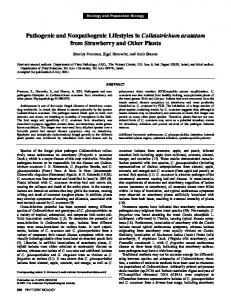
Figure 1 | Schematic of living biointerfaces. C2C12 murine myoblasts were seeded on top of L. lactis biofilms that express a FNIII7-10 fragment as a membrane protein. The construct features the Usp45 secretion peptide, GFP and the fibronectin fragment FNIII7-10 that contains both RGD (FNIII10) and PHSRN synergy sites (FNIII9). The construct is inserted into the bacterial peptidoglycan layer by the protein A anchor (spaX). This anchor protein is covalently linked to the tetrapeptides of the peptidoglycan layer. L. lactis strain forms stable biofilms with very low exopolysaccharide production, which granted exposure of FN fragments to mammalian cells. C2C12 differentiated into myoblasts triggered by the FNIII7-10 fragment displayed on the bacterial membrane.
biofilms: these bacteria firmly attach to synthetic surfaces and establish communities10,11. This results in a stable layer of bacteria on the material surface which present FNIII7-10 fragments8. Mammalian cells interact with FN via integrins, a family of transmembrane receptor proteins that anchor them to the ECM. Integrin-mediated adhesion is a complex process that involves the organization of focal adhesion clusters that link the ECM to the cytoskeleton. Focal complexes are mature adhesion sites that contains structural proteins (vinculin, paxilin, talin, tensin) and signalling molecules (FAK, focal adhesion kinase, Src)3,12,13. We have previously shown that FN null fibroblasts (FN -/-) adhered on this living biointerface based on L. lactis expressing FNIII7-10, develop focal adhesions and promote FAK-based signalling8. This work investigates the potential of this living interface based on L. lactis expressing a FN fragment as a membrane protein to direct cell differentiation by evaluating myogenic differentiation14. As previously reported8, a modified L.lactis (MG1363) was engineered to express FNIII7-10 as a membrane protein. The used construct features the Usp45 secretion peptide, GFP and the FNIII7-10 fragment conSCIENTIFIC REPORTS | 4 : 5849 | DOI: 10.1038/srep05849
taining the RGD adhesion (module III10) and PHSRN synergy site (module III9). The construct is anchored into the bacterial peptidoglycan layer by the protein A anchor (spaX) from Staphylococcus aureus. This anchor protein is covalently linked to the tetrapeptides of the peptidoglycan layer. L. lactis hardly produces any exopolysaccharide, which granted the availability of the FN fragment to interact with mammalian cells seeded on the bacterial layer (Fig. 1).
Results We have first characterised the formation of the L. lactis biofilm on glass, including the amount of FN that is available for cell interaction. Then we have addressed the stability of the biofilm after different days, that results in using a strategy to diminish bacterial metabolism and prevent uncontrolled proliferation. Afterwards, we have explored C2C12 interactions on this living interface, first in the short-term (cell adhesion, cell morphology) and then in the longterm (differentiation and related signalling). Characterization of the bacterial-expressed fibronectin. The Usp45 secretion and spaX (S. aureus protein A) anchor signals 2
www.nature.com/scientificreports
Figure 2 | Visualisation and quantification of FNIII7-10 in the bacterial membrane. (a) Low voltage SEM image of L. lactis-FN adhered to a glass surface showing the glycocalyx and the adhesion fimbriae. Scale bar size is 200 nm. (b) – (d) Western blot analysis of membrane fractions of L. lactis using protein extracts from 75, 50 and 25 mL of a stationary phase culture and 750, 500 and 250 ng of a purified FNIII7-10 fragment as standards (quantification curve shown in d). Proteins isolated from bacteria have higher molecular weight due to GFP, the Usp45 and the spaX fragments (e) Quantification of FNIII7-10 on the bacteria membrane using ELISA with HFN7.1 antibody. Biofilm activity is roughly equivalent to a surface coated with a solution of 20 mg/ mL of plasma fibronectin (300 ng/cm2). L. lactis-empty shows negligible fibronectin activity (1000 mL of bacterial culture was used both for L. lactisempty and L. lactis-FN, that correspond to a surface area coverage of 27.6 6 8.5%). (f) ELISA of bacterial biofilms (L. lactis-FN) from fresh GM17-E medium and incubated in DMEM with tetracycline 10 mg/mL for 1 and 4 days respectively. ANOVA shows no statistically significant differences between samples. All data presented as mean 6 SD.
efficiently promote the localization of FNIII7-10 in the bacterial membrane. We have confirmed the presence of FNIII7-10 by the tagged GFP, which shows fluorescent rings defining the perimeter SCIENTIFIC REPORTS | 4 : 5849 | DOI: 10.1038/srep05849
of each bacterium (Supp Fig. S1)8. Fig 2a shows bacterial morphology at high magnification by SEM. Additional SEM and AFM images are included in Supp Fig. S1. We have quantified the amount of FNIII7-10 3
www.nature.com/scientificreports
Figure 3 | Biofilm metabolism and viability. To assess the metabolism of the bacterial community inside the biofilm in conditions compatible with the culture of C2C12 cells, biofilms of L. lactis-FN and L. lactis-empty were produced. Then, GM17-E medium was changed to either DMEM, DMEM with 1% penicillin/streptomycin (P/S) or DMEM with 10 mg/mL tetracycline (TC). After 1, 2 and 4 days of culture in DMEM, DMEM1P/S and DMEM1TC, viability was tested using the LIVE/DEAD BacLight bacterial viability kit, where viable bacteria are stained in green and non-viable in red. (a) Representative images after 4 days. Scale bar size is 25 mm. (b) Image quantification shows that viability is not affected by either the use of antibiotics or the culture time. No statistically significant differences among conditions were found. Data is presented as mean 6 SD. (c) Simultaneously, bacterial metabolism was inferred by measuring the pH of the medium as a function of time, to assess the production of lactic acid. Addition of tetracycline maintained the pH stable and above 7.5. The use of P/S was enough to maintain the pH above 6.5. In antibiotic-free DMEM, the pH dropped to a minimum of 4 due to the transformation of glucose in the medium into lactic acid.
available on L. lactis membranes using two techniques that provide complementary information. First, we performed fractionation and Western blot analysis for bacterial membrane proteins and subsequent quantification using a recombinant FNIII7-10 fragment as the part of the bacteria lysate ascribed to membrane proteins (Fig. 2b). Image analysis of the Western blot bands reveal that the total amount of FNIII7-10 increases as the volume of the sample (number of colonies) does. Based on this, the approximate density of FN fragment per unit volume of bacteria is 0.98 mg/mL, which corresponds approximately to 9.8 mg/108 CFU (Fig. 2 c,d). However, considering the role of this living interface in interacting with mammalian cells, is also important to calculate the availability of FNIII7-10 after bacteria have been immobilised on a synthetic material. To do so, we have performed an enzyme-linked immunosorbent assay (ELISA) with a monoclonal antibody on high density biofilms, prepared from 1000 mL of culture (Fig. 2e). This is a well-established method to probe for structural or conformational changes in adsorbed proteins15,16. We used the monoclonal HFN7.1 antibody to probe the availability of the flexible linker between the 9th and 10th type III repeats of FN17. Figure 2e shows the amount of this integrin-binding domain on the surface of the living interface is similar to adsorbing FN from a solution of concentration 20 mg/mL on glass, a surface density of FN SCIENTIFIC REPORTS | 4 : 5849 | DOI: 10.1038/srep05849
ca. 300 ng/cm2,18,19. As we will use tetracycline containing medium (TC) to maintain a stable biofilm on the material surface during experiments with mammalian cells, and since TC might inhibit bacterial protein expression, we have assessed that there is no influence of TC on FNIII7-10 expression as a membrane protein (Fig 2f). In summary, the engineered living interface presents a high density of FNIII7-10 available on the cell surface, seeking to promote focal adhesion organisation and trigger cell fate. Biofilm formation and stability. We next examined the stability of the living interface on the material surface. This strain, once the biofilm has been established, is able to continue proliferating as long as there is a sufficient supply of nutrients. L. lactis is a homofermentative strain that metabolises any carbon source found in the medium, glucose in our experiments, to L-(1)lactic acid, in an anaerobic fashion20. This production of lactic acid leads to a decrease in the pH of the culture medium up to ,4, seriously compromising the viability of the aboveground cells. Here arises the need to control the pH of the medium, by inhibiting the bacterial metabolism. We have found that tetracycline used at low concentrations, 10 mg/mL, is enough to inhibit the bacterial replication without impacting mammalian cell metabolism21,22 (Fig 3 and 4
www.nature.com/scientificreports results on cell differentiation later in this work). Tetracycline is a broad-spectrum antibiotic that binds to the 30S subunit of the microbial ribosomes, inhibiting the protein synthesis by blocking the attachment of the charged aminoacyl-tRNA to the ribosomal A site23. Conversely, penicillin acts inhibiting the peptidoglycan cell-wall remodelling, ultimately leading to cell breakdown. As we only intend to maintain the cell population under control, it is counterproductive to promote cell lysis, and, as side effect, the modification of the biofilm morphology. Moreover, tetracycline is more effective controlling the pH at low concentrations and is nontoxic to mammalian cells21,22 (see also our results on cell behaviour afterwards). Biofilms of different densities were formed on glass and remained stable after several days on the material surface. We have effectively shown that we can control the surface density of bacteria on the material surface by controlling the seeding volume (Supp Fig. S2). We selected high-density bacterial cultures to have the highest possible L. lactis density on the material surface readily available for cell differentiation experiments. We have assessed the stability of the layer of bacteria at different time points up to 4 days. This is the time needed for myogenic differentiation experiment afterwards. Fig. 3 shows live/dead staining for L. lactis grown on glass using different media (DMEM, DMEM 1 tetracycline (TC), DMEM 1 penicillin and streptomycin, stated as P/S). After 4 days of culture bacteria viability (measured by the ration of live to total cells) is unaffected by the presence of small amounts of antibiotics in the culture medium and it is maintained at ca. 60% (Fig. 3b). The temporal evolution of the pH in the different media used is shown in Fig. 3c. Only when the culture medium is supplemented with TC, acidification does not occur and the pH remains stable for four days (Fig 3c). We suggest that this acidification of the culture medium is a consequence of the L. lactis metabolism. As said before, L. lactis is a homofermentative strain that metabolises any suitable carbon source found in the medium - glucose in our experiments - to L-(1)-lactic acid. Then, the production of L-(1)-acid lactic, with a pKa of 3.8624 must be responsible for the acidification of the culture medium. Addition of TC maintains a stable pH, which suggests that no L-lactic acid is produced and that, consequently, bacterial metabolism is inhibited. The use of antibiotics in the culture medium diminished the density of bacteria on the material surfaces, as it can be inferred from Fig. 3a and we have quantified in Supp Fig. S3. However, even if this is the case, the layer of bacteria that remains on the surface is viable and sufficient to support cell adhesion and cell differentiation, as it is shown afterwards. That is to say, even if the layer of bacteria attained on the material surface is not homogeneous (see e.g. Supp Fig S2), the attained bacterial surface involves the adequate expression of FN for mammalian cell interactions. Cell morphology, adhesion and differentiation on L. lactis. The density of the biofilm directs area and roundness of mammalian cells. We first studied cell morphology on established biofilms of different densities on glass (Supp Fig. S2). Both cell area and roundness are directly related to the biofilm density, as shown in fig. 4. C2C12 cells seeded on biofilms of different densities showed increased area (Fig. 4b), with minimal changes in cell roundness (Fig. 4c). In addition, focal adhesions were formed on cells seeded on L. lactisFN biofilms. The number of focal adhesion plaques increased with the density of bacteria on the surface (Supp Fig. S4). These results suggest that L.lactis–FN directs cell adhesion and cell morphology in the short-term, which is necessary before considering other longterm processes such as cell differentiation. We previously showed that L. lactis modified to express FNIII7-10 as a membrane protein promotes focal adhesion formation and FAK signalling at similar levels as native FN in fibroblasts8. Now, we assessed the ability of this living interface to support cell differentiation by examining myogenic differentiation25. Sarcomeric myosin SCIENTIFIC REPORTS | 4 : 5849 | DOI: 10.1038/srep05849
expression and cell bipolar alignment and fusion into myotubes, markers of myogenesis, were significantly higher on L. lactis modified with FNIII7-10 (Fig. 5a) as compared to L. lactis-empty (control bacteria that do not contain FNIII7-10). Surprisingly, myogenic differentiation was higher on the modified L. lactis than on native fibronectin and collagen type I, which represents the gold-standard substrate for myogenic differentiation (Fig. 5b)14. It is important to emphasise that these differences in myogenic differentiation are not due to differences in the number of cells on the substrates. It is known that a direct correlation between the number of cells on the surface and the level of myogenic differentiation has been found, however the number of cells on the modified bacteria is slightly lower than on the FN and Col I controls (Fig 5c). Integrin-mediated cell adhesion triggers a cascade of intracellular signals such as the p38 mitogen-activated protein kinase (p38 MAPK) pathway, which is involved in the myoblast differentiation process, by promoting the activity of several transcription factors and regulating cell cycle withdrawal26,27. An intermediary step between adhesion and downstream targets, including MAPK pathways, is the phosphorylation of FAK28; as a result, FAK phosphorylation at Tyr397 plays a central role during myoblast differentiation in 2D cultures29,30. Initially, FAK phosphorylation at Tyr397 is transiently reduced —contributing to trigger the myogenic genetic program— but it is later activated, as it is central to terminal differentiation into myotubes30. In order to study the activation of FAK on the L. lactis expressing FNIII7-10 we examined the phosphorylation of Tyr397 by Western blot. FAK phosphorylation was similar on modified L. lactis and FN-coated glass (Fig 5d e), in good correlation with the high levels of cell differentiation found in this work (Fig 5b). These results suggest that enhanced cell differentiation can be triggered by FNIII7-10 expressed on the membrane of L. lactis. The fact that the same experiments have been done with L. lactis-empty strain without significant levels of cell differentiation absolutely confirm FNIII7-10 expressed on the bacteria membrane as the trigger of differentiation.
Discussion Engineering microenvironments to provide cells with dynamic cues, as occurs in the natural extracellular matrix, is a field of intensive research seeking to direct the differentiation of stem cells and advance in strategies for tissue repair and regeneration31. Several approaches have been explored spanning from the simple functionalisation of material surfaces with cell adhesive motifs to the incorporation of protease-degradable fragments in polymer hydrogels that allow changes in the material structure as cells secrete proteases to remodel the ECM7,14,32. Dynamic surfaces have been engineered to present cell adhesive RGD sequences on-demand6,7,33. From a different perspective, dynamic cell microenvironments have been engineered to present and release growth factors in a sustainable and controlled way to stimulate and support cell differentiation34–40. We have recently shown that non-pathogenic bacteria can be genetically modified to establish a functional interface between cells and synthetic materials, and we anticipate that this living interface could be further modified to instruct cells to perform different functions in a highly regulated manner8. As an initial step and proof of concept towards this long-term goal, we engineered8 food-grade Lactococcus lactis, a gram-positive bacterium with very low production of lipopolysaccharides (LPS) - previously used for recombinant therapeutic proteins production, therapeutic drug delivery and vaccine production41 - to present a fibronectin fragment (FNIII7-10) as a membrane protein. FNIII7-10 contains RGD and the PHSRN synergy sequences and promote cell adhesion and differentiation42,43. We have previously shown that fibronectin null fibroblasts (FN -/-) adhere, spread and develop focal adhesions on this engineered L. lactis expressing FNIII7-10, in a similar way as when seeded directly on a FN coating8. 5
www.nature.com/scientificreports
Figure 4 | Morphological features of C2C12 myoblasts related to biofilm density. A 3 h culture of C2C12 cells seeded on top of biofilms of different surface density. Briefly, 10,000 cells/cm2 were seeded on top of L. lactis-FN biofilms of increasing density (covered surface area 4.6%, 11.4%, 19.1% and 27.6%), and L. lactis-empty biofilm produced with an surface density of 27.6%. Afterwards, cells were stained with a monoclonal anti-vinculin antibody (shown in red), whereas the actin cytoskeleton was stained with phallacidin (in green). Both nuclei of C2C12 cells and bacterial cells are shown in blue. (a) C2C12 cells seeded over L. lactis-empty biofilm showed no adhesion and remained in their original rounded shape. Cells seeded on L. lactis-FN biofilms showed good adhesion and well-developed focal adhesion plaques and actin cytoskeleton. Scale bar is 100 mm. (b) Quantification of cell area on the different biointerfaces suggests a correlation between cell area and the surface density of the biofilm. Area and roundness of at least 450 cells were quantified using the ImageJ software. (c), Quantification of cell roundness, defined as roundness 5 (4?Area)/(p?(major axis)2). ANOVA test (N $ 400) shows statistically significant differences between cells seeded on L.lactis-empty, FN-coated surfaces and glass.
Here, we show that this living interface that constitutively express a FN fragment is able to produce stable biofilms that support cell growth, cell adhesion and cell differentiation. The formation of the biofilm starts when some individual cells adhere to the surface, a process governed by weak non-specific forces - Van der Waals and polar Lewis acid-base interactions44,45. This initial, reversible attachment becomes irreversible after bacteria make use of several cell-wall anchored proteins (CWAP) known as adhesins. In L. lactis, the most prominent ones are the CluA sex factor and the PrtP and NisP proteinases10,46,47. These adhesins act modifying the bacterial surface properties and ultimately lead to an irreversible attachment of the bacteria to the surface. After bacteria attachment becomes irreversible, the force required to detach additional bacteria increases and more bacteria are recruited and co-aggregated to the existing ones. Afterwards, these bacteria proliferate and develop the biofilm itself. In this phase, a few hours after the initial irreversible attachment, a stable layer is formed. As L. lactis MG1363 is a strain with a low SCIENTIFIC REPORTS | 4 : 5849 | DOI: 10.1038/srep05849
production of exopolysaccharides, biofilms are usually monolayered, lacking the horizontal stratified structure found in other species48,49. Hence, the formation of L. lactis biofilms seems limited to a thin layer, usually a monolayer50, of strongly attached bacteria to almost any abiotic surface, such as polymers, glass or metallic surfaces. Usually, the adhesion of the bacterium to the substrate is mediated by the bacterial fimbriae, also called adhesion pili51. These fimbriae are also involved in the formation of the bacterial aggregates, as shown in Supp Fig. S1. Once the biofilm has been successfully established, a need to control its proliferation arises, and we found that the monolayer can be controlled to remain in a stationary phase (using low amount of tetracycline) on the material surface during the time needed to complete the myogenic differentiation process (up to 4 days). We have quantified the amount of FNIII7-10 that is available on the surface of the bacterial and verified that it promotes cell adhesion, signalling and cell differentiation. Overall, this living interface based on non6
www.nature.com/scientificreports
Figure 5 | Myogenic differentiation and cell signalling on living biointerfaces. (a) Fluorescence staining after 4 days of culture showing sarcomeric myosin-positive cells (green) and cell nuclei (red) on L. lactis-FN, L. lactis-empty, collagen I (1 mg/mL) coated glass, and FN (20 mg/mL) coated glass. Scale bar is 25 mm. (b) Myogenic differentiation as determined by the percentage of sarcomeric myosin-positive cells on the different interfaces. Higher degree of differentiation was found on L. lactis-FN compared to collagen I and FN coated glass. (c) C2C12 cell density on biofilms and control substrates (d) Representative western blot bands for pFAK and FAK on the different biointerfaces. (e) Quantification of western blot bands. ANOVA test shows statistically significant differences between each column. Data is presented as mean 6 SD.
pathogenic bacteria supports myogenic differentiation. Further genetic modification of this living interface is being currently done to engineer a dynamic system able to provide cells different temporal stimuli beyond cell adhesion, such as the delivery of growth factors and other molecules upon external demand. By engineering nonpathogenic bacteria to synthesise proteins and growth factors upon external stimuli, we aim at control the interface between synthetic biomaterials and stem cells to control self-renewal and differentiation, which can be then applied to several strategies to promote tissue repair and regeneration.
Methods Genetic modification of L. lactis. The GFP-containing plasmid pGFP-C2 (Clontech) was used to construct a vector contain ing FNIII7-10 downstream to GFP. FNIII7-10 fragment was amplified from the plasmid pET15b- FNIII7-1052. The forward and
SCIENTIFIC REPORTS | 4 : 5849 | DOI: 10.1038/srep05849
reverse primers used for the FNIII7-10 amplification were F1, with HindIII restriction site: aagcttaCCATTGTCTCCACCAACAAAC, and R1, with SalI restriction site: gtcgacttaTTCTGTTCGGTAATTAATGGAAA. The lactococcal plasmid PT1NX was used to clone the GFP- FNIII7-10 fragment using the following primers: F2, with NgomIV restriction site gccggcATGGGTAAAGGAGAAGAACTTT; and R2: TTCTGTTCGGTAATTAATGGAAA. Lactococcus lactis MG136353 containing the PT1NX plasmid was grown as standing culture, in anaerobic conditions, at 30uC in M17 medium (Oxoid, Basingstoke, UK) supplemented with glucose (0.5% v/v, Sigma) and erythromycin (10 mg/mL, Sigma-Aldrich) (hereinafter GM17-E). The plasmid extraction was carried using a protocol described elsewhere8. Competent L. lactis MG1363 cells were prepared growing the cells in rich medium supplemented with glycine (1% v/v). Cells were harvested when the culture reached an optical density of 0.6–0.8 measured at 600 nm, resuspended in glucose (0.5 M) with glycerol (10% v/v) and frozen at 280uC. Electroporation was performed at 2.5 kV using a BTX electroporator (Harvard Apparatus). In these conditions, with desalted DNA, the typical time constant was 4.5. Electroporated cells were transferred directly to 5 mL medium composed of M17, glucose (0.5 M), MgCl2 (20 mM) and CaCl2
7
www.nature.com/scientificreports (2 mM) and incubated for 2 h at 30uC. The resultant culture was centrifuged, resuspended in 50 mL of the later medium and extended on GM17-E agar plates. Biofilm formation. The protocol to establish a biofilm on the material surface has been published elsewhere54,55. Briefly, Lactococcus lactis MG1363 containing the PT1NX- FNIII7-10 -GFP plasmid8 (hereinafter L. lactis-FN) and L. lactis containing the PT1NX empty vector (L. lactis-empty) were grown as standing cultures in GM17E broth at 30uC. Then, 1 mL of each culture was used to streak fresh GM17-E agar plates (1% w/v agar). The plates were kept at 30uC for 24 h. A single colony from a GM17-E agar plate was inoculated into sterile GM17-E broth (Oxoid). The culture was grown until OD660 , 0.3, and then transferred to a P24 multiwell plate containing sterile glass coverslips in each well, using 300 mL and 1000 mL respectively in order to produce low and high areal density biofilms. The multiwell plate was sealed with Parafilm and left at 30uC in anaerobic conditions for 48 h. After that, the planktonic (non-adhered) bacteria were removed by shaking the multiwell plate at 150 rpm for 3 min followed with three washing steps with sterile ultrapure water. In the last wash, the ultrapure water was substituted with PBS supplemented with CaCl2 and MgCl2 (PBS11). The same protocol was used for empty and FN-expressing strains. Biofilm viability and metabolism. For bacterial viability studies, after 1, 2 or 4 days, biofilms were gently washed twice with sterile NaCl 0.85% w/v solution and incubated for 30 min using the BacLight LIVE/DEAD kit (Life Technologies), containing SYTO9 (5 mM) and propidium iodide (30 mM) in NaCl 0.85% w/v. Afterwards, samples were washed and mounted using BacLight Oil mounting medium (Life Technologies). Bacteria were counted using Fiji – ImageJ software56, using at least four microscope fields at 403 magnification for every sample. The viability was determined as the ratio between the viable and total number of bacteria. For metabolism analysis, biofilms were prepared following the biofilm formation protocol. Afterwards, the medium was changed to either DMEM or DMEM containing tetracycline (TC, 10 mg/mL)) or DMEM containing 1% penicillin-streptomycin (P/S). Biofilms were kept at 37uC in a humidified atmosphere under 5% CO2, and the pH was monitored as a function of time (0, 0.5, 1, 1.5, 2, 3, 4, 5, 24 and 48 h) using a pH-meter (Eutech Instruments). AFM and SEM imaging of the biofilms. For SEM imaging, bacterial biofilms were produced as described in the biofilm formation section. Freshly washed biofilms were fixed in 2.5% glutaraldehyde in phosphate buffer (PB, 0.1 M, pH 7.3) for 14 hours at 4uC. Then, biofilms were washed twice with PB and dehydrated in an ethanol/water mixture of 50%, 70%, 80%, 90%, 95% and 100% for 10 minutes each. The 100% ethanol step was repeated three times. Afterwards, samples were immersed twice in hexamethyldisilazane (Sigma) and air-dried for 10 minutes57. Samples were stored in vacuum until imaging in the microscope. Prior to imaging, samples were sputtercoated with a 1 nm layer of platinum and observed in a Zeiss Ultra 55 Field Emission SEM (Zeiss Microscopy, Germany) with 1 kV of gun acceleration voltage. For AFM, a culture in stationary phase was washed twice with sodium acetate (100 mM, pH 4.8) and three millilitres of the resulting bacterial suspension with the same bacterial density as the original culture were filtered through a porous polycarbonate membrane with a mean pore size of 0.8 mm (Millipore). The membrane was gently washed with the same sodium acetate solution to remove the non-trapped bacteria, while avoiding dewetting58. The wet sample was imaged in a Multimode AFM equipped with a NanoScope IIIa controller from Veeco (Manchester, UK) in QNM mode. Si cantilevers from Veeco were used, with a force constant of 0.7 N/m. Drive amplitude was 100 mV and amplitude setpoint was 250 mV. Surface density of FN - ELISA. 3 mL from a 24 h standing culture of L. lactis-FN and L. lactis-empty grown in sterile GM17-E, with a bacterial density of 107 cfu/mL, were filtered through a porous polycarbonate membrane with a mean pore size of 0.8 mm (Isopore, Millipore). The membrane was washed twice with PBS11 to remove the non-trapped bacterial cells. Another set of membranes was coated with 20, 10, 5 and 2.5 mg/mL of human plasma fibronectin (Sigma) in PBS11 for 1 h at RT, and washed twice with PBS11. Uncoated membranes were used to assess the absence of any background signal in the polycarbonate membranes. Membranes were then blocked with 1% bovine serum albumin (Sigma) in PBS11 for 1 h at RT, and incubated for 1 h at 37uC with monoclonal mouse HFN7.1 anti-fibronectin antibody. The primary antibody dilution was 154000 from a 32 mg/mL antibody stock. After that, samples were washed twice with PBS11/Tween 20 (0.5% v/v) and incubated with ALP-conjugated donkey anti-mouse secondary antibody (Jackson Immunoresearch) for 1 h at RT. Samples were washed again twice with PBS11/ Tween 20 (0.5% v/v), and 400 mL of 4-methylumbelliferyl phosphate (0.6 mM, Sigma) in borax/NaOH pH 10 buffer was added. After 45 min at 37uC, the supernatant was transferred to a P96 multiwell plate. Fluorescence was measured at 365/460 nm (excitation/emission wavelengths) with a Perkin-Elmer Victor3 Multilabel Counter (Perkin Elmer). Surface density of FN - Western blot. Three aliquots of 1.5, 1 and 0.5 mL from a 24 h standing culture of both L. lactis-FN and L. lactis-empty strains in GM17-E were lysed using a modified acetone/SDS protocol59. Briefly, cells were harvested by centrifugation at 7000 g for 5 min, washed twice with PBS without Mg21 and Ca21 and centrifuged again. The pellet was treated with 1 mL of acetone at -20uC for 10 min, with occasional vigorous vortexing. The extract was centrifuged at 7000 g for 5 min and carefully dried under a gentle stream of N2 until a porous, dry white powder was obtained. Proteins were solubilised by incubating the previously obtained
SCIENTIFIC REPORTS | 4 : 5849 | DOI: 10.1038/srep05849
powder with 100 mL of SDS (1% w/v) for 20 min at RT, with occasional vortexing. Debris were pelleted by centrifugation at 7000 g for 5 min. 20 mL of the supernatant was mixed with 20 mL of 2x Laemmli buffer and boiled for 5 min at 95uC, and samples were loaded on 10% SDS-PAGE gels. Simultaneously, 750, 500 and 250 ng of a purified fragment of FNIII7-10 were loaded in the same gel as controls. Proteins were transferred to a positively charged PVDF membrane (GE Healthcare) using a semidry transfer cell system (Bio-Rad). Membranes were blocked using 5% skimmed milk in PBS for 1 hour at RT and then incubated with the MAB1937 antibody (Millipore) directed against the 8th type III repeat in human FN diluted 151000 in PBS containing Tween 20 (0.1% v/v) and skimmed milk (2% w/v), overnight, at 4uC. After several washes with PBS/Tween 20 (0.1% v/v), the blot was incubated using horseradish peroxidase-conjugated anti-mouse antibody (GE Healthcare) diluted 1550,000 in PBS/Tween 20 (0.1% v/v)/skimmed milk (2% w/v) for 1 h at RT. Membranes were washed several times with Tween 20 (0.5% v/v) in PBS. The immunoreactive bands were visualized using the ECL Plus Western blot kit (GE Healthcare). C2C12-biofilm co-culture. C2C12 cells (American Type Culture Collection) were maintained in DMEM medium with 20% Foetal Bovine Serum (FBS) and 1% penicillin-streptomycin. Then, they were seeded over freshly washed biofilms at 20.000 cells per cm2. The culture was maintained 3 h at 37uC in a humidified atmosphere under 5% CO2 to ensure initial adhesion to the biofilm. After 3 h, the medium was changed to DMEM with 1% ITS (insulin-transferrin-selenium, Life Technologies) and tetracycline (10 mg/mL, Sigma-Aldrich) to maintain the proliferation of the bacteria under control and prevent the acidification of the culture medium by lactic acid production. The culture was kept for 4 days at 37uC and 5% CO2 in a humidified atmosphere. Immunofluorescence. Cultures were fixed at 4uC with 70% ethanol, 37% formaldehyde and glacial acetic acid (205251) for 15 min and then blocked with 5% goat serum in PBS11 for 1 h at RT. The samples were incubated with MF-20 antibody against sarcomeric myosin (Developmental Studies Hybridoma Bank, University of Iowa, USA) at 15250 dilution in PBS11 and 5% goat serum, followed by two washes with PBS11/Tween 20 (0.5% v/v) and another blocking step with 5% goat serum in PBS11 for 10 min at RT. Then were washed twice with PBS11/ Tween 20 (0.5% v/v) and incubated with rabbit anti-mouse Cy3-conjugated secondary antibody (Jackson Immunoresearch) at 15200 dilution in PBS11 with 5% goat serum. Samples were then washed three times with PBS11/Tween 20 (0.5% v/ v), mounted with Vectashield-DAPI, and observed under an epifluorescence microscope (Nikon Eclipse 80i). Images from the fluorescence microscope (DAPI channel - nuclei, and Cy3 channel - sarcomeric myosin) of the C2C12 culture were acquired at 103 magnification, transformed to an 8-bit grayscale bitmap (Fiji - ImageJ software) and segmented using the Trainable Weka Segmentation plugin to create a binary mask, for both DAPI and Cy3 channels. Total nuclei per image were counted using the particle analysis command. Then, the segmented DAPI channel image was subtracted from the Cy3 channel segmented image, and the remaining nuclei were counted and assigned to non-differentiated cells. The fraction of differentiated cells was calculated subtracting the non-differentiated nuclei from the total nuclei count. For vinculin and actin visualisation, a slightly different protocol was used. Cell fixation was performed using formalin for 30 min at 4uC. Then cells were blocked with 1% BSA in PBS11. A monoclonal anti-vinculin antibody (Sigma), diluted 15400 in BSA 1%/PBS11 was used as primary antibody. The incubation lasted 1 hour at RT. Then, cells were washed twice with PBS11/Tween 20 (0.5% v/v), and incubated with rabbit anti-mouse Cy3 conjugated (Jackson Immunoresearch), 15200 dilution, with FITC-conjugated phallacidin (Life Technologies) diluted 15100 in 1% BSA/PBS11/Tween 20 (0.5% v/v). The secondary antibody was incubated 1 hour at RT in absence of light. Cells were washed three times with PBS/Tween 20 (0.5% v/v) and mounted with Vectashield-DAPI (Vector Laboratories). FAK phosphorylation. After 3 hours of culture, cells were lysed with RIPA buffer (Tris-HCl 50 mM, 1% Nonidet P-40, 0.25% Na deoxycholate, 150 mM NaCl, 1 mM EDTA) supplemented with protease inhibitor cocktail tablets (Complete, Roche). Proteins were concentrated using Microcon YM-30 Centrifugal Filter devices (Millipore) as manufacturer described. To determine FAK protein expression and its Tyr397 phosphorylated form (pFAK), concentrated samples were subjected to 7% SDS-PAGE gel electrophoresis. Proteins were transferred to a positively charged PVDF membrane (GE Healthcare) using a semidry transfer cell system (Bio-Rad) and blocked by immersion in skimmed milk (5% w/v) in PBS for 1 h at room temperature. The blot was incubated with anti-FAK antibody (Upstate) and anti-pFAK antibody (Millipore) diluted 152500 in PBS containing Tween 20 (0.1%) and skimmed milk (2%). After several washes with PBS/Tween 20 (0.1% v/v), the blot was incubated in horseradish peroxidase-conjugated antibody (GE Healthcare) diluted 1550000 in PBS/Tween 20 (0.1% v/v) and milk (2% w/v) for 1 hour at room temperature. After several washes with PBS containing Tween (0.1% v/v) and milk (2% w/v), immunoreactive bands were visualized using Supersignal West-femto Maximum Sensitivity Substrate (Thermo Scientific). Statistical analysis. All data presented in this paper are shown as mean 6 SD. Figure 4b and 4c (cell area and cell roundness): A D’Agostino & Pearson omnibus normality test was performed, finding that the data do not follow a Gaussian
8
www.nature.com/scientificreports distribution. Then, a non-parametric ANOVA followed by a Kruskal-Wallis (Dunn) multiple comparison test was performed, with p 5 0.05. Figure 5b and 5e, corresponding to the C2C12 differentiation assay and cell density respectively: A D’Agostino & Pearson omnibus normality test was done, finding that the data follows a Gaussian distribution. Then, a one-way ANOVA with p 5 0.05 followed by a Tukey multiple column comparison test was performed. 1. Sipe, J. D. Tissue engineering and reparative medicine. Ann N Y Acad Sci 961, 1–9 (2002). 2. Griffith, L., Naughton, G. & Naughton, G. Tissue engineering--current challenges and expanding opportunities. Science (80-) 295, 1009 (2002). 3. Grinnell, F. Focal adhesion sites and the removal of substratum-bound fibronectin. J Cell Biol 103, 2697–2706 (1986). 4. Lutolf, M., Gilbert, P. & Blau, H. Designing materials to direct stem-cell fate. Nature 462, 433–441 (2009). 5. Petrie, T., Raynor, J., Dumbauld, D., Lee, T. & Jagtap, S. Multivalent IntegrinSpecific Ligands Enhance Tissue Healing and Biomaterial Integration. Sci Transl Med 2, 45ra60 (2010). 6. Weis, S., Lee, T. T., del Campo, A. & Garcı´a, A. J. Dynamic cell-adhesive microenvironments and their effect on myogenic differentiation. Acta Biomater 9, 8059–66 (2013). 7. Todd, S. J., Scurr, D. J., Gough, J. E., Alexander, M. R. & Ulijn, R. V. Enzymeactivated RGD ligands on functionalized poly(ethylene glycol) monolayers: surface analysis and cellular response. Langmuir 25, 7533–7539 (2009). 8. Saadeddin, A. et al. Functional living biointerphases. Adv Healthc Mater 2, 1213–8 (2013). 9. Aota, S., Nomizu, M. & Yamada, K. M. The short amino acid sequence Pro-HisSer-Arg-Asn in human fibronectin enhances cell-adhesive function. J Biol Chem 269, 24756–61 (1994). 10. Habimana, O. et al. Positive role of cell wall anchored proteinase PrtP in adhesion of lactococci. BMC Microbiol 7, 36 (2007). 11. Mercier, C. et al. Positive role of peptidoglycan breaks in lactococcal biofilm formation. Mol Microbiol 46, 235–43 (2002). 12. Garcı´a, A. J. Get a grip: integrins in cell-biomaterial interactions. Biomaterials 26, 7525–9 (2005). 13. Hynes, R. O. Integrins: Bidirectional, Allosteric Signaling Machines. Cell 110, 673–687 (2002). 14. Garcı´a, A. J., Vega, M. D. & Boettiger, D. Modulation of cell proliferation and differentiation through substrate-dependent changes in fibronectin conformation. Mol Biol Cell 10, 785–98 (1999). 15. Ugarova, T. P. et al. Conformational transitions in the cell binding domain of fibronectin. Biochemistry 34, 4457–4466 (1995). 16. McClary, K. B., Ugarova, T. & Grainger, D. W. Modulating fibroblast adhesion, spreading, and proliferation using self-assembled monolayer films of alkylthiolates on gold. J Biomed Mater Res 50, 428–439 (2000). 17. Schoen, R. C., Bentley, K. L. & Klebe, R. J. Monoclonal antibody against human fibronectin which inhibits cell attachment. Hybridoma 1, 99–108 (1982). 18. Keselowsky, B. G., Collard, D. M. & Garcı´a, A. J. Surface chemistry modulates fibronectin conformation and directs integrin binding and specificity to control cell adhesion. J Biomed Mater Res A 66, 247–59 (2003). 19. Salmero´n-Sa´nchez, M. et al. Role of material-driven fibronectin fibrillogenesis in cell differentiation. Biomaterials 32, 2099–105 (2011). 20. Oliveira, A. P., Nielsen, J. & Fo¨rster, J. Modeling Lactococcus lactis using a genome-scale flux model. BMC Microbiol 5, 39 (2005). 21. Rivadeneira, J., Di Virgilio, A. L., Audisio, M. C., Boccaccini, A. R. & Gorustovich, A. A. Evaluation of antibacterial and cytotoxic effects of nano-sized bioactive glass/collagen composites releasing tetracycline hydrochloride. J Appl Microbiol 1–9 (2014). doi:10.1111/jam.12476. 22. Bacon, J. A., Linseman, D. A. & Raczniak, T. J. In vitro cytotoxicity of tetracyclines and aminoglycosides in LLC-PK(1), MDCK and Chang continuous cell lines. Toxicol In Vitro 4, 384–8 (1990). 23. Connell, S. R., Tracz, D. M., Nierhaus, K. H. & Taylor, D. E. Ribosomal protection proteins and their mechanism of tetracycline resistance. Antimicrob Agents Chemother 47, 3675–3681 (2003). 24. Dawson, R. Data for biochemical research (Oxford University Press, Oxford, 1989). 25. Sabourin, L. A. & Rudnicki, M. A. The molecular regulation of myogenesis. Clin Genet 57, 16–25 (2001). 26. Mancini, A. et al. Regulation of myotube formation by the actin-binding factor drebrin. Skelet Muscle 1, 36 (2011). 27. Sastry, S. K. et al. Quantitative changes in integrin and focal adhesion signaling regulate myoblast cell cycle withdrawal. J Cell Biol 144, 1295–1309 (1999). 28. Chatzizacharias, N. A., Kouraklis, G. P. & Theocharis, S. E. Disruption of FAK signaling: A side mechanism in cytotoxicity. Toxicology 245, 1–10 (2008). 29. Clemente, C. F. M. Z., Corat, M. A. F., Saad, S. T. O. & Franchini, K. G. Differentiation of C2C12 myoblasts is critically regulated by FAK signaling. Am J Physiol Regul Integr Comp Physiol 289, R862–R870 (2005).
SCIENTIFIC REPORTS | 4 : 5849 | DOI: 10.1038/srep05849
30. Quach, N. L. & Rando, T. A. Focal adhesion kinase is essential for costamerogenesis in cultured skeletal muscle cells. Dev Biol 293, 38–52 (2006). 31. Kirschner, C. M., Alge, D. L., Gould, S. T. & Anseth, K. S. Clickable, Photodegradable Hydrogels to Dynamically Modulate Valvular Interstitial Cell Phenotype. Adv Healthc Mater (2014) doi:10.1002/adhm.201300288. 32. Khetan, S. et al. Degradation-mediated cellular traction directs stem cell fate in covalently crosslinked three-dimensional hydrogels. Nat Mater 12, 458–65 (2013). 33. Ulijn, R. V. Enzyme-responsive materials: a new class of smart biomaterials. J Mater Chem 16, 2217 (2006). 34. Makarenkova, H. et al. Differential interactions of FGFs with heparan sulfate control gradient formation and branching morphogenesis. Sci Signal 2, ra55 (2009). 35. Silva, A. K., Richard, C., Bessodes, M., Scherman, D. & Merten, O. W. Growth factor delivery approaches in hydrogels. Biomacromolecules 10, 9–18 (2009). 36. Lutolf, M. P. & Hubbell, J. A. Synthetic biomaterials as instructive extracellular microenvironments for morphogenesis in tissue engineering. Nat Biotechnol 23, 47–55 (2005). 37. Hahn, M. S., Miller, J. S. & West, J. L. Three-dimensional biochemical and biomechanical patterning of hydrogels for guiding cell behavior. Adv Mater 18, 2679–1 (2006). 38. Moon, J. J., Hahn, M. S., Kim, I., Nsiah, B. A. & West, J. L. Micropatterning of poly(ethylene glycol) diacrylate hydrogels with biomolecules to regulate and guide endothelial morphogenesis. Tissue Eng A 15, 579–585 (2009). 39. Phelps, E. A., Landazuri, N., Thule, P. M., Taylor, W. R. & Garcı´a, A. J. Bioartificial matrices for therapeutic vascularization. Proc Natl Acad Sci U S A 107, 3323–3328 (2010). 40. Patterson, J., Martino, M. M. & Hubbell, J. A. Biomimetic materials in tissue engineering. Mater Today 13, 14–22 (2010). 41. Cortes-Perez, N. G., da Costa Medina, L. F., Lefevre, F., Langella, P. & BermudezHumaran, L. G. Production of biologically active CXC chemokines by Lactococcus lactis: evaluation of its potential as a novel mucosal vaccine adjuvant. Vaccine 26, 5778–5783 (2008). 42. Cutler, S. M. & Garcı´a, A. J. Engineering cell adhesive surfaces that direct integrin a5b1 binding using a recombinant fragment of fibronectin. Biomaterials 24, 1759–1770 (2003). 43. Rico, P., Gonza´lez-Garcı´a, C., Petrie, T. A., Garcı´a, A. J. & Salmero´n-Sa´nchez, M. Molecular assembly and biological activity of a recombinant fragment of fibronectin (FNIII7–10) on poly(ethyl acrylate). Colloids Surfaces B Biointerfaces 78, 310–316 (2010). 44. Van Oss, C. J., Good, R. J. & Chaudhury, M. K. The role of van der Waals forces and hydrogen bonds in ‘‘hydrophobic interactions’’ between biopolymers and low energy surfaces. J Colloid Interface Sci 111, 378–390 (1986). 45. Van Loosdrecht, M. C., Lyklema, J., Norde, W., Schraa, G. & Zehnder, A. J. Electrophoretic mobility and hydrophobicity as a measured to predict the initial steps of bacterial adhesion. Appl Environ Microbiol 53, 1898–1901 (1987). 46. Godon, J.-J., Jury, K., Shearman, C. A. & Gasson, M. J. The Lactococcus lactis sexfactor aggregation gene cluA. Mol Microbiol 12, 655–663 (1994). 47. Giaouris, E., Chapot-Chartier, M.-P. P. & Briandet, R. Surface physicochemical analysis of natural Lactococcus lactis strains reveals the existence of hydrophobic and low charged strains with altered adhesive properties. Int J Food Microbiol 131, 2–9 (2009). 48. Gulot, E. et al. Heterogeneity of diffusion inside microbial biofilms determined by fluorescence correlation spectroscopy under two-photon excitation. Photochem Photobiol 75, 570–578 (2002). 49. Habimana, O., Meyrand, M., Meylheuc, T., Kulakauskas, S. & Briandet, R. Genetic features of resident biofilms determine attachment of Listeria monocytogenes. Appl Environ Microbiol 75, 7814–7821 (2009). 50. Rieu, A. et al. Listeria monocytogenes EGD-e biofilms: no mushrooms but a network of knitted chains. Appl Environ Microbiol 74, 4491–4497 (2008). 51. Oxaran, V. et al. Pilus biogenesis in Lactococcus lactis: molecular characterization and role in aggregation and biofilm formation. PLoS One 7, e50989 (2012). 52. Petrie, T. A., Capadona, J. R., Reyes, C. D. & Garcı´a, A. J. Integrin specificity and enhanced cellular activities associated with surfaces presenting a recombinant fibronectin fragment compared to RGD supports. Biomaterials 27, 5459–70 (2006). 53. Schotte, L. Secretion of biologically active murine interleukin-10 by Lactococcus lactis. Enzyme Microb Technol 27, 761–765 (2000). 54. Burmølle, M. et al. Enhanced biofilm formation and increased resistance to antimicrobial agents and bacterial invasion are caused by synergistic interactions in multispecies biofilms. Appl Environ Microbiol 72, 3916–3923 (2006). 55. Zaidi, A. H., Bakkes, P. J., Krom, B. P., van der Mei, H. C. & Driessen, A. J. M. Cholate-stimulated biofilm formation by Lactococcus lactis cells. Appl Environ Microbiol 77, 2602–10 (2011). 56. Schindelin, J. et al. Fiji: an open-source platform for biological-image analysis. Nat Methods 9, 676–82 (2012). 57. Araujo, J. C. Comparison of hexamethyldisilazane and critical point drying treatments for SEM analysis of anaerobic biofilms and granular sludge. J Electron Microsc (Tokyo) 52, 429–433 (2003). 58. Dufreˆne, Y. F. Atomic force microscopy and chemical force microscopy of microbial cells. Nat Protoc 3, 1132–8 (2008).
9
www.nature.com/scientificreports 59. Bhaduri, S. Modification of an acetone-sodium dodecyl sulfate disruption method for cellular protein extraction from neurotoxigenic Clostridium botulinum. Foodborne Pathog Dis 9, 172–4 (2012).
Additional information Supplementary information accompanies this paper at http://www.nature.com/ scientificreports Competing financial interests: The authors declare no competing financial interests.
Acknowledgments The support from ERC through HealInSynergy 306990 is acknowledged.
Author contributions A.R.N. and M.S.S. designed the research. A.R.N. performed it and prepared all figures. A.R.N., P.R., A.S., A.J.G. and M.S.S. analysed the data. A.R.N. and M.S.S. wrote the paper.
SCIENTIFIC REPORTS | 4 : 5849 | DOI: 10.1038/srep05849
How to cite this article: Rodrigo-Navarro, A., Rico, P., Saadeddin, A., Garcia, A.J. & Salmero´n-Sa´nchez, M. Living biointerfaces based on non-pathogenic bacteria to direct cell differentiation. Sci. Rep. 4, 5849; DOI:10.1038/srep05849 (2014). This work is licensed under a Creative Commons Attribution-NonCommercialNoDerivs 4.0 International License. The images or other third party material in this article are included in the article’s Creative Commons license, unless indicated otherwise in the credit line; if the material is not included under the Creative Commons license, users will need to obtain permission from the license holder in order to reproduce the material. To view a copy of this license, visit http:// creativecommons.org/licenses/by-nc-nd/4.0/
10