Hindawi Publishing Corporation Disease Markers Volume 35 (2013), Issue 5, Pages 543–549 http://dx.doi.org/10.1155/2013/948569
Clinical Study Local and Systemic Inflammatory Responses to Experimentally Induced Gingivitis Shaneen J. Leishman,1 Gregory J. Seymour,2,3 and Pauline J. Ford1 1
The University of Queensland, School of Dentistry, Brisbane QLD 4072, Australia The University of Queensland, School of Medicine, Herston QLD 4006, Australia 3 Sir John Walsh Research Institute, University of Otago, Dunedin 9054, New Zealand 2
Correspondence should be addressed to Shaneen J. Leishman;
[email protected] Received 14 June 2013; Revised 10 September 2013; Accepted 30 September 2013 Academic Editor: Fabrizia Bamonti Copyright © 2013 Shaneen J. Leishman et al. This is an open access article distributed under the Creative Commons Attribution License, which permits unrestricted use, distribution, and reproduction in any medium, provided the original work is properly cited. This study profiled the local and systemic inflammatory responses to experimentally induced gingivitis. Eight females participated in a 21-day experimental gingivitis model followed by a 14-day resolution phase. Bleeding on probing and plaque index scores were assessed before, during, and after resolution of gingival inflammation, and samples of saliva, GCF, and plasma were collected. Samples were assessed for biomarkers of inflammation using the BioPlex platform and ELISA. There were no significant changes in GCF levels of cytokines during the experimental phase; however, individual variability in cytokine profiles was noted. During resolution, mean GCF levels of IL-2, IL-6, and TNF-𝛼 decreased and were significantly lower than baseline levels (𝑃 = 0.003, 𝑃 = 0.025, and 𝑃 = 0.007, resp.). Furthermore, changes in GCF levels of IL-2, IL-6, and TNF-𝛼 during resolution correlated with changes in plaque index scores (𝑟 = 0.88, 𝑃 = 0.004; 𝑟 = 0.72, 𝑃 = 0.042; 𝑟 = 0.79, 𝑃 = 0.019, resp.). Plasma levels of sICAM-1 increased significantly during the experimental phase (𝑃 = 0.002) and remained elevated and significantly higher than baseline levels during resolution (𝑃 < 0.001). These results support the concept that gingivitis adds to the systemic inflammatory burden of an individual.
1. Introduction There is currently widespread interest in the relationship between periodontal disease and cardiovascular disease (CVD); however, despite this, the relationship remains to be established as a causal one, and longitudinal and mechanistic studies are required to strengthen the evidence available. The anatomical proximity of the bacterial biofilm to the periodontal vasculature facilitates the systemic dissemination of bacteria and host products [1]. Thus, it is proposed that local inflammation within the oral cavity, paralleled by transient bacteraemia, adds to the systemic inflammatory burden of an individual, promoting systemic disease. In the case of CVD, circulating inflammatory mediators may activate the vascular endothelium, enhancing the development of atherosclerosis (reviewed in Seymour et al. [2]). Indeed, cardiovascular (CV) risk has been associated with increased levels of interleukin-1 (IL-1), IL-6, tumour necrosis factoralpha (TNF-𝛼), soluble intercellular adhesion molecule-1
(sICAM-1), and acute-phase proteins such as C-reactive protein (CRP) and fibrinogen [3–6]. Furthermore, plasma levels of inflammatory markers have been shown to be stronger predictors of future CV events than low density lipoprotein (LDL) levels [7]. Periodontitis patients have increased systemic levels of inflammatory markers (IL-6, CRP, haptoglobin, and leukocytosis) [8–11] and exhibit dyslipidemia and endothelial dysfunction [11, 12], all of which can be reduced by periodontal therapy [13, 14]. Complete resolution of periodontitis by extraction of all teeth has also been shown to result in significant reductions in a number of CV risk factors such as CRP, plasminogen activator inhibitor-1 (PAI-1), and fibrinogen, particularly in nonsmokers [15]. While a number of studies have investigated the systemic effects of periodontitis, relatively less is known regarding the systemic effects of gingivitis. Gingivitis is inflammation of the gingival tissues only and, unlike periodontitis, does not involve attachment or alveolar bone loss. Clinical signs
544 include gingival redness, swelling, and bleeding upon probing of the gingival sulcus during clinical examination. It is initiated by the local accumulation of bacteria (dental plaque) on periodontal surfaces [16], but the host inflammatory response to the bacterial challenge is an important determinant of disease severity (reviewed in Gemmell et al. [17]). Gingivitis is ubiquitous and has the potential to contribute significantly to the systemic inflammatory burden of an individual. As gingivitis is rapidly inducible and reversible [16] it is an appropriate model to assess the longitudinal effects of oral inflammation on systemic inflammation. The gingival crevicular fluid (GCF) is an inflammatory exudate produced as a result of inflammation within the gingival tissues and flows continually into the gingival sulcus. This flow is increased during gingivitis and periodontitis. The GCF is enriched with bacterial and host products and, therefore, reflects the inflammatory changes occurring within the gingival tissues. Within the oral cavity, GCF mixes with secretions from the salivary glands and adds to the whole saliva. Gingivitis is associated with increased GCF levels of inflammatory mediators including IL-1𝛽 [18–22]; however, despite this, serum changes in cytokines are yet to be reported [19, 23, 24]. However, studies have shown endotoxemia and an increased peripheral leukocyte response to the accumulation of dental plaque [23, 25, 26]. The aim of the present study, therefore, was to use the full-mouth experimental gingivitis model to investigate whether the accumulation of dental plaque led to a systemic inflammatory response that could be detected within oral fluids.
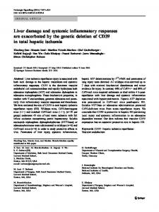
2. Materials and Methods 2.1. Subjects. Eight nonsmoking, systemically healthy female students of the University of Queensland (25.5 ± 7.1 years; range 17–41 years), with no history of periodontal disease, participated in the study. Exclusion criteria included subjects requiring antibiotic cover for dental treatment, those who had antibiotics in the preceding 3 months, or those who were pregnant. Institutional ethics committee approval was obtained to carry out the study. A written explanation of the purpose of the study was provided, and signed informed consent, according to the Helsinki Declaration, was obtained from each subject prior to entry into the study. 2.2. Clinical Study Design. Gingival inflammation was induced in subjects using the experimental gingivitis model developed by L¨oe et al. [16] (Figure 1(a)). At baseline (day 0), subjects underwent a clinical assessment at which time bleeding on probing (BOP) across the full mouth was used to assess gingival inflammation. BOP was examined at six sites per tooth and expressed as a percentage of the total number of sites examined. The plaque index of Silness and L¨oe [27] was used to assess full-mouth plaque accumulation. Subsequently, subjects were asked to abstain from all oral hygiene procedures, including chewing gum, for a period of 21 days. At day 21, subjects underwent a second clinical assessment, received a professional prophylaxis, and were instructed to resume normal hygiene practices. Subjects also
Disease Markers received an Oral-B Triumph powered toothbrush (Procter & Gamble, Cincinnati, OH, USA) to facilitate the resolution of gingival inflammation. Fourteen days later (day 35), subjects underwent a final clinical assessment to confirm the restoration of gingival health. To standardise the sample collection procedure, all participants were requested to provide a diet summary for the 24 h period prior to the appointment. All subjects were given morning appointments and asked not to eat or drink anything except water after 9 p.m. on the previous day. Finally, subjects were asked to floss and brush their teeth 2 h prior to each appointment, except prior to attending on day 21. 2.3. Collection of GCF, Saliva, and Plasma Samples. At each visit, samples of peripheral venous blood, saliva, and GCF were collected. GCF was collected from sites that reflected the gingival status, either the upper premolars or lower incisors. A Millipore filter paper strip with 1 mm width was inserted into the gingival sulcus (Figure 1(b)), left for 30 s (or replaced when almost saturated up to a total sample time of 30 s) and placed in a sterile tube containing 180 𝜇L of buffer (10 mM NaH2 PO4 and 150 mM NaCl, at pH 7.2). Strips visibly contaminated by bleeding were discarded and another sample collected. Unstimulated saliva (2–4 mL) was collected from each individual by allowing saliva to pool in the floor of the mouth followed by expectoration into sterile specimen tubes and then was aliquoted. GCF and saliva samples were stored at −20∘ C before being processed. 2.4. Preparation of GCF, Saliva, and Plasma Samples. GCF and saliva samples were centrifuged (800 g for 10 min) and the supernatants were retained and stored at −80∘ C. Plasma was isolated from peripheral blood following Ficoll-Paque (Pharmacia LKB, Uppsala, Sweden) gradient centrifugation. 2.5. Assessment of Inflammatory Mediators. GCF, saliva, and plasma samples were assessed for levels of cytokines (IL-1𝛽, IL-2, IL-4, IL-5, IL-6, IL-10, IL-12(p70), IL-13, IFN-𝛾, and TNF-𝛼) using a BioPlex cytokine assay kit in combination with the BioPlex manager software (Bio-Rad, Hercules, CA, USA) as well as for CRP and sICAM-1 using high-sensitivity enzyme linked immunosorbent assay (ELISA) kits (Bender MedSystems, Burlingame, CA, USA) according to the manufacturers’ instructions. Results are reported as standard curve units (cytokines: pg/mL; CRP and sICAM-1: ng/mL) [22]. 2.6. Statistical Analysis. It has been reported that methods based on the 𝑡-distribution are more suitable for the analysis of continuous data from small samples than rank (nonparametric) methods [28]. As such, repeated measures general linear models with Bonferroni adjustment for multiple comparisons were used to test for differences in clinical indices and mediator levels over time. Specific changes were examined between baseline and peak of induction (day 21), between days 21 and 35 (resolution of disease), and between day 35 and baseline. For two subjects, the analysis of saliva using the BioPlex system was not possible due to technical reasons. Therefore, the statistical analysis of
Disease Markers
545 Induction phase
Day 0
Clinical assessment Samples collected
Resolution phase
21
Clinical assessment Samples collected
35
Clinical assessment Samples collected
(a)
(b)
Figure 1: (a) Study design and (b) GCF collection.
saliva, with regard to cytokine levels, was performed on six subjects. Pearson correlations were used to examine the associations between clinical indices and mediator levels. Statistical analyses were carried out using SPSS v19.0 (SPSS Inc, Chicago, IL, USA).
3. Results 3.1. Clinical Assessment. The mean BOP and plaque index scores for each visit are shown in Table 1. At baseline, six of the eight subjects had naturally occurring gingivitis (>10% BOP). The results of the general linear model indicated that there was a significant change in BOP over time (𝑃 = 0.001) (Table 1). Gingival inflammation was significantly induced at day 21 with a 3.1-fold increase in BOP (𝑃 = 0.004) compared with baseline (Table 1). Following prophylaxis and resumption of oral hygiene procedures at day 21, plaque index scores decreased 7.2-fold (𝑃 < 0.001; student’s paired 𝑡-test) and BOP decreased 4.9-fold (𝑃 = 0.003) at day 35 to levels lower than baseline (𝑃 = 0.005) (Table 1). 3.2. GCF Mediator Levels during Experimental Gingivitis. Five of the twelve mediators in the GCF demonstrated changes in levels over time: IL-2 (𝑃 = 0.037), IL-6 (𝑃 = 0.012), and TNF-𝛼 (𝑃 = 0.03) at the 5% significance level and IL-1𝛽 (𝑃 = 0.070) and IFN-𝛾 (𝑃 = 0.063) at the 10% significance level (Table 2). There were no significant changes in the GCF levels of cytokines during the induction phase; however, not surprisingly, individual variability in cytokine profiles was noted during this time. At day 35, following the resolution of gingivitis, mean levels of IL-2, IL-6, and TNF-𝛼 decreased 20.0-fold (𝑃 = 0.062; 10% significance level), 16.0-fold (𝑃 = 0.054; 10% significance level), and 16.1-fold (𝑃 = 0.064; 10% significance level), respectively, in concordance with improvement in clinical indices and were significantly lower than baseline levels (𝑃 = 0.003, 𝑃 = 0.025, and 𝑃 = 0.007, resp.) (Table 2). Furthermore, changes in the GCF levels of IL-2, IL-6, and TNF-𝛼 during the resolution phase correlated with changes in plaque index scores (𝑟 = 0.88, 𝑃 = 0.004; 𝑟 = 0.72, 𝑃 = 0.042; 𝑟 = 0.79, 𝑃 = 0.019, resp.). Mean levels of IL1𝛽 and IFN-𝛾 at day 35 were also significantly lower than baseline levels (𝑃 = 0.004 and 𝑃 = 0.014, resp.) (Table 2).
There were no significant changes in the GCF levels of IL4, IL-5, IL-10, IL-12(p70), or IL-13 over time. GCF levels of sICAM-1 and CRP were low or undetectable (Table 2). 3.3. Salivary Mediator Levels during Experimental Gingivitis. Saliva showed higher levels of cytokines than found in GCF; however, these levels did not change over time. As found in GCF, salivary levels of sICAM-1 were low or undetectable (Table 3). 3.4. Plasma Mediator Levels during Experimental Gingivitis. sICAM-1 was the only mediator in the plasma that demonstrated significant changes in levels over time (𝑃 < 0.001) (Table 4). At day 21, following the cessation of oral hygiene procedures, mean levels of sICAM-1 increased 2.6-fold (𝑃 = 0.002), in concordance with increases in clinical indices and remained elevated and significantly higher than baseline levels during the resolution phase (𝑃 < 0.001) (Table 4). All subjects had detectable plasma levels of CRP at baseline. During induction, four of the eight subjects had increased levels of CRP, and all of these subjects had reduced levels at day 35, following the resolution of gingivitis. Plasma levels of cytokines, with the exception of IL-5, were low or undetectable (Table 4).
4. Discussion In the literature there is strong evidence for an association between periodontitis and systemic inflammation, but relatively less is known regarding the systemic effects of gingivitis. This study is one of only two reported studies [25] to employ the full-mouth experimental gingivitis model to investigate the systemic effects of dental plaque. Furthermore, it is the only full-mouth experimental gingivitis model to have simultaneously investigated the inflammatory responses within the GCF and saliva. One limitation of previous experimental gingivitis models has been the use of the split mouth design [19, 24]. This design is suitable for the investigation of local responses to the accumulation of dental plaque but is not suitable when investigating systemic effects. A previous study has shown an association between serum sICAM-1 levels and clinical signs of BOP in periodontitis patients.
546
Disease Markers Table 1: Clinical indices at baseline, peak of induction (day 21), and at resolution (day 35) of experimental gingivitis.
Plaque Index BOP (%)
Baseline — 12.01 (1.28)
Day 21 2.14 (0.12) 37.19 (5.90)
Day 35 0.30 (0.07) 7.64 (1.20)
𝐹 statistic 𝑇(7) = 17.76∗ (1.03, 7.21) = 28.79
𝑃 value