Loricate larvae (Scalidophora) from the Middle Cambrian of Australia ANDREAS MAAS, DIETER WALOSZEK, JOACHIM T. HAUG & KLAUS J. MÜLLER Maas, A., Waloszek, D., Haug, J.T. & Müller, K.J., 2009:12:24. Loricate larvae (Scalidophora) from the Middle Cambrian of Australia. Memoirs of the Association of Australasian Palaeontologists 37, 281-302. ISSN 0810-8889. We report a new species of Cambrian Scalidophora, Orstenoloricus shergoldii gen. et sp. nov., represented by 15 apparently larval specimens from Australia. The tubular anterior region bears approximately 10 or 14 sharp accordion-like ring folds that are finely overprinted by a zigzag pattern. The longer posterior region appears more firmly sclerotised, has a vase-shaped body and is composed of 20 longitudinal plates. This design strongly resembles that of the trunk of socalled loricate larvae of extant Vinctiplicata, one of the scalidophoran taxa among cycloneuralian Nemathelminthes, combining Priapulida and Loricifera. The construction and fabric of the annulated region suggest that it was flexible and could be expanded and invaginated. In addition to those striking features, the fossil larvae expose a pair of socket-like structures at the transition between the annulated region and the lorica that originally appear to have given rise to robust spine- or seta-like outgrowths. Priapulid larvae lack such robust spines. Our specimens fall into at least two size classes, indicating the presence of more than a single larval stage. One of the specimens with a small lorica but a very long annulated region bears a second pair of sockets on the same side as the anterior setae or spines and in about the last fifth of the lorica. This set of possibly very robust setae is not found in priapulid larvae, but a similar set is characteristic for the so-called ‘Higgins larvae’ of Loricifera. Comparisons between the fossils and all available larval types of Scalidophora form the basis of a discussion of the affinities of the fossil larvae with Priapulida and Loricifera. The general design of the annulated region and the lorica and the presence of spines imply a close relationship of the new species with Loricifera. It also implies that the lorica and the multiply folded neck region represent ground-pattern characters of the Vinctiplicata and possibly some other Cambrian taxa. Recent molecular phylogenetic interpretations of the relationships of Loricifera with Nematomorpha heavily affect all in-group relationships within cycloneuralians. These new fossil larvae might therefore stimulate a reconsideration of the morphology and the evolution of characters and structural systems in the discussion of the phylogeny within the Cycloneuralia and also in general. The new taxon adds more to the knowledge of larval and adult Nemathelminthes in the Cambrian, which seem to have been an important faunal component at that time. Maas, A. (
[email protected]), Waloszek, D., Haug, J. T., Biosystematic Documentation, University of Ulm, Helmholtzstrasse 20, D-89075 Ulm, Germany Müller, K. J., Steinmann Institute for Palaeontology, University of Bonn, Nussallee 8, 53115 Bonn, Germany. Received 10 August 2009. Keywords: Phosphatisation, 3D preservation, Priapulida, Loricifera, Scalidophora, Cycloneuralia, loricate larvae, constructional and functional morphology, systematic relationships, phylogeny, evolution.
PRIAPULIDA are a species-poor group of worm-shaped, bottom living to burrowing marine animals (see van der Land & Nørrevang 1985). The 19 modern species range from tiny forms of no more than 2 mm (Kirsteuer 1976 for Tubiluchus corallicola van der Land, 1968) to animals almost 40 cm in length (Shirley & Storch 1999 for Halicryptus higginsi Shirley & Storch, 1999). The systematic relationships of Priapulida have been debated for many years, often without a
phylogenetic approach (e.g. Shapeero 1961). Yet, the more recent interpretation that Priapulida, the minute meiofaunal Loricifera and the similarly small and meiofaunal Kinorhyncha together represent the monophylum Scalidophora (Lemburg 1995) has found considerable acceptance (Ax 2001; Neuhaus & Higgins 2002; Dong et al. 2004). A close relationship of Priapulida and Loricifera as Vinctiplicata (Lemburg 1999) is assumed. Scalidophora is considered the sister
AAP Memoir 37 (2009)
282
A
B
Fig. 1. Recent suggested relationships within Nemathelminthes. A. Scheme proposed by Ax (2001, see also Ahlrichs 1995 and Lemburg 1999). B. Scheme showing the suggestions of Sørensen et al. (2008), with the close alliance of loriciferans to nematomorphs rather than to priapulids.
taxon to Nematoida within the Cycloneuralia. Cycloneuralia and Gastrotricha are united in the Nemathelminthes (cf. Ahlrichs 1995; Ax 2001; see Fig. 1A). Adrianov & Malakhov (1995) postulate a close relationship of Scalidophora and Nematomorpha. This long-held view was challenged recently, when Sørensen et al. (2008) concluded, on the basis of molecular data, that Loricifera should rather be closely allied with Nematomorpha (Fig. 1B). The consequences of this could not be more far-reaching and affect not only the situation of the different higher taxa and their monophyletic status, but also have severe consequences for the interpretation of morphology, developmental paths of features and structural complexes, evolution of anatomical features and life habits. In other words, character state polarities and plausibility of morphological development using the parsimony principle and biological sense are all affected. Cycloneuralia have a cuticle produced by their epidermis. It may be very flexible and contains chitinous matter in the inner layer (Carlisle 1959; Shapeero 1961; Rieger & Rieger 1977; Merriman 1981). It is moulted several times during growth. Parts may also be more firmly sclerotised, as, for example, in the plated forms of the loricate larvae of several priapulids or loriciferans (e.g. Kristensen & Gad 2004 for a representative of Nanaloricidae). A shared feature of scalidophoran species is a retractile pharynx or proboscis, which is armed with rings of mouthward pointing toothlike structures. Well developed pharyngeal teeth and a special post-pharyngeal body region, called the ‘introvert’, similarly armed, but with backward oriented tooth-like structures termed scalids, are exclusive to scalidophorans. The scalids are innervated and may bear gland openings. They occur in a huge number of varieties in the different in-group taxa and are arranged in staggered (i.e. alternating) rows. The pharynx and introvert can be invaginated, which is achieved by specific musculature (e.g. Land & Nørrevang 1985). The introvert serves for locomotion in that the scalids hook onto the surface when the introvert everts
(for a description of the movement see Hammond 1970; Huang et al. 2004a, b). During invagination the pharynx is extruded. This system is also found in the larvae of Loricifera and Priapulida and the young and adults of Kinorhyncha. The trunk portions of living scalidophorans are quite different from one another. Kinorhyncha have a tubular, plated trunk, with a high degree of bilateral symmetry. The tail ends in a conical to weakly bifid end piece with two caudolateral setae. Loricifera have a loricate, rigid trunk throughout postembryonic development. Various structures along the body, mostly on the ventral side, indicate bilateral symmetry. The trunk of Priapulida varies from sac-shaped (i.e. more or less circular in cross section; Land 1970; Calloway 1982) to very elongate and is commonly extended into caudal outgrowths of varying design. Its bilateral symmetry is less distinct than in the other taxa, but is still evident in internal features, such as the ventral nerve cord and paired gonads. Priapulida may be important faunal elements even in anoxic mud (e.g. Halicryptus spinulosus, see Janssen & Oeschger 1992) or hypersaline and brackish waters. Yet they are absent in many regions and are considered to be generally rare (cf. Calloway 1982). Of the 19 extant species, most are meiofaunal to interstitial forms (e.g., members of Meiopriapulus, Maccabeus, Tubiluchus; e.g. Por & Bromley 1974; Morse 1981; Higgins & Storch 1991), while other species are macrobenthic and centimetres long (e.g., Storch 1991; Higgins et al. 1993; Shirley & Storch 1999). This record may be biased by the species’ preferential mud bottom habitat, variously cryptic life strategy, small size and transparency when meiofaunal. Development includes a larval stage with a more firmly sclerotised trunk termed the lorica, but the sclerotisation is abandoned during later growth (Hammarsten 1913; Land 1970). In some species the hatchling does not have a lorica, it is therefore termed pre-lorica larva (Kirsteuer 1976), and it is only the succeeding larval stages, which possess a lorica. Conversely, Higgins & Storch (1991)
AAP Memoir 37 (2009) reported direct development in Meiopriapulus fijiensis Morse, 1981, i.e. a loricate larva does not occur. Loriciferans are no larger than 500 µm. Only about 20 species are described, and they are marine meiofaunal organisms. The trunk cuticle of the animals is divided into several axially oriented plates that articulate with each other, forming the characteristic lorica as described above. The introvert is, plesiomorphically, armed by scalids arranged in rows, but one can distinguish different types of scalids serving various functions (Kristensen 1983, 1991, 2002, 2003). The mouth is located on the tip of a loriciferan-specific mouth cone, which can be intruded into the trunk. It seemingly functions like a pipette to suck on small, mainly unicellular organisms. Development may include a larva (‘Higgins larva’) with structures such as a pair of terminal outgrowths for locomotion (termed ‘toes’), which are missing in adults. The life of Loricifera may also include a parthenogenetically reproducing cycle or other specialisations, which we do not discuss here (see Higgins & Kristensen 1986; Gad 2005a, b; Heiner & Kristensen 2009). Both Loricifera and Kinorhyncha have no fossil record. In contrast to this, several Cambrian taxa have been described and assigned to Priapulida. Conway Morris (1977) noted five taxa in his monograph on Middle Cambrian scalidophoran fossils from the Burgess Shale, British Columbia, Canada. These are: Ottoia Walcott, 1911 (type and only species, O. prolifica Walcott, 1911), not unlike the extant Halicryptus and with posterior hook rings like Maccabeus and Meiopriapulus; Selkirkia Walcott, 1911 (type species, S. columbia Conway Morris, 1977; two other species described); Louisella Walcott, 1911 (type species, L. pedunculata Walcott, 1911), with rows of ventral structures as in Tubiluchus; Ancalagon Conway Morris, 1977 (type species, Ottoia minor Walcott, 1911); Fieldia Walcott, 1912 (type species, F. lanceolata Walcott, 1912). In addition, Conway Morris (1977) mentioned two further species: Scolecofurca rara Conway Morris, 1977, based on a single specimen, and the type species of Lecythioscopa Conway Morris, 1977, Canadia simplex Walcott, 1931, as probable “archaeo-priapulids”. Their status, however, remains obscure. Since then, more fossil putative priapulids have been described from different lagerstätten, mainly from the Lower Cambrian Chengjiang biota of China. These include Maotianshania cylindrica Sun & Hou, 1987 and Cricocosmia
283
jinningensis Hou & Sun, 1988 (Conway Morris & Robison 1986, 1988; Sun & Hou 1987; Hou & Sun 1988; Hou et al. 1991; Wills 1998; Han et al. 2004; Huang et al. 2004a, b; Hou et al. 2006; Huang et al. 2006; see Maas et al. 2007a for an overview of the species from Chengjiang). These species are represented by thousands of specimens, which makes Nemathelminthes one of the most significant faunal components of the Chengjiang biota (Maas et al. 2007a). Only the record of the palaeoscolecids, usually assigned to Priapulida (e.g. Müller & Hinz-Schallreuter 1993; Ivantsov & Wrona 2004), extends into the Ordovician, possibly even into the Silurian (e.g. Mikulic et al. 1985; Boogaard 1988, 1989). This group of rather long, large worms is characterised by an annulated cuticle subdivided into fine plate-like structures of varying sizes (Fig. 2A, B). However, it is at best unclear whether these taxa really are referable to the Priapulida (or the Nematomorpha, see Hou & Bergström 1994), since the shared features mentioned (e.g., scalids, introvert, lorica), if recognised at all, are most likely plesiomorphies, retained either from the scalidophoran or vinctiplicatan ground pattern (see Maas et al. 2007a for a detailed discussion of this matter). In consequence, these fossil taxa may, at best, be considered as scalidophorans or cycloneuralians. Only a single later record, from the Carboniferous Mazon Creek fauna, of Priapulites konecniorum Schram, 1973, which, according to Land & Nørrevang (1985), is very similar to the living species of Priapulus, may well be a member of the Priapulida. Despite the uncertainty of their in-group affinities, it is readily apparent that the Cambrian fossil scalidophorans show a high morphological diversity, which parallels that of extant priapulids. Likewise it seems probable that the former also may have had different life styles, some even unknown among extant taxa. For instance, species of Selkirkia from the Burgess Shale constructed tubes, a mode of life unknown in extant species (Conway Morris 1977). Cycloneuralian larvae are generally small and delicate. It is, therefore, not very likely that such small animals would be found in the fossil record. Conversely, a large amount of palaeoscolecid material is known from various Cambrian lagerstätten, especially from the Middle Cambrian of Australia (Fig. 2A, B; Müller & Hinz-Schallreuter 1993). Embryonic stages of a cycloneuralian have been described as two different species of Markuelia Valkov, 1984 (Dong et al. 2004; Donoghue et al. 2006b, b). In addition to these, specimens of a hitherto undescribed species of Markuelia have been found in the same locality as the larvae described herein
284
AAP Memoir 37 (2009)
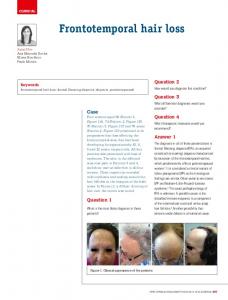
Fig. 2. Examples of nemathelminth and arthropod fragments extracted from Middle Cambrian limestone rocks at Mt. Murray, Duchess Embayment, Queensland. A. Fragment of Austroscolex spatiolatus Müller & Hinz-Schallreuter, 1993 (CPC 39944, spec. 8371, sample 7339). Note the annulated surface with one annulus bearing an abaxial row of large and an abaxial row of significantly smaller button-shaped outgrowths. B. aff. Hadimopanella oezguli Gedik, 1977, cuticle fragment with larger button-shaped structures and finer ornaments surrounding these (CPC 23019, spec. 8288, sample 7324, depicted by Müller & Hinz-Schallreuter 1993, text-fig. 7D, E). C. Still undescribed species of Markuelia Valkov, 1984 (CPC 39946, spec. 9261, sample 7313, depicted by Maas et al. 2007b, fig. 7A [as UB W 133]). D. Post-mandibular limb of an unknown phosphatocopine (CPC 30717, sample 7407, depicted by Walossek et al. 1993, fig. 4A, B). E. Type-a larva (CPC 30711, sample 7323, depicted in Walossek et al. 1993, fig. 1A). F. Multi-annulated limb fragment, possibly representing an exopod of a non-crustacean (CPC 30719, sample 7324, depicted by Walossek et al. 1993, fig. 5A, B). G. Shergoldana australiensis Maas, Waloszek, Haug & Müller, 2007 (CPC 23065, sample 7323, depicted by Maas et al. 2007b, figs. 1–5 [as UB W 277]).
(Fig. 2C; Donoghue et al. 2006b; see also Haug et al. this volume). This locality has also yielded various arthropod remains (Fig. 2D–F). Another exceptional record of an immature form from the same locality is a single larval specimen assigned to the species Shergoldana australiensis Maas, Waloszek, Haug & Müller, 2007b (Fig. 2G). All these immature forms and the palaeoscolecids are three-dimensionally preserved as secondarily phosphatised cuticle of the animals, which can be isolated from their enclosing limestone by using mild acetic acid (see Müller 1985). This ‘Orsten’ type of preservation seems to be restricted only to small fossils but not, however, to specific regions (e.g. Maas et al. 2006). Therefore it was not too surprising to find more small animals in the residues (Walossek et al. 1993) and even some resembling the loricate larvae of priapulids. Here we describe and illustrate several specimens of this type of fossil, which most likely represent larval semaphoronts. The material consists of small specimens possessing a vase-shaped, lorica-like trunk, strikingly similar to that of the meiofaunal priapulid Tubiluchus corallicola Land, 1968. Closer inspection, however, uncovered detailed features that make a relationship to priapulids less likely but indicates a closer affinity to
the Loricifera. For systematic clarity we have decided to assign the larvae to the new species Orstenoloricus shergoldii gen. et sp. nov. The other Middle Cambrian larva from Australia, the early semaphoront, Shergoldana australiensis, has clearly different features, but the phylogenetic interpretation of our new finds do not conflict with those drawn by Maas et al. (2007b). The documentation of the larvae of the new species will be followed by a comparison between them and the larvae of priapulids and loriciferans. For this we constructed models of the lorica of the new form and extant larvae using van der Land’s comprehensive description of Tubiluchus corallicola. Our aim was to understand the functionality of such a body design, barely altered since the Cambrian, and likewise, to understand the possible life habits of the fossil species. The comparisons also aim at testing whether the form displayed by the new fossil larvae and by Tubiluchus corallicola, which is regarded as sister species to all other extant priapulids, reflects a plesiomorphic design among priapulid larvae or is plesiomorphic for Vinctiplicata or even Scalidophora. Lastly, we try to identify whether our fossil has more in common with the one or other vinctiplicatan in-group. The discovery of
AAP Memoir 37 (2009)
Fig. 3. Scheme of the two types of larvae in the material and lengths obtained from them. A. Common type. The neck region is much shorter than the lorica. The anal region (ar) is relatively narrow. B. Second type represented by the single, differing specimen (see also Fig. 10). The neck region comprises more folds than in the common type and is more or less as long as the lorica. As in the common type, anterior spines (asp) are present, additionally a pair of posterior spines (psp) is present anterior to the broader anal region (ar).
these larvae of a new fossil species within the cycloneuralians, and the unexpected larval type demonstrated by Shergoldana australiensis, adds significantly to the ongoing discussion of cycloneuralian and scalidophoran phylogeny, and to the problem of where such a specific larva type such as the loricate larva might first have occurred. The recent molecular phylogenetic interpretation of the relationships of loriciferans with nematomorphs by Sørensen et al. (2008) has a strong bearing on all in-group relationships within cycloneuralians. Our study aims, therefore, to stimulate a reconsideration of morphology and the evolution of characters and structural systems in the discussion of any phylogeny, rather than simply to accept molecular studies without reservation. The new taxon adds to the still incomplete record of nemathelminths in the Cambrian, which apparently were a very important faunal component at that time, possibly even more so than today.
285
MATERIAL AND METHODS Material The material, 15 specimens in all (CPC 39929– 39943), was extracted from limestone collected from outcrop one kilometre north of Mt. Murray (E 139° 58’ 27.6’’ S 21° 48’ 50.4’’). This is a small hill at the margin of the Georgina Basin, south of the city of Mt. Isa in the Duchess Embayment, Queensland, Australia. The material was collected by Raimond Below and Dieter Waloszek during a field trip guided by the late John Shergold in 1986 (more details given by Müller & Hinz 1992; Müller & Hinz-Schallreuter 1993) and includes a series of other elements (Fig. 2A–G). Eleven specimens are from one sample (no. 7324), which also yielded arthropod appendage fragments including a piece of an exopod of possibly a ‘trilobitoid’ arthropod (cf. Walossek et al. 1993) and an undetermined palaeoscolecid. Four more samples (nos. 7325, 7342, 7339 and 7374) were from other beds of the same outcrop and yielded single specimens only. In samples 7339 and 7374 only, the specimens co-occurred with palaeoscolecids (Fig. 2A). Preservation As in the Swedish ‘Orsten’ material, only the integumental surface (= cuticle) is preserved in these Middle Cambrian fossils from Australia. Accordingly, most of the specimens are hollow or filled with secondary phosphatic matter, which may even be an effect that occurred during the preparation process. In all, the Australian material is not as well preserved as the Swedish ‘Orsten’ material. A few specimens may represent steinkerns, a possibility with the poorly preserved type-A larvae from the same Australian locality (Walossek et al. 1993) or from the Lower Ordovician of Newfoundland, Canada (Roy & Fåhræus 1989). Most of the Australian specimens are contorted by collapsing prior to phosphatisation. The surface generally appears as a coarse, irregular phosphatic layer, possibly not quite reflecting the original uppermost cuticular layer (epicuticle). Surface details are, therefore, only rarely preserved and restricted to small areas. The quality difference from the Swedish material could be due to differences in the sedimentary/ early diagenetic conditions, but this is unclear. Processing To extract phosphatised fossils, the rock was etched with 15% acetic acid (process described by Müller 1985; Müller & Walossek 1985). The residues were graded in size during the dissolution process on two sieves. The described specimens have been picked from only the finest fraction.
286
AAP Memoir 37 (2009)
AAP Memoir 37 (2009)
287
Fig. 5. Morphometrical data. A. Lorica length (x) against neck length (y). B. Total length (x) against lorica length (y). Note that in the two proportions specimen CPC 39929 (Fig. 4A) (filled circle) is clearly different from the rest. All other specimens CPC 39930–39943 (open circles) appear to be a single unit (enclosed by stippled line), although most likely represent several developmental stages.
Other methods The isolated specimens were glued on stubs for electron microscopy study. They were originally coated with a layer of gold about 20 years ago. Before studying we recoated the stubs using a gold-palladium mixture. The coating process stopped automatically after reaching a 20 nm thick layer. The material was studied under a Zeiss DSM 962 scanning electron microscope, which provides digital grey-scale images of 1024 × 1024 pixel size. Computer-aided image processing and graphics were made using ADOBE Photoshop CS™ and ADOBE Illustrator™. Some images are fusion products of two images stitched manually in Photoshop. Terminology Terminology follows, as far as possible, Land (1970) and other workers on the scalidophoran groups (e.g. Malakhov and co-authors; Lemburg 1999 and references therein; Fig. 3A, B). Yet, in several cases we found that the terminology in use for the different taxa not only differs between authors or contains inconsistencies (see also Neuhaus & Higgins 2002), but also has apparently been adopted from other animal groups, such as arthropods. For example, the use of ‘head’ for the introvert, ‘thorax’ or ‘abdomen’
for the region behind the introvert (herein termed neck), have been avoided. Lastly, we also regard it as inadvisable to use the term ‘appendages’ for certain outgrowths of the scalidophorans, even if they are mobile setae (e.g., Gad 2004) or toes, and apply other terms (e.g. setae or scalids) to avoid confusion. We prefer to use the terms mountain folds (instead of primary ridges) and valley folds (instead of secondary ridges), commonly used in the paper folding technique of origami. RESULTS General aspects In gross morphology all available specimens of the new Australian species are rather similar to each other. All specimens consist of two major regions. The anterior one is annulated and comprises between nine and eleven or, in one case, 14 ring-shaped folds. It is referred to as the neck region in the following (Fig. 3A) and is about as wide as the posterior region. The latter is an elongate sac-like trunk formed by plate-like structures and interpreted as a lorica. Preservation varies between specimens. Well developed plates occur only in a few specimens; in some they are visible while in others, only ridges are obvious (cf. Fig. 4A–O). This may not be a preservational artefact but
Fig. 4 (opposite). Orstenoloricus shergoldii gen. et sp. nov. SEM micrographs. A. CPC 39929, spec. 8372, having two sets of spines (arrows), the first pair is located at the rim around the anterior end of the lorica, a putative second pair being located posteriorly on an abaxial ridge where the lorica merges into the terminal area. B–O. Specimens having only the anterior pair of spines (arrowed in some images). B. CPC 39930, spec. 8287. C. CPC 39931, spec. 8373. D. CPC 39932, spec. 8661. E. Holotype CPC 39933, spec. 8663. F. CPC 39934, spec. 8664. G. CPC 39935, spec. 8665. H. CPC 39936, spec. 8880. I. CPC 39937, spec. 9000. J. CPC 39938, spec. 9052. K. CPC 39939, spec. 9053. L. CPC 39940, spec. 9054. M. CPC 39941, spec. 9055. N. CPC 39942, spec. 9056. O. CPC 39943, spec. 9751. Arrows point to anterior spines. Scale bars: 50 µm.
AAP Memoir 37 (2009)
288
CPC 39929 39930 39931 39932 39933 39934 39935 39936 39937 39938 39939 39940 39941 39942 39943
sample 7339 7324 7374 7324 7324 7324 7324 7324 7342 7324 7324 7324 7324 7324 7325
tl 820 700 900 950 480 650 570 840 700
ne 440 200 250 300 140 210 170 270 280
lo 380 500 650 650 320 480 440 400 570 470 700 600 710 420
max. ø ne 190 180 270 230 225 120 190
max. ø lo 250 170 240 180 130 175 -
210 200
160 180 200 170 -
Table 1. Measurements of specimens at hand. Specimen unlike the others (CPC 39929) highlighted by grey background. All values in µm; tl = total length, ne = length of neck; lo = length of lorica; max. ø ne = maximum diameter of neck; max. ø lo = maximum diameter of lorica.
may indicate that the plates were less distinct in some speciens. An explanation could be that they were less sclerotised at the time of death of these specimens, perhaps after moulting. Very rarely are remains of a more anterior region preserved. These indicate the original presence of more structures at the front – in the case of priapulids this would be the introvert and pharynx. All specimens are, more or less, collapsed, but it is very likely that both neck and lorica were almost circular in cross section originally. Morphometric measurements (Table 1) uncovered slight differences between the specimens (and there are a few morphological differences, too). The results indicate there are two different morphotypes in our material (Table 1). One is characterised by having a lorica generally twice as long as the neck (Fig. 5A), i.e. the proportion of total length to lorica length is about 1.5 to 1 (Fig. 5B). This (Fig. 4) covers a large proportion of the size range of the material, with lengths from 480 µm to 950 µm. Likewise the lorica lengths range from 320 to 710 µm. This indicates that, although growth parameters are similar, the larger first morphotype may contain specimens of more than a single developmental stage. Since morphology also separates a single small specimen as a second morphotype (Fig. 4A), while there are also significant similarities, we describe this larva separately.
Body of the loricate larvae – morphotype I Neck. The anterior part of the larvae is characterised by 9–11 annular folds. We term this region the ‘neck’ to emphasise the similarity to a corresponding region in the larvae of priapulids and loriciferans (Fig. 6A, B). The neck was most likely circular in cross-section originally and lacked any signs of bilateral symmetry. Fewer folds in some specimens are merely due to incomplete preservation, whereas a twelfth fold may belong to structures in front of the folded neck. In those eight specimens with the maximum number of neck folds, the neck tapers in diameter to approximately two thirds of the maximum width (Fig. 6C). The annular folds are arranged accordion-like with sharp mountain folds and even slopes leading to distinct valley folds. Moreover the mountain folds are overprinted by a zigzag pattern, and a fine fold runs downward from each inflection of the zigzag to the valley fold (Fig. 6D–F). In several specimens this is less distinct or even effaced due to the coarse preservation. Functional aspects of the neck. The varying degree of flattening of the folds points to the possibility of considerable elongation of the neck. Contraction was also possible, perhaps to a maximum when the mountain folds approached each other. The finer folds and zigzag pattern indicate that
AAP Memoir 37 (2009)
289
Fig. 6. Neck region and spines at transition to lorica (termed anterior spines). A–D. Holotype CPC 39933. E. CPC 39938. F. CPC 39930. G. CPC 39936. H. CPC 39938. I, J. CPC 39931. Arrows point to the anterior spine (or its socket).
290
AAP Memoir 37 (2009)
Fig. 7. Analogue to the fold pattern of the neck region of the fossil larvae, using the fabric type ‘crinkle’. A. 10 annuli of the sleeve of a jumper made of this fabric. B, C. Several annuli rolled inwards (arrows), in lateral (B) and frontal (C) view. D. Overstretched condition with annuli and secondary fold pattern smoothed. E. Sleeve curved (arrow) to test torsion and lateral flexure.
the neck was able to curve to some degree, but it was also able to increase or decrease in diameter or possibly even to invaginate. We tested this by choosing an analogous system present in the folds of a certain fabric called ‘crinkle’. The model in Figure 7 is part of the sleeve of a jumper made of this fabric (about 10 folds shown; Fig. 7A), demonstrating the possibility of invagination (Fig. 7B, C), extreme stretching (Fig. 7D), and curving (Fig. 7E). In this model case the secondary pattern is even more complicated since it has a starshaped overprint. Yet, it demonstrates as much shape stability – folds sustain the shape virtually automatically – as it permits high flexibility. The neck region lacks any joints, as developed in arthropods, but can be as much stretched (in life this would be performed by the haemolymph of the primary body cavity) as it can be compressed (in life by musculature). Transition zone to the lorica. If present, the posteriormost annulus forms the transition to the lorica and seems to have a less distinct zigzag pattern. Spine-like outgrowths arise from its
posterior slope (Fig. 6G–I) pointing posteriorly and outwardly. These spines have been recognised on eight of the fourteen specimens, while in three more specimens they are less clear. Generally, only the sockets and/or proximal portions of the spines are preserved – approximately 25 µm in diameter (Fig. 6H), the longest spine piece available (CPC 39931, Fig. 4C) is about 30 µm long. Their robustness suggests an original length of more than 50 µm. It remains unclear if these spines were articulated. More than two spines could not be verified in any specimen, usually only one could be clearly identified. It seems likely that such spines occur only on one side, but regrettably we could not confirm with certainty, since we cannot easily remove such fragile phosphatised specimens from the SEM stubs without risking destruction. Lorica. The trunk is an elongate, sac-like portion. Its surface is subdivided into 20 axially oriented plates (Figs. 8A–D, 9B), which seem to be more strongly sclerotised than the neck region. We term it the lorica because of its similarity to the
Fig. 8 (opposite). Views of the lorica of the described fossils. A–C. CPC 39935 (cf. Fig. 4G). A. Complete view. B, C. View of posterior end from different angles. Note the distinct star-like pattern with mountain and valley folds of the lorica plates. D–F. CPC 39940 (cf. Fig. 4L). D. Frontal view of the lorica. Plates or lorica numbered. E. Postero-lateral view. F. Close-up of the terminal end including the putative anal region. G. CPC 39934. Arrows point to the terminal slit.
AAP Memoir 37 (2009)
291
292
AAP Memoir 37 (2009) manner (Fig. 8E), which is however interpreted as an artefact caused by shrinkage, possibly because this region was softer than the rest of the lorica. Surface structures such as tubuli (a feature of priapulids) have not been observed. Preservation differs among the specimens, and only in some cases are the plates distinct. They are most clearly preserved in CPC 39935 (Figs 4G, 8A–C). In other cases, the plates are rather indistinct, and only their borders, mostly the mountain folds, are marked (e.g. Fig. 6I, J). This could be explained by the death of the individual in a different part of the moult interphase, such that the cuticle in some specimens is more rigid than in others. Another preservational effect is that all specimens have apparently collapsed after death. The main part was most likely circular in cross section, while the posterior end was more flattened. Terminally the plates merge into a small blunt area devoid of folding, where the lorica plates merge at a transverse ridge. A slit-like depression is located in the centre of this area in at least five of the larvae (Fig. 8E, F), and is interpreted as the anal opening. The lorica of O. shergoldii tapers gently into a bluntly rounded end posteriorly (Fig. 8F).
Fig. 9. Redrawing of a paper model of a lorica made of 20 plates with a blade-like design. A. Single plate of the lorica of Orstenotubulus shergoldii gen. et sp. nov. with one straight and one curved (bladeshaped) side. B. Profile of loricae inflated. C. Cross section of a lorica in inflated condition (valley folds up, introvert invaginated), numbers counting all folds (redrawn from Malakhov & Adranov 1995). D. Profile of loricae in deflated condition (cf. Land 1970) mf: mountain fold, vf: valley fold. E. Cross section of a lorica in deflated condition (valley folds down, introvert extruded).
loricate larvae of priapulids and loriciferans. The 20 individual plates of the lorica are positioned axially along the trunk in one dextral and one sinistral row, abutting each other (Figs 8A, 9B, D). This connective margin represents the valley fold. The opposite axial side is curved. The margin curves anteriorly slightly inward, then is gently convex and finally curves gently inward again posteriorly. The curved margins of two adjacent plates are again connected with each other, the connective margin being the mountain fold. Accordingly, the single plates attain a kind of knife-blade shape (Fig. 9A). In CPC 39940 (Fig. 4L) and CPC 39942 (Fig. 4N) the cuticle is folded in the middle of the lorica in an accordion-like
Functional aspects of the lorica. The blade-like shape of the plates of the lorica has a peculiar effect. When the valley folds are folded inward – i.e. when the anterior body was evaginated – the mountain folds are sharply raised (Fig. 9D, E). When the valley folds pop up to the level of the mountain folds – a situation that refers to the invagination of the anterior body – the lorica widens to the maximum of the curved valley folds (Fig. 9B, C). The anterior constriction hinders the lorica from further widening and keeps it in shape, which is more sac- or vase-shaped. Virtually the same mechanism has been described by Land (1970) for the loricate larvae of the extant Tubiluchus corallicola (see Fig. 9 showing our paper model, produced to test this mechanism). Other surface structures. Apart from the sockets of possibly long spines or setae we did not find any other surface structures. However, detection of fine surface structures is rather unlikely because of the rather coarse preservation of all specimens. It is even possible that the top layer, the so-called epicuticle, is not preserved at all. As a consequence, all epicuticular structures would be lacking. In the Swedish ‘Orsten’ arthropods, particularly crustaceans, we know that all fine structures are epicuticular, while only larger structures are visible on underlying cuticular layers (cf. Maas et al. 2006).
AAP Memoir 37 (2009)
293
Figure 10. SEM images of CPC 39929, specimen set off from the rest by its possession of a second pair of spines in the posterior fifth of the lorica, but on the same side as the anterior pair (possibly the ventral side). A. Side view. Note the well-defined anterior spines (asp). B. Top view. The relative length of the neck (ne) compared to the lorica (lo) is apparent in this view. C. Detail view, highlighting the lorica with the anterior and posterior spines (psp). D. Terminal view. Above the anal region the bases of the posterior spines are apparent in this view. E. Detail of D. Base of left posterior spine.
294
Features of CPC 39929 (morphotype II) One specimen, CPC 39929 (Figs. 4A, 10A–E), differs from all other specimens, even those of a similar size, in three aspects. These differences are: firstly, in the relationship between neck length and lorica length; secondly, in the relatively large anal area, starting at an abaxial ridge; and thirdly, in the presence of a pair of sockets of possibly posteriorly and outwardly pointing setae or spines on this abaxial ridge in addition to the anterior set of setae or spines (Fig. 10B, C). Although the neck, which has about 14 folds – compared to approximately 10 in all other specimens – is much flattened and stretched, it is longer than the relatively short lorica, while in all other specimens the neck is roughly half as long as the lorica (Table 2, Fig. 5A, B). Even assuming that the neck could be expanded to some degree (see also Fig. 7A, D), this still would not conform to the relationships of the other specimens. The sockets of the posterior spines are rather prominent (Fig. 10D), and the amorphous infilling suggests the spines were rather robust (Fig. 10E). Similar sets of spines or setae occur in loriciferan larvae, but not in priapulids, and they mark the ventral side of the animal. Hence it is possible that this holds true also for this specimen, as well as the others, which possess only the anterior set of spines. A fourth, less distinct difference from the rest of the material is that the anal region is more constricted and the terminal (anal?) end less slitlike (Fig. 10D). Summary In summary, several features of the material studied are noteworthy: 1) The presence of one specimen (CPC 39929), which is generally similar but differs in some specific features from all other specimens; 2) a large plasticity in length within the larger set of specimens (CPC 39930–39943) with a difference in lorica lengths of more than 100%; 3) an anterior region, termed the neck, made of about 10 fine annular folds with an overprinted pattern of a fine zigzag curvature of the mountain folds and fine folds running from the points of change down toward the valley folds; 4) a system of 20 plates, standing at an angle against each other, which results in 10 mountain folds and 10 valley folds; the cross section is circular, with the folds standing up star-like around the lorica; 5) the presence of posteriorly and slightly outwardly pointing spines, possibly only on one side of the animal: a pair of possibly robust anterior spines at the annulus between neck and lorica, and, in one specimen (CPC 39929), another pair of even more prominent spines nesting in
AAP Memoir 37 (2009) sockets at the transition from the lorica plates into the anal region; 6) the size range of neck and lorica together is between 500 and 900 µm; estimating additional 200 µm for an introvert leads to estimates of body lengths of the larvae between 700 µm and 1,100 µm. SYSTEMATIC PALAEONTOLOGY NEMATHELMINTHES Gegenbaur, 1859 (= Aschelminthes Grobben, 1910) CYCLONEURALIA Ahlrichs, 1995 (= Introverta Nielsen, 1995) SCALIDOPHORA Lemburg, 1995 VINCTIPLICATA Lemburg, 1999 Orstenoloricus shergoldii gen. et sp. nov. Holotype and horizon. CPC 39933 (spec. 8663), total length 950 µm, possibly a late larval semaphoront of the species (Fig. 4E). Locality: Monastery Creek Formation, approximately 1 km north of Mt Murray 139° 58’ 27.6’’ E, 21° 48’ 50.4’’ S, Duchess Embayment, Queensland, Australia (sample 7323). Age: late Templetonian, Middle Cambrian. The specimen is housed in the Commonwealth Palaeontological Collection (CPC) of Geoscience Australia, Canberra. Additional material. Thirteen additional paratype specimens CPC 39930–39932 (Fig. 4B–D) and CPC 39934–39943 (Fig 4F–O), ranging from 380–900 µm in total length (Tab. 2). Another, morphologically slightly different, fourteenth paratype specimen CPC 39929 (Fig. 4A) is here also treated as belonging to the same species. All paratype specimens are from the same horizon as the holotype. Etymology. Generic name derived from the specific type of preservation and the form of the trunk of the only semaphoront known so far. Species name after the late John H. Shergold, dedicated Australian palaeontologist and leader of the field trip in 1986, during which the rock sample containing the fossils was collected. Diagnosis. Body tubular, made of at least two regions. Anterior region (neck) of 10 ring-shaped folds with a fine subordinate zigzag-like folding of the ridges (mountain folds) and fine ridges running anteriorly and posteriorly down the slopes of the ring folds toward the centre of the valleys (valley folds). Posterior region developed as a better sclerotised sac-like structure with specifically sclerotised cuticle, the lorica. Lorica made of 20 dextral and sinistral elongate plates
AAP Memoir 37 (2009)
295
Fig. 11. Illustrations of larval loricae of extant Vinctiplicata. A. Tubiluchus corallicola Land, 1968 (Priapulida) redrawn from Land (1968). B. Priapulus caudatus Lamarck, 1816 (Priapulida), dorso-lateral view, redrawn from Land (1970). C. Priapulus tuberculatospinosus Baird, 1868 (Priapulida), lateral view, redrawn from Land (1970). D, E. Halicryptus spinulosus Siebold, 1849 (Priapulida) in dorsal/ventral view (D) and lateral view (E) simplified after Storch & Higgins (1991). F. Higgins larva of Pliciloricus pedicularis Gad, 2005a (Loricifera) simplified after Gad (2005a).
with one straight and one curved long margin. Posterior tapered to a blunt end. Up to two pairs of spine-like setae on ventral side, one at the transition between neck and lorica, another one close to the posterior end of the lorica. Remarks. This semaphoront of Orstenoloricus shergoldii is significantly different from the single larva of the possible scalidophoran species Shergoldana australiensis from the same locality. The only known specimen of S. australiensis is not only a fraction of the length (120 µm) of the new form, but it also has completely different body regions: The putatively post-oral annulated region of S. australiensis is made of spiral folds, and the trunk is made of two parts, a region with twelve five-sided plates from which posterodistally pointing and slightly curved spines arise, flanked by smaller secondary spines, and a short bifurcate region. The entire trunk is covered with fine setulae or denticles. The spine-bearing plates clearly resemble the pharyngeal teeth of scalidophorans, but they are turned through 180 degrees. A lorica is not present. Also, the S. australiensis larva ends in a short bifid end, therefore is most likely complete, (possibly the plesiomorphic state within cycloneuralians and nemathelminths in general; see Maas et al. 2007b). It seems unlikely to us that the two taxa are successive stages of the same species. Species of Markuelia are known only from a late embryonic, possibly hatchling stage from Cambrian rocks from, e.g., China and Australia
(Fig. 2C; Dong et al. 2004; Donoghue et al. 2006a, b; Haug et al. this volume). The hatchling is a scalidophoran worm of at least 2 mm length. Its affinities are unclear. It cannot be interpreted as an early embryonic stage of any known Cambrian scalidophoran including Shergoldana australiensis (see Maas et al. 2007b) and the new species O. shergoldii, in particular because the latter are much smaller than the Markuelia embryo. DISCUSSION Comparisons with other vinctiplicatans Lorica and neck region in priapulids. The larvae of Orstenoloricus shergoldii gen. et sp. nov. have a lorica comprising 20 connected plates, which stand in pairs against each other at an angle. This results in the formation of valley and mountain folds. Land (1970) described such a plate system in detail for the extant priapulid Tubiluchus corallicola, named and numbered it precisely and also described the mechanism in operation when the anterior body – neck plus introvert and pharynx – is intruded and extruded. Yet, most authors have not clearly referred to mountain and valley folds but sometimes only refer to ridges (e.g. Land 1968, who partly described the T. corallicola larva plates correctly [his figs. 2, 3] but partly not [his fig. 12]); though sometimes mentioning thicker and thinner ones. Larvae of Tubiluchus corallicola have a strikingly similar lorica (Kirsteuer 1976; Land 1982; Higgins & Storch 1989) to that of O. shergoldii including
296
the anterior constriction and mountain and valley folds, but in T. corallicola larvae the lorica is swollen and vase-shaped (term of Sanders & Hessler 1962) and equipped with fine tubuli and many sensory structures (e.g. flosculi). As in O. shergoldii, the lorica of T. corallicola is radially symmetrical (Adrianov & Malakhov 2001a). In larvae of other priapulid species, the number of plates might be significantly smaller than 20. Lemburg (1999) argues that in Eupriapulida, the lorica consists of one large plate ventrally and dorsally, connected by a series of 6 plates laterally, making only 14 plates in all. In fact the rigid dorsal and ventral plates originated from four plates each, which had fused during evolution. The loricae of these eupriapulid larvae have a dorso-ventrally flattened, rather box-like appearance with a rectangular cross-section (Land 1970; Higgins et al. 1993; Adrianov & Malakhov 2001a). In particular, the six lateral plates allow widening of the lorica and therefore enable the introvert to be invaginated. Accordingly, even in the presence of the rigid ‘megaplates’ it is still possible for the widening mechanism to operate. The loricae themselves are bilaterally symmetrical, which is also supported by a dorsoventral flattening of the loricae. Comparison of the larvae of O. shergoldii and T. corallicola (Fig. 11A) with the more strongly sclerotised box-form larvae mentioned above, reveals that in older literature the similarity went unnoticed because of the lack of recognition of the plating arrangement. More strongly sclerotised dorsal and ventral plates (Fig. 11B–E) were mostly counted as single plates only. Since priapulid workers apparently counted these plates differently, their descriptions of a variety of plates or folds in the various priapulid larvae was illusory. The lorica design was long seen as a clear difference between T. corallicola, considered to reflect the plesiomorphic condition (e.g. Land & Nørrevang 1985), and the species referred to Eupriapulida Lemburg, 1999. The modification of the lorica in the form of the fusion of plates into larger units leads automatically to a less flexible lorica, but which is counteracted by flexible sides – and a stronger bilateral symmetry. Another similarity between T. corallicola and O. shergoldii is in the arrangement of the tubuli (in T. corallicola) and similar outgrowths (in O. shergoldii) on the mountain folds. In the ‘box larvae’ of the Eupriapulida these are located in the posterior region of the lorica in two abaxial rows (2 × 4 tubuli; Fig. 11B, C), while they are arranged around the lorica in T. corallicola in five circlets (Fig. 11A). The neck region of O. shergoldii is also very similar to that of T. corallicola. However, the neck
AAP Memoir 37 (2009) of O. shergoldii has about ten folds, while that of T. corallicola has two in the youngest to eight folds in the oldest larvae (Kirsteuer 1976). Other taxa have an inconspicuous neck region without any folds but additional abaxial plates immediately anterior to the lorica (Lemburg 1999) that aid in the closure of the lorica when the introvert is withdrawn. Both of these characters are regarded as eupriapulid autapomorphies (Lemburg 1999). With this, the assumption of Land & Nørrevang (1985) that priapulid larvae are rather uniform, is shown to be in error here because there are at least three different larval types, the T. corallicola type and two different types among those having megaplates. In addition, the terminal region of the loricae of O. shergoldii is very similar to all fossil and extant priapulid larvae, in tapering to a blunt end and containing a slit-like opening. The ‘Higgins larva’ of Loricifera is similar to this but is characterised by a pair of terminal outgrowths termed toes, which only occur within Loricifera. While the lorica is lost at metamorphosis to the adult priapulid, it remains the characteristic design of adult Loricifera. Since the lorica may reduce the flexibility and motility of the trunk, as known in adults, such larvae locomote either by using their introvert scalids for hooking onto the substrate and pulling themselves along, as in priapulids, or by the development of mobile, propelling ‘toes’ as in loriciferan larvae. Lorica and neck region in loriciferans. Loricifera also have loricate larvae – their adult stages retain this feature. Here the plates are called plicae, and they have a minimum of 22 plates. Lemburg (1999) states that there are only 20 plates and given that there are also twenty plates in priapulid larvae, he postulates the possibility of 20 plates for the ground pattern of Loricifera. The largest number is developed in the earliest larvae, called the ‘Higgins larvae’. The lorica ranges from being rather radially symmetrical (Fig. 12B, C; Gad, 2005b) to bilaterally symmetrical (cf. Gad, 2004). The bilateral symmetry may be stronger on one side than on the other; one side is specialised with fusion of the lorica plates in various ways during ontogeny, while the other side may still display a more radially symmetrical condition (e.g. Higgins & Kristensen 1986; Gad 2004). The system of fusion is never the same as in priapulids, so it is most likely a matter of convergence. Both the larvae of O. shergoldii and those of Loricifera have seta- or spine-like structures on one side (ventral in extant forms), but the number and arrangement of the plates is different. Like the larvae of Orstenoloricus shergoldii and Tubiluchus corallicola, Loricifera have a
AAP Memoir 37 (2009)
297
Fig. 12. Scanning electron micrographs of scalidophoran larvae. A. Pre-loricate larva of the priapulid Tubiluchus sp. (reproduced by kind permission of D. Scharf). B. Loricate larva of Priapulus caudatus Lamarck, 1816 (reproduced by kind permission of S.A. Wennberg, Uppsala; from Wennberg 2008, fig. 3). C. Higgins larva of the loriciferan Armoloricus elegans Kristensen & Gad, 2004 (reproduced by kind permission of R.M. Kristensen, Copenhagen).
prominent neck region (often named thorax), which is also made of annular folds, a zigzag pattern and subordinate crossing folds (Fig. 11F). However, in contrast to O. shergoldii, there is only a maximum of 6 folds, fewer than that occurring in O. shergoldii and in Priapulida. In the Loricifera it seems that only the pliable neck is infolded (as described above, see also Fig. 7B, C), when the introvert is invaginated. Other aspects. The size differences in the material suggest the presence of several size classes, possibly of different developmental stages. There are hardly any comparable data because the number of instars within priapulids is poorly known. Kirsteuer (1976) specified six larval stages for Tubiluchus corallicola, and the size increase between the first and last instar is reported to be about 300%. Within priapulids, there may be a pre-loricate stage, as in T. corallicola (Kirsteuer 1976), as well as loricate larvae which moult several times. However, the number of moults may vary and in some cases, direct development also occurs (e.g., in Meiopriapulus fijiensis, see Higgins & Storch 1991). In contrast to priapulids, Loricifera have evolved very complicated life cycles including different forms of paedogenetic and parthenogenetic reproductive cycles (see above). It is therefore difficult to determine
the ground pattern for these taxa. Therefore, it remains difficult for us to assess if the unusual larva in our material, CPC 39929 (Fig. 4A), is the smallest stage preceding the others, or if it belongs to another species. If conspecific, one would have to assume that the larva changes the proportions of its parts and loses the posterior set of spines when moulting to the next stage. Significance for systematic relationships within scalidophorans The specimens of Orstenoloricus shergoldii gen. et sp. nov. possess some characters that can be discussed in a phylogenetic context with regard to the larvae of other vinctiplicatans: 1. Orstenoloricus shergoldii has a prominent neck with a number of folds (Fig 6A–F). Within priapulids such a distinct neck region with a fold system occurs only in the larvae of the taxon Tubiluchus (Fig. 12A). Members of the Eupriapulida do not have this feature, which is interpreted as an autapomorphy of that taxon (Lemburg 1999). Within Loricifera a prominent neck region occurs in the Higgins larvae (Fig. 12B, C). In accordance with Lemburg (1999), we regard the folded neck region of O. shergoldii as a plesiomorphic character, being part of the ground pattern of Vinctiplicata (Fig. 13). 2. The presence of a lorica in O. shergoldii
298
AAP Memoir 37 (2009)
Fig. 13. Argumentation scheme of the different possible positions of the new larva within the Scalidophora (cf. Lemburg 1999: Kinorhyncha as the sister taxon to the Vinctiplicata comprising Loricifera and Priapulida), and the putative design of the lorica in the ground patterns (in the stem species) at the appropriate nodes.
(Fig. 4A–O) is, in accordance with other members of the Vinctiplicata, a character regarded as an autapomorphy of that taxon (Lemburg 1999). Therefore, O. shergoldii is at least a member of this taxon, the lorica being a plesiomorphic feature (Fig. 13). 3. The lorica of O. shergoldii consists of 20 blade-like plates (cf. Fig. 8A–D). The same number has been included in the ground pattern of Vinctiplicata by Lemburg (1999). Our data on O. shergoldii support Lemburg’s (1999) interpretation. The condition in O. shergoldii would, hence, be plesiomorphic (Fig. 13). 4. The loricate trunk of O. shergoldii is radiallysymmetrical. This is in accordance with that of the larvae of species of Tubiluchus (Adrianov & Malakhov 2001a), while eupriapulids have a boxlike lorica. Since radially-symmetrical loricae also occur within loriciferans, it seems most parsimonious that a radially-symmetrical lorica represents the ground pattern character for the larvae of Vinctiplicata (see also Lemburg 1999). Deviation from this pattern seems to be derived. Examples of this are the fusion of four ventral and four dorsal plates in in-group Priapulida or
the merging of a different number of lorica plates as exemplified by various loriciferan larvae. The condition present in O. shergoldii would accordingly represent a plesiomorphy (Fig. 13). 5. The bluntly rounded end of the trunk of O. shergoldii (Fig. 8F) with the slit-like opening is equivalent to that of priapulid and loriciferan larvae and is interpreted as another plesiomorphic character of the Vinctiplicata (Fig. 13). 6. The larvae of O. shergoldii most likely possessed a pair of robust spines or setae located at the transition between the anterior annulated neck region and the lorica (Figs. 6I, J, 10A, B). These spines seem to occur only on one side. Priapulid larvae lack such robust setae. Their tubuli are much softer and do not occur at the transition but on the mountain folds of the lorica. In the oldest larva of Tubiluchus corallicola they are arranged in five circlets, comprising, from the anterior to the posterior, 5, 4, 5, 2 and 2 tubuli respectively (for oldest larva known, see Kirsteuer 1976; cf. Fig. 11A–C). Wennberg et al. (2009) reported tubuli in the hatchling, pre-loricate larva of Priapulus caudatus, which are located at the transition between introvert and neck, so being
AAP Memoir 37 (2009) located differently to the abovementioned spines or setae and most likely non-homologous. Larval Loricifera, on the other hand, possess one to three pairs of setae, restricted to the ventral side (Fig. 12B, C), exactly at the position described above for O. shergoldii (cf., e.g., Higgins & Kristensen 1986; Gad, 2004; Gad & Martínez Arbizu, 2005). Accordingly, the transitional lorica-neck setae are a shared character of Loricifera and O. shergoldii. Such structures do not occur outside Vinctiplicata. If interpreting them as plesiomorphy, i.e. as an autapomorphy of Vinctiplicata, one has to assume they have been lost in the priapulid ground pattern. Interpretation of close relationship of O. shergoldii and the Loricifera is regarded as more parsimonious (Fig. 13). 7. The larvae of O. shergoldii possess another pair of spines or setae located on the transition between lorica and posterior circumanal region (Fig. 10C, D). These spines occur on the same side as the spines on the anterior lorica area. Again, spines on that area are lacking among priapulids. The tubuli located on the mountain folds of the lorica are mentioned above (Fig. 11A–C). However, in Loricifera one or two pairs of setae with distinct sockets occur exactly in the same position; i.e., the postero-dorsal and postero-lateral setae (Fig. 12B, C; Higgins & Kristensen 1986; Gad 2005b), the ‘toes’ might even be interpreted as an additional pair of strongly modified posterior setae. Whether one accepts the hypothesis of toes being modified setae, the presence of setae with distinct sockets at the transition of the lorica to the circumanal region both in O. shergoldii and Loricifera can be interpreted as another shared character uniting them (Fig. 13). 8. The frontal area is unknown in O. shergoldii. The presence of an introvert with radially arranged scalids and a possibly protrusible pharynx with radially arranged teeth, possible autapomorphies of Scalidophora, can be assumed. This assumption is based on the probable systematic position of O. shergoldii within the Vinctiplicata, evident from other characters discussed above. In summary, a number of characters suggest that O. shergoldii is a member of the Vinctiplicata. A close relationship with the Loricifera rather than with Priapulida is evident from the presence of a characteristic pair of anterior and posterior setae or spines with distinct sockets anterior and posterior to the lorica. Since toes are most likely not developed, it is suggested that O. shergoldii represents the sister taxon to the Loricifera, i.e. belonging to the stem lineage of modern forms (Fig. 13). The morphology of O. shergoldii supports the monophyly of the Scalidophora and Vinctiplicata
299
and stresses characters mentioned by Lemburg (1999) as autapomorphies for these taxa. The molecular analysis accomplished by Sørensen et al. (2008) that resulted in the hypothesis of a close relationship of Loricifera and Nematomorpha cannot be supported. Such an assumption would have the consequence of convergent development of all autapomorphic characters of the taxa Scalidophora and Vinctiplicata (sensu Lemburg 1999; Fig. 13), or it would imply that all these characters are plesiomorphies developed by the stem species of Cycloneuralia. This does not seem very parsimonious. In our view, the material of O. shergoldii demonstrates that it is possible to trace characters from the Cambrian to today and that these characters can be used to establish phylogenies and to reconstruct evolutionary pathways. Based on the phylogenetic considerations above, we also find support for assigning specific Cambrian loricate scalidophorans of up to 10 mm in length, such as Palaeopriapulites parvus Hou, Bergström, Wang, Feng & Chen, 1999 and Sicyophorus rara Luo & Hu in Luo, Hu, Chen, Zhang & Tao, 1999 (see Maas et al. 2007b) to the Vinctiplicata. However, the material available is not good enough to allow a more precise positioning. These and other scalidophoran species from the Cambrian, known from vast numbers of specimens, indicate that Scalidophora were an important faunal component in the Cambrian, and this seems to hold not only for centimetre long animals but also for the much smaller specimens presented here. They and the larger fossil scalidophorans from Lower to Middle Cambrian faunas of the Chengjiang and Burgess-Shale type document the long record and conservative design, life habits and dominance of the benthic to infaunal predatory scalidophorans. Even the morphology of the larvae of this group was established early. Specimens in the three-dimensional ‘Orsten’ preservation, as they are presented here, provide a new opportunity to advance the knowledge of this group of Bilateria. CONCLUSIONS The conservatism exhibited in larval design from the Cambrian to the Recent is very striking. This is also true for the functional system of the lorica. When the introvert is invaginated, the lorica expands to increase its volume. The lorica plates then lie horizontally with the valley folds rising up to the curvature of the mountain folds. This simple ‘trick’ of enhancing flexibility of a cuticular body wall was apparently invented by the Middle Cambrian as shown by the lorica of O. shergoldii. Although ‘Orsten’ sediments are generally
300
presumed to reflect soft-bottom conditions, similar types of larvae to those described here have not been reported from any other locality. Similarly, only the Australian locality has produced palaeoscolecid remains. This could be due to the patchy distribution of these taxa or to taphonomic processes. The possibly allochthonous embedding of the phosphatised fossils in the Middle Cambrian of the Duchess Embayment precludes interpretation of the faunal association and environment. Nevertheless, such material also provides a new opportunity to advance our knowledge of the preservation of phosphatised organisms under varying sedimentary conditions. Phosphatisation seems to limit the size range of fossils discovered, but this may be overcome with finds in new localities with differing sedimentary conditions. ACKNOWLEDGEMENTS We thank John Laurie (Canberra), for kindly improving the language and helpful comments on the manuscript. Nigel Hughes (Riverside) and an anonymous reviewer gave invaluable suggestions for further improvement. Sofia Wennberg (Uppsala), Reinhardt M. Kristensen (Copenhagen) and David Scharf provided photographs. Thanks to Ralf Janssen (Uppsala) for discussion of priapulid development. Thanks are particularly due to the Zentrale Einrichtung Elektronenmikroskopie, University of Ulm, for kindly permitting the use of their SEM, a Zeiss DSM 962. The study has been supported by the Deutsche Forschungsgemeinschaft, Bonn, grant number WA 754/11–1. All illustrated specimens with CPC numbers are deposited in the Commonwealth Palaeontological Collection of Geoscience Australia, Canberra. REFERENCES
A drianov , A.V. & M alakhov , V.V., 1995. The phylogeny and classification of the phylum Cephalorhyncha. Zoosystematica Rossica 3, 181–201. Adrianov, A.V. & Malakhov, V.V., 2001. Symmetry of priapulids (Priapulida). 2. Symmetry of larvae. Journal of Morphology 247, 111–121. Ahlrichs, W.H., 1995. Ultrastruktur und Phylogenie von Seison nebaliae (Grube 1859) und Seison annulatus (Claus 1876): Hypothesen zu phylogenetischen Verwandschaftsverhältnissen innerhalb der Bilateria. Cuvillier, Göttingen, 310 p. Ax, P., 2001. Das System der Metazoa III. Spektrum, Heidelberg, 283 p. Boogaard, M. van den, 1988. Some data on Milaculum Müller, 1973. Scripta Geologica 88, 1–25. Boogaard, M. van den, 1989. Isolated tubercles of some Palaeoscolecida. Scripta Geologica 90, 1–12.
AAP Memoir 37 (2009) Calloway, C.B., 1982. Priapulida. 941–944 in Parker, S.P. (ed.), Synopsis and classification of living organisms 1. McGraw-Hill, New York Carlisle, D.B., 1959. On the exuvia of Priapulus caudatus Lamarck. Arkiv för Zoologi 12, 79–81. Conway Morris, S., 1977. Fossil priapulid worms. Special Papers in Palaeontology 20, 1–159, 30 pls. Conway Morris, S. & Robison, R.A., 1986. Middle Cambrian priapulids and other soft-bodied fossils from Utah and Spain. University of Kansas Paleontological Contributions 117, 1–22. Conway Morris, S. & Robison, R.A., 1988. More softbodied animals and algae from the Middle Cambrian of Utah and British Columbia. University of Kansas Paleontological Contributions 121, 1–22. Dong X., Donoghue, P.C.J., Cheng H. & Liu J., 2004. Fossil embryos from the Middle and Late Cambrian of Hunan, South China. Nature 427, 237–240. Donoghue, P.C.J., Bengtson, S., Dong X., Gostling, N.J., Huldtgren, T., Cunningham, J.A., Yin C., Yue Z., Peng F. & Stampanoni, M., 2006a. Synchrotron X-ray tomographic microscopy of fossil embryos. Nature 442, 680–683. Donoghue, P.C.J., Kouchinsky, A., Bengtson, S., Cunningham, J., Dong X., Repetski, J.E., Valkov, A. K. & Waloszek, D., 2006b. Fossilized embryos are widespread but the record is temporally and taxonomically biased. Evolution & Development 8, 232–238. G ad , G., 2004. A new genus of Nanaloricidae (Loricifera) from deep-sea sediments of volcanic origin in the Kilinailau Trench north of Papua New Guinea. Helgoland Marine Research 58, 40–53. Gad, G., 2005a. Giant Higgins-larvae with paedogenetic reproduction from the deep sea of the Angola Basin – evidence for a new life cycle and for abyssal gigantism in Loricifera? Organisms, Diversity & Evolution 5, Supplement 1, 59–75. Gad, G., 2005b. A parthenogenetic, simplified adult in the life cycle of Pliciloricus pedicularis sp. n. (Loricifera) from the deep sea of the Angola Basin (Atlantic). Organisms, Diversity & Evolution 5, Supplement 1, 77–103. Gad, G. & Martínez Arbizu, P., 2005. First description of an Arctic Loricifera – a new Rugiloricus species from the Laptev Sea. Marine Biology Research 1, 313–325. Hammarsten, O., 1913. Beiträge zur Entwicklung von Halicryptus spinulosus (von Seibold). Zoologischer Anzeiger 41, 501–505. Hammond, R.A. 1970. The burrowing of Priapulus caudatus. Journal of Zoology 162, 469–480. Han J., Shu D., Zhang Z. & Liu J., 2004. The earliestknown ancestors of recent Priapulomorpha from the Early Cambrian Chengjiang Lagerstätte. Chinese Science Bulletin 49, 1860–1868. Haug, J.T., Maas, A., Waloszek, D., Donoghue, P.C.J.
AAP Memoir 37 (2009) & Bengtson, S., this volume. A new species of Markuelia from the Middle Cambrian of Australia. Memoirs of the Association of Australasian Palaeontologists 37, 303-313. Heiner, I. & Kristensen, R.M., 2009. Urnaloricus gadi nov. gen. et nov. sp. (Loricifera, Urnaloricidae nov. fam.), an aberrant Loricifera with a viviparous pedogenetic life cycle. Journal of Morphology 270, 129–153. H iggins , R.P. & K ristensen , R.M., 1986. New Loricifera from Southeastern United States Coastal Waters. Smithsonian Contributions to Zoology 438, 1–70. Higgins, R.P. & Storch, V., 1989. Ultrastructural observations of the larva of Tubiluchus corallicola (Priapulida). Helgoländer Meeresuntersuchungen 43, 1–11. Higgins, R.P. & Storch, V., 1991. Evidence for direct development in Meiopriapulus fijiensis (Priapulida). Transactions of the American Microscopical Society 110, 37–46. Higgins, R.P., Storch, V. & Shirley, T.C., 1993. Scanning and Transmission Electron Microscopical Observations on the Larvae of Priapulus caudatus (Priapulida). Acta Zoologica 74, 301–319. Hou X. & Bergström, J., 1994. Palaeoscolecid worms may be nematomorphs rather than annelids. Lethaia 27, 11–27. Hou X., Bergström, J. & Yang J., 2006. Distinguishing anomalocaridids from arthropods and priapulids. Geological Journal 41, 259–269. H ou X., R amsköld , L. & B ergström , J., 1991. Composition and preservation of the Chengjiang fauna – a Lower Cambrian soft-bodied biota. Zoologica Scripta 20, 395–411. Hou X. & Sun W., 1988. Discovery of Chengjiang fauna at Meishucun, Jinning, Yunnan. Acta Palaeontologica Sinica 27, 1–12, pls 1–4. Huang, D., Chen J. & Vannier, J., 2006. Discussion on the systematic position of the Early Cambrian priapulomorph worms. Chinese Science Bulletin 51, 243–249. Huang D., Vannier, J. & Chen J., 2004a. Recent Priapulidae and their Early Cambrian ancestors: comparison and evolutionary significance. Geobios 37, 217–228. Huang D., Vannier, J. & Chen J., 2004b. Anatomy and lifestyles of Early Cambrian priapulid worms exemplified by Corynetis and Anningvermis from the Maotianshan Shale (SW China). Lethaia 37, 21–33. I vantsov , A.Y. & W rona , R., 2004. Articulated palaeoscolecid sclerite arrays from the Lower Cambrian of eastern Siberia. Acta Geologica Polonica 54, 1–22. J ansen , H.H. & O eschger , R., 1992. The body wall of Halicryptus spinulosus (Priapulida) – ultrastructure and changes induced by hydrogen
301 sulfide. Hydrobiologia 230, 219–230. Kirsteuer, E., 1976. Notes on adult morphology and larval development of Tubiluchus corallicola (Priapulida), based on in vivo and scanning electron microscopic examinations of specimens from Bermuda. Zoologica Scripta 5, 239–255. Kristensen, R.M., 1983. Loricifera, a new phylum with Aschelminthes characters from the meiobenthos. Zeitschrift für zoologische Systematik und Evolutionsforschung 21, 163–180. Kristensen, R.M. 1991. Loricifera – A general biological and phylogenetic overview. Verhandlungen der Deutschen Zoologischen Gesellschaft 84, 231–246. Kristensen, R.M., 2002. An introduction to Loricifera, Cycliophora, and Micrognathozoa. Integrative and Comparative Biology 2, 641–651. Kristensen, R.M., 2003. Comparative morphology: Do the ultrastructural investigations of Loricifera and Tardigrada support the clade Ecdysozoa? 467–477 in Legakis, A., Sfenthourakis, S., Polymeni, R. & Thessalou-Legaki, M. (eds.), The New Panorama of Animal Evolution. Proceedings XVIII International Congress of Zoology, Athens, 2000. Pensoft, Sofia. Kristensen, R.M. & Gad, G., 2004. Armorloricus, a new genus of Loricifera (Nanaloricidae) from Trezen ar Skoden (Roscoff, France). Cahiers de Biologie Marine 45, 121–156. Land, J. van der, 1968. A new aschelminth, probably related to the Priapulida. Zoologische Mededelingen 42, 239–250. Land, J. van der, 1970. Systematics, zoogeography, and ecology of the Priapulida. Zoologische Verhandlingen 112, 1–118, pls. 1–5. Land, J. van der, 1982. A new species of Tubiluchus (Priapulida) from the Red Sea. Netherlands Journal of Zoology 32, 324–335. Land, J. van der & Nørrevang, A., 1985. Affinities and intraphyletic relationships of the Priapulida. 261273 in Conway Morris, S., George, J.D., Gibson, R. & Platt, H.M. (eds.), The origins and relationships of lower invertebrates. The Systematic Association Special Volume 28. Clarendon Press, Oxford L emburg , C., 1995. Ultrastructure of the sense organs and receptor cells of the neck and lorica of Halicryptus spinulosus larva (Priapulida). Microfauna Marina 10, 7–30. Lemburg, C., 1999. Hypothesen zur Phylogenie der Priapulida und deren Bedeutung für die Evolution der Nemathelminthes. Cuvillier, Göttingen, 275 p. Maas, A., Braun, A., Dong X., Donoghue, P., Müller, K.J., Olempska, E., Repetski, J.E., Siveter, D.J., Stein, M. & Waloszek, D., 2006. The ‘Orsten’ – more than a Cambrian Konservat-Lagerstätte yielding exceptional preservation. Palaeoworld 15, 266–282. M aa s , A ., H u a n g D., C h e n J ., W a l o s z e k ,
302 D. & B raun , A. 2007a. Maotianshan-Shale nemathelminths - Morphology, biology, and the phylogeny of Nemathelminthes. Palaeogeography, Palaeoclimatology, Palaeoecology 254, 288–306. Maas, A., Waloszek, D., Haug, J.T. & Müller, K.J., 2007b. A possible larval roundworm from the Cambrian ‘Orsten’ and its bearing on the phylogeny of Cycloneuralia. Memoirs of the Association of Australasian Palaeontologists 34, 499–519. Merriman, J.A., 1981. Cuticular structures of the priapulid Halicryptus spinulosus: a scanning electron microscopical study. Zoomorphology 97, 285–295. Mikulic, D.G., Briggs, D.E.G. & Kluessendorf, J., 1985. A new exceptionally preserved biota from the Lower Silurian of Wisconsin, U.S.A. Philosophical Transactions of the Royal Society of London B 311, 75–85. Morse, M.P., 1981. Meiopriapulus fijiensis n. gen., n. sp.: an interstitial priapulid from coarse sand in Fiji. Transactions of the American Microscopical Society 100, 239–252. M üller , K.J., 1985. Exceptional preservation in calcareous nodules. Philosophical Transactions of the Royal Society of London B 311, 67–73. Müller, K.J. & Hinz, I., 1992. Cambrogeorginidae fam. nov., soft-integumented problematica from the Middle Cambrian of Australia. Alcheringa 16, 333–353. M üller , K.J. & H inz -S challreuter , I., 1993. Palaeoscolecid worms from the Middle Cambrian of Australia. Palaeontology 36, 549–592. Müller, K.J. & Walossek, D., 1985. Skaracarida, a new order of Crustacea from the Upper Cambrian of Västergötland, Sweden. Fossils and Strata 17, 1–65. Neuhaus, B. & Higgins, R.P., 2002. Ultrastructure, biology, and phylogenetic relationships of Kinorhyncha. Integrative and Comparative Biology 42, 619–632. Por, F.D. & Bromley, H.J., 1974. Morphology and anatomy of Maccabeus tentaculatus (Priapulida: Seticoronaria). Journal of Zoology 173, 173–197. Rieger, G.E. & Rieger, R.M., 1977. Comparative fine structure study of the gastrotrich cuticle and aspects of cuticle evolution within the aschelminthes. Zeitschrift für Zoologische Systematik und Evolutionsforschung 15, 81–124. Roy, K. & Fåhræus, L.E., 1989. Tremadocian (Early Ordovician) nauplius-like larvae from the Middle Arm Point Formation, Bay of Islands, western Newfoundland. Canadian Journal of Earth Sciences 26, 1802–1806. Sanders, H.L. & Hessler, R.R., 1962. Priapulus atlantisi and Priapulus profundus. Two new species of Priapulids from bathyal and abyssal depths of the North Atlantic. Woods Hole Oceanographic
AAP Memoir 37 (2009) Institution Contributions 1268, 125–130. Schram, F.R., 1973. Pseudocoelomates and a nemertine from the Illinois Pennsylvanian. Journal of Paleontology 47, 985–989. S hapeero , W.L., 1961. Phylogeny of Priapulida. Science 133, 879–880. Shirley, T.C. & Storch, V., 1999. Halicryptus higginsi n.sp. (Priapulida): a giant new species from Barrow, Alaska. Invertebrate Biology 118, 404–413. Sørensen, M.V., Hebsgaard, M.B., Heiner, I., Glenner, H., Willerslev, E. & Kristensen, R.M., 2008. New data from an enigmatic phylum: evidence from molecular sequence data supports a sister-group relationship between Loricifera and Nematomorpha. Journal of Zoological Systematics and Evolutionary Research 46, 231–239. Storch, V., 1991. Chapter 8. Priapulida. 333–350 in Harris, F.H. (ed.), Microscopic Anatomy of Invertebrates Volume 4: Aschelminthes. Wiley-Liss, New York. S torch , V. & H iggins , R.P. 1991. Scanning and Transmission Electron Microscopic Observations on the Larva of Halicryptus spinulosus (Priapulida). Journal of Morphology 210, 175–194 Sun W. & Hou X., 1987. Early Cambrian worms from Chengjiang, Yunnan, China: Maotianshania gen. nov. Acta Palaeontologica Sinica 26, 303–305, pls. 1–2. W alcott , C.D., 1911. Cambrian Geology and Paleontology II. Middle Cambrian annelids. Smithsonian Miscellaneous Collections 57, 109134. W alcott , C.D., 1912. Cambrian Geology and Paleontology II. Middle Cambrian Branchiopoda, Malacostraca, Trilobita and Merostomata. Smithsonian Miscellaneous Collections 57, 145228. Walcott, C.D., 1931. Addenda to descriptions of Burgess Shale fossils. Smithsonian Miscellaneous Collections 85, 1-46. Walossek, D., Hinz-Schallreuter, I., Shergold, J.H. & Müller, K.J., 1993. Three-dimensional preservation of arthropod integument from the Middle Cambrian of Australia. Lethaia 26, 7–15. W e n n b e r g , S.A., 2008. Aspects of priapulid development. Acta Universitatis Uppsaliensis. Digital Comprehensive Summaries of Uppsala Dissertations from the Faculty of Science and Technology 451, 1–45. Wennberg, S.A., Janssen, R. & Budd, G.E., 2009. Hatching and earliest larval stages of the priapulid worm Priapulus caudatus. Invertebrate Biology 128, 1–15. Wills, M.A., 1998. Cambrian and Recent disparity: the picture from priapulids. Paleobiology 24, 177–199.