Does prophylaxis impair GDQ, or can it help by reducing the number of fits? The value they quote for GDQ of children without subsequent fits2 is not a fair ...
Correspondence 665 Guignard JP, Torrado A, Mazouni SM, Gautier E. Renal function in respiratory distress syndrome. J Pediatr 1976; 88: 845-50. 4 Feldman H, Guignard JP. Plasma creatinine in the first month of life. Arch Dis Child 1982; 57: 123-6. 5 Trompeter RS, Al-Dahaan J, Haycock GB, Chik G, Chantler C. Normal values for plasma creatinine concentrations related to maturity in normal term and preterm infants. IntJPediatr Nephrol 1983 (in press).
3
R S TROMPEIrFR AND G B HAYCOCK Evelina Children's Department,
400-
320 Thoracic gas volume corrected to endexpiration (ml)
Guy's Hospital, London SE] 9RT
0
280
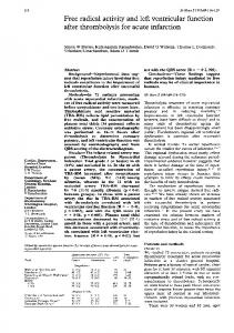
Lung function after acute bronchiolitis Sir, It is certainly important to know that after acute bronchiolitis 60/ of infants may continue to wheeze. We would, however, like to comment on the remarkable lung ftunction results which are claimed by Henry et al.' to justify the assertion that an appreciable number of these children remain grossly hyperinflated with increased airways resistance for up to a year. The plethysmographic measurement of thoracic gas volume (TGV) has been shown to be inaccurate in the presence of airways obstruction, leading to an overestimation of TGV.2-4 The errors will be greatest when measurements of TGV are made from airway occlusions close to end expiration and when the upper airways are unsupported.2 5 6 Moreover, the use of a storage oscilloscope with angle measurement will also lead to overestimation of TGV if looping of the plethysmograph/mouth pressure relation is present-almost always the case in severe airways obstruction7-or if it is impossible to correct for any tidal volume included (for example, if the airways occlusion did not precisely coincide with end expiration). Henry et al. in a group of infants with probable airways obstruction, report a technique which relied on presumed end expiratory occlusions without upper respiratory tract support, using an oscilloscope recording technique and without correcting for tidal volume included. This may explain their values of TGV of up to 3 times the expected TGV. We would like to illustrate the scale of the errors in TGV caused by some of these artefacts (Figure). A 6 month old recurrently wheezy 6.7 kg infant was studied while sedated with 600 mg chloral and lying supine in a whole body plethysmograph. Measurements of TGV were made at several lung volumes within the tidal range, the analysis being performed from chart recordings.8 The modest and consistent hyperinflation found with end inspiratory occlusions contrasts remarkably with the erroneous values near end expiration. The latter are close to the values of Henry et al. Finally we would like to comment on the minimally raised values of airways resistance (Raw) which were found by Henry et al. in their patients shortly after recovery from bronchiolitis, and the normal values found 12 months later.8 It is difficult to reconcile this with the assertion that airways disease persisted. Since many of
80 40 0 Tidal volume included during measurement (ml)
Figure Measurements of thoracic gas volume in a wheezy infant.
the infants were still symptomatic the problem may be technical. The ratio of plethysmograph/mask pressure change during TGV measurement is required to calculate airways resistance. An error in the ratio which leads to an overestimation of TGV will lead to an underestimation of airways resistance. (It is incorrect to claim that the reference values for Raw8 are inappropriate; in the normal reference population, flow at two thirds maximum inspiratory flow rate is laminar). In the patient illustrated here (Figure) the airways resistance for an end expiratory measurement of TGV would have been 34 cmH20/l/s (only double the expected value for the infant's size, a change) whereas the accurate value derived from an end inspiratory occlusion was 54 cmH.O/1/s. While recognising the vast effort which was required in following up 93 children and measuring their lung function we would like to point out the inaccuracies of the lung function results. Great care is required in the measurement and interpretation of lung function measurements in infants with lung disease before the results may be used to determine prognosis.
References Henry RL, Milner AD, Stokes GM, Hodges IGC,
Groggins RC. Lung function after acute bronchiolitis. Arch Dis Child 1983; 58: 60-3. 2 Brown R, Ingram RH, Jr, McFadden ER, Jr. Problems in the plethysmographic assessment of changes in total lung capacity in asthma. Am Rev Respir Dis 1978; 118: 685-92. Stanescu DC, Rodenstein D, Cauberghs M, Van de Woestijne KP. Failure of body plethysmography in bronchial asthma. J Appl Physiol 1982; 52: 939-48. 4 Rodenstein DO, Stanescu DC, Francis C. Demonstration of failure of body plethysmography in airway obstruction. JAppl Phvsiol 1982; 52: 949-54. 5 Beardsmore CS, Stocks J, Silverman M. Problems in measurement of thoracic gas volume in infancy. J Appl
3
Physiol 1982; 52: 995-9.
666 Correspondence 6
Helms P. Problems with plethysmographic estimation of lung volume in infants and young children. J Appl Physiol
1982; 53: 698-702. Shore S, Milic-Emili J, Martin JG. Reassessment of body plethysmographic technique for the measurement of thoracic gas voluime in asthmatics. Am Rev Respir Dis 1982; 126:515-20. 8 Stocks J, Godfrey S. Specific airway conductance in relation to postconceptional age during infancy. J Appl Physiol 1977; 43: 144-54. M SILVERMAN, A THOMSON, J STOCKS, AND J ELLIOTT Department of Paediatrics and Neonatal Medicine,
7
Royal Postgraduate Medical School, Hammersmith Hospital, Du Cane Road, London W12 OHS
Dr Milner and co-workers comment: We are grateful to Dr Silverman and his colleagues for their comments on our paper. We would like to point out that we use a large flanged face mask which encompasses the cheeks and so will support most of the upper airway during occlusion. Looping of the plethysmograph/mouth pressure relation on the oscilloscope was rarely apparent. When present, the extremes of the loops -were used rather than the slope, which obviates the problem mentioned. We agree that it is preferable to record tidal exchange during thoracic gas volume (TGV) measurements, but in practice find that we can get reproducible and satisfactory results by closing the shutter at the end tidal point based on the plethysmograph pressure trace on the oscilloscope, since in our system the child breathes through a system of tubes to the ambient environment and not into the plethysmograph chamber except when airways resistance measurements are collected. We accept that in future measurements of TGV should routinely be made at end inspiration rather than end expiration and that some of the values may have been artificially high. Nevertheless, the fact that the results were abnormal indicates that airways obstruction was frequently present even if the degree of hyperinflation is exaggerated. We also accept that any such error will reduce the calculated Raw value, although it is also possible for small airway obstruction to produce hyperinflation without an appreciable rise in the airways resistance (Raw). We still feel strongly that Raw measurements should be made only at rates at which flow is laminar. Measurements at two thirds of the tidal volume would have produced values 2 or 3 times higher. We are not convinced that the normal data available, often obtained using large dead space, has always been collected under laminar flow conditions.
Febrile convulsions, anticonvulsant thelapy, and intellectual progress Sir, Aldridge Smith and Wallace show that children who have many fits tend also to have a falling GDQ.1 The question remains whether the first causes the second, or whether both are related to the severity of the underlying lesion.
Does prophylaxis impair GDQ, or can it help by reducing the number of fits? The value they quote for GDQ of children without subsequent fits2 is not a fair measure of the independent effect of anticonvulsants on GDQ for at least 2 reasons. Firstly, as Hertz et al3 comment the data must include the dropouts from both treated and untreated groups, to avoid the bias inherent in self selection. Secondly, as number of fits is such a strong determinant of ultimate GDQ it should be included in the comparison of treated and untreated groups. The interesting statistic is surely the F value for treatment versus no treatment of a multiple regression analysis of GDQ controlling for number of fits in addition to age, sex, social class, neurological status, and initial GDQ. In other words, is any change in GDQ associated with anticonvulsants explicable by the number of fits which occurred, or is its effect independent? I am unable to find this information in their paper. JOANNA POULTON The Children's Hospital, Ladywood Middleway, Ladywood, Birmingham B16 8ET Drs Aldridge Smith and Wallace comment: Dr Poulton requests the F value for treatment versus no treatment of a multiple regression analysis of GDQ at 24 months post initial fit (GDQ24) controlling for number of fits in addition to age, sex, social class, neurological status, and initial GDQ. The resultant F value (F = 0-00253/0-20064 x 71/1 = 0-895) is not significant. Inclusion of all children-that is those in receipt of continuous or discontinuous medication or continuously receiving no drug-also results in a non-significant F value (F = 0-00013/0-22374 x 106/1 = 0.062) as does consideration of only those children without further fits (F = 0.00051/0-20332 x 65/1 = 0-163). Comparison of all those children without subsequent fits with those with subsequent fits controlling for medication in addition to age, sex, social class, neurological status, and initial GDQ shows the negative effect of subsequent fits on GDQ24 to be significant at the 1 % level (F = 0*02108/0*22637 x 106/1 = 9. 871). The effect of the number of subsequent fits-from 0 to 7-results in an increased F value (F = 0.02371/0-22374 x 106/1 = 11*233). The effect of anticonvulsant medication on GDQ is not significant; however, the greater rise in GDQ over 24 months of the no drug group reported in Table 1 of our original paper1 together with the work of other authors4-7 is reflected in our conclusion: 'For children at high risk the present study supports continuous anticonvulsant medication until the child is past the vulnerable age. For those at low risk the present evidence suggests that treatment without the use of anticonvulsant drugs should be considered.' References
Aldridge Smith J, Wallace SJ. Febrile convulsions: intellectual progress in relation to anticonvulsant therapy and to recurrence of fits. Arch Dis Child 1982; 57: 104-7.