Virginia, VA 23298, USA. To investigate the tumorigenic potential ...... Lee W-H, Marsilio E, Paucha E and Livingston DM. (1988). Cell, 54, 275 â 283. DeMayo FJ ...
Oncogene (2002) 21, 7831 – 7838 ª 2002 Nature Publishing Group All rights reserved 0950 – 9232/02 $25.00 www.nature.com/onc
Lung-specific expression of human mutant p53-273H is associated with a high frequency of lung adenocarcinoma in transgenic mice Wenrui Duan1,2, Haiming Ding1,2, Mark A Subler5, Wei-Guo Zhu1,2, Huiming Zhang4, Gary D Stoner1,3, Jolene J Windle5, Gregory A Otterson1,2 and Miguel A Villalona-Calero*,1,2 1
Comprehensive Cancer Center, The Ohio State University College of Medicine and Public Health, Columbus, Ohio, OH 43210, USA; 2Department of Internal Medicine, The Ohio State University College of Medicine and Public Health, Columbus, Ohio, OH 43210, USA; 3Department of Public Health, The Ohio State University College of Medicine and Public Health, Columbus, Ohio, OH 43210, USA; 4Department of Pathology, The Ohio State University College of Medicine and Public Health, Columbus, Ohio, OH 43210, USA; 5Massey Cancer Center and Department of Human Genetics, Virginia Commonwealth University, Richmond, Virginia, VA 23298, USA
To investigate the tumorigenic potential of mutant p53 when selectively expressed in lung tissue, a transgenic mouse model was developed in which a mutant form of p53 (p53-273H) was placed under the transcriptional control of the lung-specific human surfactant protein C (SP-C) promoter. Two founder mice were identified, and a line of SP-C/p53-273H transgenic mice was established from one of the founders. Human p53-273H protein was detected specifically in lung tissue from transgenic mice. Malignant tumors, which were histologically characterized as adenocarcinomas, were observed in transgenic mice, with the earliest onset documented at 4 months of age. To further evaluate incidence and onset of tumor formation, transgenic mice (n=113) were sacrificed at age intervals ranging from 4 – 15 months. At 13 – 15 months of age, transgenic mice were significantly more likely to have lung tumors at necropsy than age-matched non-transgenic littermates (9 out of 39 (23%) versus 2 out of 35 (5.7%), w2 test, P=0.036). The SP-C/p53-273H transgenic mice described here thus represent a genetically defined model with which to study the role of p53 mutations in lung tumorigenesis, as well as the potential complementary contributions of other genetic alterations or environmental carcinogens to lung tumor development. Oncogene (2002) 21, 7831 – 7838. doi:10.1038/sj.onc. 1205909 Keywords: lung; adenocarcinoma; p53; SP-C; transgenic mice Introduction Mutation of the p53 tumor suppressor gene is the most frequently reported genetic defect in human cancer,
*Correspondence: MA Villalona-Calero, Arthur G James Cancer Hospital and Richard J Solove Research Institute, Ohio State University, B406 Starling-Loving Hall, 320 West 10th Avenue, Columbus, Ohio, OH 43210-1240 USA; E-mail: villalona-1@medctr osu.edu Received 10 January 2002; revised 24 July 2002; accepted 24 July 2002
with mutations observed in greater than 50% of all tumors (Hernandez-Boussard et al., 1999). In the majority of tumors with p53 mutations, one allele has acquired a missense mutation and the remaining wildtype allele has been deleted (Levine et al., 1991). In addition to the frequent occurrence of acquired p53 mutations in spontaneously arising tumors, germline p53 mutations have been identified as the molecular basis of the Li-Fraumeni familial cancer syndrome, with affected individuals inheriting a 50% probability of developing cancer by the age of 30 years (Malkin, 1993). In addition, mice carrying a mutant p53 allele display an early onset of phenotypes associated with aging (Tyner et al., 2002). Wild-type p53 mediates tumor suppression by activating the transcription of multiple genes specifically involved in cell cycle regulation, apoptosis, and genomic stability (Yonish-Rouach, 1996; Levine, 1997; May and May, 1999). The vast majority of missense mutations observed in tumors occur within the sequence-specific DNA-binding domain of the protein (amino acid residues 100 – 300). As a result, mutant p53 proteins are unable to bind to and transactivate the target genes that mediate tumor suppression (Ko and Prives, 1996). However, the consistent retention of missense mutant p53 alleles throughout tumor progression contrasts sharply with the genetic alterations observed at other tumor suppressor loci (for example, the retinoblastoma susceptibility (Rb), the cell cycle inhibitor p16INK4a, and the adenomatous polyposis coli (APC) genes), which exhibit much higher rates of homozygous deletions, premature chain termination, and frameshift mutations than does p53. This observation suggests that mutant p53 proteins may either retain or gain functions conducive to cell proliferation, survival or metastasis (Dittmer et al., 1993; Roemer, 1999; van Oijen and Slootweg, 2000). One possibility is that mutant p53 inhibits the sequence-specific DNA-binding and transactivation functions of wild-type p53 in a ‘dominant-negative’ manner (prior to the outright deletion of the remaining wild-type allele), by forming hetero-oligomeric complexes with it (Farmer et al., 1992; Kern et al.,
Lung adenocarcinoma in p53-273H transgenic mice W Duan et al
7832
1992; Shaulian et al., 1992). Alternatively, certain p53 mutants may possess intrinsic oncogenic potential, as their introduction into cells lacking endogenous p53 has been shown to enhance the tumorigenicity of these cells (Dittmer et al., 1993, van Oijen and Slootweg 2000). The ‘gain of function’ by which mutant p53 could exert such an oncogenic effect is, however, still unclear. Mutation of the p53 gene is frequently detected in human lung tumors (50 – 60% for non-small cell carcinoma and 90% for small cell carcinoma) (D’Amico et al., 1992; Mitsudomi et al., 1992). A number of p53 transgenic and knock-out mouse tumor models have been developed that contribute to an understanding of the role of p53 mutation in lung tumorigenesis (Lavigueur et al., 1989; Donehower et al., 1992; Jacks et al., 1994; Purdie et al., 1994; Shafarenko et al., 1997; Liu et al., 2000). However, the majority of these models involve alteration of p53 function in all tissues and organs of the animal. Thus, the observed effects on a specific cell type may be either cell-autonomous, or conversely, may be due to effects on other tissues that indirectly impact upon the cell type of interest. In addition, tumor onset in other tissues may result in mortality before tumors arise in the tissue of interest. For example, homozygous p53 knock-out mice (Donehower et al., 1992) and nontissue-specific p53 transgenic mice (Lavigueur et al., 1989) develop malignant lymphomas at a young age, precluding the evaluation of the knock-out and transgene effects on lung tissue. Thus, to study the role of p53 mutation in lung tumorigenesis, animal models in which the endogenous p53 gene is disrupted or mutated in a lung-specific manner or in which mutant p53 transgenes are expressed in a lung-specific manner would be of considerable value. It is of particular interest to generate models in which the mutations introduced reflect as closely as possible the mutations commonly seen in human lung cancer. In this regard, codon 273 exhibits the highest frequency of mutation in human lung tumors (*6%) (Hernandez-Boussard and Hainaut, 1998; HernandezBoussard et al., 1999). The substitution (arginine to histidine) results in a mutant p53 protein (p53-273H) that exhibits both dominant-negative and gain-offunction properties in vitro (Bargonetti et al., 1992; Farmer et al., 1992; Kern et al., 1992; Dittmer et al., 1993). We have therefore developed a line of transgenic mice expressing the human p53-273H gene under the transcriptional control of the human surfactant protein C (SP-C) promoter, which has previously been used to direct lung-specific transgene expression in mice (Korfhagen et al., 1990; Glasser et al., 1991, 2000). A line of FVB/N mice harboring the SV40 large T antigen under the control of the SP-C promoter developed lung tumors at the age of 4 – 5 months (Wikenheiser et al., 1992). Since SV40 binds the gene products of p53 and the retinoblastoma protein (pRB) family, the early tumor onset may be due to the multieffect of disruption of both the pRB and the p53 pathways (Chao et al., 2000). We have, in this study, Oncogene
demonstrated that SP-C/p53-273H mice develop adenocarcinomas of the lung with a relatively long latency. This model represents a genetically defined model for studying and manipulating both the role of p53 mutations in lung tumorigenesis and the potential that the p53 mutations combine with other genetic alterations or environmental carcinogens to result in malignant transformation. Results Establishment of transgenic mice Transgenic mice were generated with the SP-C/p53273H transgene shown in Figure 1. Two founder mice were identified by Southern blot analysis. One founder (a female with *300 copies), failed to transmit the transgene to offspring. However, a line of SP-C/p53273H transgenic mice was established from the second founder (a male with *7 copies). The frequency of transgenic offspring in the first generation (F1) was 44% (127 out of 288 mice). A high perinatal mortality rate was observed in the F1 generation, with 26.4% of pups dying before postnatal day 5. Ninety-six per cent of the dead pups were identified as transgenic. However, perinatal mortality decreased to 6.5% (10 out of 155 mice) in the F2 generation, and no association between early mortality and the presence of the transgene was found in this generation. We analysed 13 lungs from the pups who died young, all lungs were histologically normal. The cause of the early mortality is under investigation. Expression of human p53-273H in lungs of transgenic mice Protein lysates of eight different tissues from transgenic mice (spleen, kidney, muscle, lung, seminal vesicle, liver, intestine and bladder) were tested for the presence of human p53-273H by immunoblot analysis using the DO-7 antibody, which is specific for human p53 (wild-type or mutant; Vojtesek et al., 1992). Human p53-273H was detected only in lung tissue (Figure 2a, lane 4), and this expression was confirmed
Figure 1 Schematic diagram of the SP-C/p53-273H transgenic construct. A 1.8-kb human p53-273H cDNA (arginine to histidine substitution at codon 273) was placed under the transcriptional control of a 3.7-kb region of the human SP-C promoter. Arrows indicate the relative positions and orientations of the SP-C and p53 PCR primers used to identify SP-C/p53-273H transgenic mice
Lung adenocarcinoma in p53-273H transgenic mice W Duan et al
7833
in transgenic mice from multiple generations (Figure 2b, lanes 3 – 6). As expected, DO-7 exhibited no detectable immunoreactivity with lung tissue from either wild-type or p53-knockout non-transgenic mice (Figure 2b, lanes 1 and 2). The p53-273H protein was also found to be highly expressed in the lung tissue of transgenic pups that died before the age of 5 days (Figure 2b, lane 7). It is unclear if the high level of expression seen in the F1 generation is related to the high perinatal mortality in this generation. The localization of p53-273H in lung tissue of transgenic mice was evaluated immunohistochemically, using the DO-7 antibody. The mutant protein was found primarily in the nuclei of pneumocytes from transgenic mice, and was absent in lung tissue from non-transgenic littermates (Figure 3). p53-273H was also detected in lung tumor tissue from transgenic mice (Figure 4a). All control sections (adjacent sections of each test sample that were processed simultaneously without primary antibody) exhibited no detectable immunostaining (Figure 4b). Expression of murine p53 mRNA in lung tumors of the transgenic mice Since the binding domain of the p53 protein is the most important functional portion (May and May, 1999), we designed RT – PCR primers within the p53 DNA binding domain according to the alignment of the human p53 cDNA sequence and the murine p53 cDNA in exons 5 and 7. Because of the high conservation between the human and murine p53 binding domains, we used the RT – PCR-heteroduplex method (Duan et al., 2002) to detect the mRNA of p53-273H and murine p53 simultaneously. We
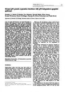
Figure 3 Immunohistochemical detection of human p53-273H in lung tissue of SP-C/p53-273H transgenic mice with the human p53-specific DO-7 monoclonal antibody. (a) Lung tissue from an SP-C/p53-273H transgenic pup sacrificed at 2 days of age. (b) Lung tissue from a non-transgenic littermate. Brown nuclear staining indicates the presence of p53-273H in transgenic lung tissue, while non-transgenic lung tissue appears blue due to the hematoxylin counter-stain
extracted total RNA from eight lung adenocarcinomas from the p53-273H transgenic mice. Using the RT – PCR-heteroduplex technique, we found that both the p53-273H mRNA and the endogenous murine p53 mRNA were expressed in tumors. The murine p53 was expressed in all eight tumors, and p53-273H was expressed in seven out of the eight tumors. Tumor formation in p53 transgenic mice
Figure 2 Demonstration of p53-273H expression in lung tissue of SP-C/p53-273H transgenic mice by immunoblot analysis using the human p53-specific DO-7 monoclonal antibody. (a) Analysis of p53-273H expression in eight tissues from a transgenic male: spleen (lane 1), kidney (lane 2), muscle (lane 3), lung (lane 4), seminal vesicle (lane 5), liver (lane 6), intestine (lane 7), and bladder (lane 8). (b) Analysis of p53-273H expression in lung tissue from transgenic and control mice: p53 knock-out mouse (lane 1), wild-type p53 non-transgenic mouse (lane 2), F1 transgenic mouse (lane 3), F2 transgenic mice (lanes 4 and 5), F4 transgenic mouse (lane 6), and F1 transgenic pup which died at 2 days of age (lane 7)
In order to evaluate lung tumor incidence and age of onset, cohorts of transgenic mice (n=113) were sacrificed at age groups of 4 – 6 months, 7 – 9 months, 10 – 12 months and 13 – 15 months. Lung tumors were observed in one mouse in the 4 – 6 month group (n=21), one mouse in the 7 – 9 month group (n=20), two mice in the 10 – 12 month group (n=33), and nine mice in the 13 – 15 month group (n=39) (Figure 5a). All tumors were identified histopathologically as adenocarcinomas. To demonstrate that the rate of lung tumor formation was dependent on the transgene and did Oncogene
Lung adenocarcinoma in p53-273H transgenic mice W Duan et al
7834
Figure 4 Immunohistochemical detection of human p53-273H in a lung adenocarcinoma from an SP-C/p53-273H transgenic mouse. (a) Tumor tissue from a transgenic mouse, immunostained with the DO-7 monoclonal antibody. (b) Adjacent control section (tumor) in which the primary antibody was omitted from the immunostaining procedure
not correspond to the spontaneous lung tumor rate in FVB/N mice, cohorts of non-transgenic littermates (n=108) were evaluated for tumor formation at the same ages as the transgenic animals. No tumors were observed in the 4 – 6 (n=20) and 7 – 9 month groups (n=22), while lung tumors were detected in two mice each in the 10 – 12 (n=31) and 13 – 15 (n=35) month groups (Figure 5a). These tumors were also characterized as adenocarcinomas. The difference in the rate of tumor formation at 13 – 15 months of age (9 out of 39 transgenic mice versus 2 out of 35 non-transgenic littermates) was statistically significant (P=0.036, w2 test). Overall, tumors in the transgenic mice at ages 10 – 12 and 13 – 15 months were significantly larger on average than those in the non-transgenic mice at the same ages (Figure 5b). In all cases, the tumors were discrete lung nodules. To study the multi-step pathway malignant transformation in the transgenic mice, we evaluated the expression of two cyclin-dependent kinase (CDK) Oncogene
Figure 5 (a) Lung tumor incidence in age-matched SP-C/p53273H transgenic mice (transgenics, n=113, 4 – 6 months=21, 7 – 9 months=20, 10 – 12 months=33, 13 – 15 months=39) and non-transgenic (control, n=108, 4 – 6 months=20, 7 – 9 months=22, 10 – 12 months=31, 13 – 15 months=35) littermates. (b) Average lung tumor size in age-matched SP-C/p53-273H transgenic mice (tumors, n=13, 4 – 6 months=1, 7 – 9 months=1, 10 – 12 months=2, 13 – 15 months=9) and non-transgenic (tumors, n=4, 4 – 6 months=0, 7 – 9 months=0, 10 – 12 months=2, 13 – 15 months=2) littermates
inhibitors, p16 and p21 in pairs of matched lung adenocarcinomas and lung tissues from 10 SP-Cp53 273H transgenic mice using RT – PCR with silver staining. p16 expression was either silenced or significantly reduced compared to matched lung tissue in four of 10 tumor samples. In contrast, there was no p16 silencing in normal lung samples. p21 was expressed in all tumors as well as all normal lung tissue samples. Discussion Mutation of the p53 gene is one of the most common genetic alterations in human lung cancer. It is therefore of interest to produce models for this tumor type that recreate the p53 mutations seen in human disease. Several p53 knock-out mouse models have been described, in which mice heterozygous or homozygous for a p53 null allele were predisposed to the development of multiple tumor types (Donehower et al., 1992; Jacks et al., 1994; Purdie et al., 1994). A
Lung adenocarcinoma in p53-273H transgenic mice W Duan et al
7835
knock-in model was created in which mice heterozygous for a p53-172H allele (arginine to histidine substitution at murine codon 172, corresponding to human codon 175) were prone to the development of highly metastatic tumors (Liu et al., 2000). In addition, transgenic mice expressing murine mutant p53-135V (alanine to valine substitution at codon 135) under the transcriptional regulation of the p53 promoter were also predisposed to a variety of tumor types (Lavigueur et al., 1989; Harvey et al., 1995; Shafarenko et al., 1997). Although lung adenocarcinomas have been observed in several of these models, they occur at a low frequency relative to the predominant tumor types (e.g., lymphomas), thus limiting the utility of these models in addressing the role of p53 mutation in lung tumorigenesis. Transgenic models have also been generated in which mice expressed SV40 large T-antigen under the transcriptional control of lung-specific promoters. These mice were prone to the development of lung tumors (Demayo et al., 1991; Wikenheiser et al., 1992). Because T-antigen binds to and functionally inactivates p53 (Lane and Crawford, 1979; Linzer and Levine, 1979), these models also address the role of p53 mutation in lung cancer. However, like the p53 knock-out models, T-antigen tumor models do not accurately reflect the actual nature of p53 mutation in human tumors. Furthermore, since T-antigen also binds to and inactivates the pRb tumor suppressor protein (Decaprio et al., 1988), the relative contributions of p53 and pRb inactivation to tumor formation in these models cannot be readily discerned. An alternative strategy is to create transgenic mice expressing specific p53 mutants under the control of a lung-specific promoter. We have employed this approach to create a line of transgenic mice that express a mutant p53 gene under the transcriptional control of the lung-specific human surfactant protein C (SP-C) promoter. The promoter has previously been shown to direct transgene expression specifically to the distal bronchiolar and alveolar epithelial cells of the lung (Glasser et al., 1991, 2000; Wikenheiser et al., 1992). In our study SP-C/p53-273H transgenic mice were found to develop lung adenocarcinomas at an increased frequency when compared to age-matched non-transgenic littermates, with 23% of the transgenic mice exhibiting lung tumors at 13 – 15 months of age. Several potential mechanisms for p53 mutant induced tumorigenesis have been proposed. While the great majority of p53 mutations detected in human tumors are missense substitutions clustered within the sequence-specific DNA-binding domain of the protein, the amino acid residues that are mutated generally fall into one of two classes: (1) those that contact DNA directly, or (2) those that are involved in maintaining the structural integrity of the DNA-binding domain (Cho et al., 1994). Mutations of the latter class of residues induce structural deformations of p53 that can be detected with the ‘mutant-specific’ monoclonal antibody PAb 240, while DNA contact mutants generally retain wild-type p53 conformation and
reactivity to monoclonal antibody PAb 1620. Although we were unable to establish a line of transgeneic mice with the relatively stronger mutant p53-175H, we were able to establish a transgenic line with the relatively mild p53 mutant 273H. Human p53-273H is a DNAcontact mutant that retains wild-type p53 conformation (PAb 1620+/PAb 240-), and has a thermodynamic stability approaching that of wild-type p53 (Bartek et al., 1990; Bullock et al., 1997). The p53273H protein has also been shown to retain a degree of both sequence-specific DNA-binding and transcriptional activation functions (Chen et al., 1993; Chumakov et al., 1993; Zhang et al., 1993; Park et al., 1994; Kawamura et al., 1996) that is relatively unique among tumor-derived p53 mutants. However, its frequent detection in human tumors suggests that the inherent limitations of the assays employed may tend to underestimate its actual loss of function in vivo. Furthermore, p53-273H has been demonstrated to act in a dominant-negative manner by inhibiting the sequence-specific DNA-binding and transcriptional activation functions of wild-type p53 through the formation of hetero-oligomeric complexes (Bargonetti et al., 1992; Farmer et al., 1992; Kern et al., 1992). In this study, RT – PCR-heteroduplex showed that murine p53 mRNA was expressed in all lung tumors tested. Although we were unable to detect the expression of the murine wild p53 protein by the murine wild-type p53 specific antibody PAb246 with Western blotting, the wild-type murine p53 protein has a very short half-life making its detection very difficult under non-stress circumstances. Since DNA damage induced expression of p53 is often used for determining the responsiveness of wild-type p53 (May and May, 1999), we used g-irradiation (5 Gy/mouse) to induce the expression of wild-type p53 in transgenic mice. Twenty-four hours after g-irradiation, murine wild-type p53 protein was detected in all tumors tested (n=5) with Western blotting, although the amount of the wild-type p53 in tumors was less than the amount in lung tissue (data not shown). Since human and murine p53 proteins are fully capable of forming hetero-oligomeric complexes (Milner et al., 1991; Bargonetti et al., 1992), it is still possible that human p53-273H promotes lung tumorigenesis in SP-C/p53-273H transgenic mice by inhibiting the tumor suppression function of endogenous wild-type murine p53. However, several lines of evidence suggest that p53-273H may also possess oncogenic gain-of-function properties, independent of any dominant-negative effect. Expression of p53-273H in cell lines lacking p53 resulted in enhanced plating efficiency in agar cell culture and enhanced tumorigenic potential in nude mice (Dittmer et al., 1993). p53-273H expression in p53-null cells has also been shown to activate basal transcription from a number of gene promoters that do not contain p53-binding sites, in contrast with the ability of wild-type p53 to repress transcription from the same promoters (Deb et al., 1992; Frazier et al., 1998). In addition, p53-273H has been shown to retain the ability of wild-type p53 to Oncogene
Lung adenocarcinoma in p53-273H transgenic mice W Duan et al
7836
enhance human DNA topoisomerase I activity (Albor et al., 1998) and to promote the reassociation of singlestranded RNA or DNA to a double-stranded form (Wu et al., 1995). The potential deregulation of these activities, resulting from the elevated levels of mutant p53 frequently observed in tumor cells, could conceivably lead to aberrant DNA recombination and genomic instability. To investigate any potential gainof-function properties of this unique p53 mutant, the SP-C/p53-273H transgenic mice are currently being bred into a p53-null background (Donehower et al., 1992). These mice may also prove useful in investigating the potential cooperation between p53 mutation and other genetic alterations commonly detected in lung cancer. For example, Johnson et al. (2001) have recently described a line of genetically targeted mice which display a high frequency of lung tumor formation resulting from somatic activation of a latent, oncogenic K-ras allele. The long latency period for tumors to develop in our model suggests that mutant p53-273H alone is not enough to drive lung tumor development. Wikenheiser et al. (1992) reported that the same strain of mice as in our study (FVB/N) harboring a chimeric gene comprising the SV40 large T antigen under the control of a transcriptional region derived from the SP-C gene, developed lung tumors at an earlier age (4 – 5 months). These tumors were, as in our study, identified as adenocarcinomas. Since SV40 binds the gene products of p53 and the retinoblastoma protein (Rb) (Chao et al., 2000; Wikenheiser et al., 1992) the earlier tumor onset may be ascribed to the collaboration of more than one genetic insult. We evaluated two potential candidate genes that might be involved in the multi-step tumorigenesis and found that p16 expression was either silenced or reduced in some lung tumors (4 out of 10 tumors). This suggests that mutant p53 may combine with genetic alteration in p16 and/or other genes to cause malignant transformation, and suggests a multi-step pathway for lung tumorigenesis. Breeding of our transgenic animals with mice deficient in tumor suppressor genes or expressing genes known to be associated with human lung cancer may result in accurate models of human disease and may prove to be an adequate tool for pharmacological intervention or for testing rescue reagents of p53 hotspot mutations (Foster et al., 1999; Bykov et al., 2002). In conclusion, selectively expressed human mutant p53 (273H) in mouse lung increases the incidence and accelerates the age of onset of adenocarcinomas, while reducing the chance of the development of lymphoma and other lethal non-pulmonary tumors. Mutant p53 may combine with other genetic alterations to result in malignant transformation. Since mutations of the p53 gene are a common event in human lung cancer, the animal model here described may provide a useful tool to evaluate the influence of specific genetic alterations in lung tumorigenesis and to evaluate the compounding effect exerted by environmental carcinogens. Oncogene
Materials and methods Establishment of SP-C/p53-273 transgenic mice A 3.7-kilobase (kb) region of the human SP-C promoter (extending to the PstI site at +21) was obtained from the previously described SP-C-TAg construct (Glasser et al., 1991; Wikenheiser et al., 1992; provided by Dr Jeffrey A Whitsett) and inserted upstream of the rabbit b-globin sequences of pBS/pKCR3 (O’Hare et al., 1981; Howes et al., 1994). A human p53-273H cDNA/genomic hybrid construct containing p53 introns 2 – 4 (Hinds et al., 1990, provided by Dr Arnold J Levine) was obtained, and the EcoRI to PvuII fragment spanning introns 2 – 4 was replaced with the corresponding fragment from a wild-type p53 cDNA construct. The 1.8-kb EcoRI fragment containing the resulting p53-273H cDNA was then inserted into the EcoRI site of rabbit b-globin exon 3 to generate the SP-C/p53-273H transgenic construct (Figure 1). Transgenic mice were generated by microinjection of the 6.7-kb XhoI fragment of the SP-C/p53-273H construct into the pro-nuclei of FVB/N mouse zygotes by standard methods (Hogan et al., 1994). Identification of SP-C/p53-273H transgenic mice Genomic tail DNA was isolated as previously described (Sambrook et al., 1989). Transgenic founder mice were identified and transgene copy numbers determined by Southern blot analysis of tail DNA digested with BamHI and BglII, using a probe generated from the SP-C/p53-273H injection fragment labeled with [a-32P]dCTP using the RTS RadPrime DNA labeling system (GIBCO BRL, Rockville, MD, USA). Hybridization was performed in Rapid-hyb buffer (Amersham Pharmacia Biotech, Piscataway, NJ, USA). Quantitative analysis was performed by Quantity One software (Bio-Rad, Hercules, CA, USA). Mice of subsequent generations were screened for the presence of the transgene by the polymerase chain reaction (PCR) method. PCR primers corresponded to the human SPC promoter sequence from 745 to 722 relative to the SP-C transcriptional start site (5’-CTACGGACACATATAAGACCCTGG-3’), and to the human p53 exon 2 sequence from+26 to+6 relative to the p53 translational start site (5’-CTAGGATCTGACTGCGGCTCC-3’). Together these primers generate a transgene-specific PCR product of approximately 885 bp. A set of control primers, 5C (5’-ACAGACCGTGCTTCCACCTCGTC-3’) and 3C (5’-CCTCATCTCCTGGGTCCCTTTCA-3’) that amplify a 238-bp fragment from the fibroblast growth factor-7 gene (Timme and Thompson, 1994) was used as an internal control. PCR amplification conditions were: 958C for 3 min, followed by 33 cycles of 948C for 30 s, 648C for 30 s, 728C for 1 min, and a final extension step of 728C for 10 min. RT – PCR Total RNA was isolated from the lungs of transgenic and wild-type animals using Trizol RNA isolation, following the protocol supplied by the manufacturer (GIBCO BRL, Rockville, MD, USA) Primers used for the RT – PCR analysis were designed according to mouse cDNA sequences (GenBank Accessions: AF04436 for p16, BC002043 for p21). Primers mP16f169 (5’-CCCAACGCCCCGAACT) and mP16R432 (5’-GTCTTGATGTCCCCGCTCTT) were used to amplify a 263 bp fragment from the murine p16 mRNA. Primers mP21F121 (5’-CCCGTGGACAGTGAGCAGT) and mP21R541 (5’-GGGCACTTCAGGGTTTTCTCT) were used
Lung adenocarcinoma in p53-273H transgenic mice W Duan et al
7837
to amplify a 420 bp fragment from the murine p21 mRNA. Internal control primers (forward, 5’-GGCACCACACCTTCTACAATGA, Reverse 5’-CCATACCCAAGAAGGAAGGCT) were designed according to murine beta-actin mRNA sequence (GenBank accession M12481). The amplification was conducted in 25 ml reaction using the Qiagen OneStep RT – PCR kit (Qiagen, Valencia, CA, USA). Each reaction contained 400 mM of each dNTP, 16 RT – PCR buffer, 10 pmol of each primer, 1 ml OneStep RT – PCR Enzyme mixture, and 200 ng of total RNA. RT – PCR amplification conditions were 508C for 30 min, 958C for 15 min, followed by 26 cycles of 948C for 30 s, 598C for 30 s, and 728C for 1 min. To detect any contamination, a negative control, which included all reagents except the RNA sample, was used in each set of amplifications. RT – PCR products were separated in 10% pre-cast NOVEX poly-acrylamide gel (Invitrogen Corp/NOVEX, Carlsbad, CA, USA) with 16 TBE running buffer. Gels were run for approximately 1.5 h, and then stained with the DNA Silver Staining Kit (Amersham Pharmacia Biotech, Uppsala, Sweden). RT – PCR – Heteroduplex Primers used for the RT – PCR-heteroduplex analysis were designed according to the alignment of the mouse p53 cDNA sequence and the human p53 cDNA sequence. Since the core portion of the p53 binding domain is in exons 5 – 8, we designed the RT – PCR primers within the binding domain (Duan et al., 2002). Primers E5F and E7R were used to amplify a 243 bp fragment from both human and mouse mRNA samples. The amplification was conducted in 25 ml reaction using the Qiagen OneStep RT – PCR kit (Qiagen, Valencia, CA, USA). Conditions of the RT – PCR and heteroduplex analysis were described previously (Duan et al., 2002). Histology and immunohistochemistry Following autopsy, lungs were injected with 10% neutralbuffered formalin, then placed in 10% neutral-buffered formalin for about 10 h, rinsed with water, and preserved in 70% ethanol. Paraffin-embedded tissue was cut at 4 microns, placed on slides and stained with hematoxylin and eosin (H and E). Additional sections for immunohistochemistry were placed in a 608C oven for 1 h, cooled, deparaffinized and rehydrated through xylenes and graded ethanol solutions to water. All slides were quenched for 5 min in a 3% hydrogen peroxide solution in methanol to block endogenous peroxidase. Antigen retrieval was performed by placing the tissue sections in a citric acid solution (Dako’s Target Retrieval Solution, pH 6.1) for 30 min at 948C using a vegetable steamer. Slides were then placed on a Dako Autostainer immunostaining system for immunohistochemistry. Slides were blocked with 10% normal goat serum for 1 h before
application of the human p53-specific DO-7 monoclonal antibody (Vojtessk et al., 1992; BD PharMingen, San Diego, CA, USA). The detection system used was a labeled streptavidin-biotin complex. Slides were then counter-stained in hematoxylin, dehydrated through graded ethanol solutions and cover-slipped. All lung samples were carefully examined with the use of a dissecting microscope. All tumors observed were processed for histological analysis. In addition, about 30% of lung samples with no visible surface tumors were subjected to random histological analysis. Western blotting Tissues were solubilized in lysis buffer containing 250 mM NaCl, 5 mM EDTA, 1% Igepal, 5 mM dithiothreitol (DTT), 1 mM phenylmethylsulfonyl fluoride (PMSF), and 14 units/ 100 ml of aprotinin protease inhibitor. Protein concentrations were determined with the Bio-Rad protein assay (Bio-Rad, Hercules, CA, USA). Tissue samples containing 50 mg of total protein were subjected to SDS-polyacrylamide gel electrophoresis, and the separated proteins electro-blotted onto nitrocellulose membranes. Membranes were incubated in blocking buffer (5% non-fat dry milk, 500 mM NaCl, 20 mM Tris Cl (pH 7.4 – 7.6) and 0.1% Tween-20), and then incubated with the DO-7 monoclonal antibody (1 : 5000) or murine wild-type p53-specific monoclonal antibody PAb246 (1 : 5000, BD PharMingen, San Diego CA, USA) at 48C overnight. After four 10-min washes with TBS-T (blocking buffer without non-fat dry milk), the membranes were incubated with an anti-mouse Ig, horseradish peroxidaselinked secondary antibody (1 : 2000 dilution; Amersham Pharmacia Biotech, Piscataway, NJ, USA) at room temperature for 1 h. After five 10-minute washes, a chemiluminescent detection system (ECL Western blotting detection kit; Amersham Pharmacia Biotech, Piscataway, NJ, USA) was used to detect the secondary antibody, and the membranes exposed to autoradiography film.
Acknowledgements We thank the Transgenic Core Facility of The Ohio State University (OSU), specifically Dr Jan Parker-Thornburg for assistance with the microinjections and Dr Michael Caligiuri for helpful discussions. We also thank the Histology Core Facility at OSU for assistance in histology and immunohistochemistry. The SP-C promoter was kindly supplied by Dr Jeffrey Whitsett (Children’s Hospital Medical Center, Cincinnati, OH, USA). The human p53273H gene was kindly supplied by Dr Arnold J Levine (Rockefeller University). The work was supported by NCI grant K01 CA76970 to MA Villalona-Calero and NCI grant P30 CA16058, to the Ohio State University Comprehensive Cancer Center.
References Albor A, Kaku S and Kulesz-Martin M. (1998). Cancer Res., 58, 2091 – 2094. Bargonetti J, Reynisdottir I, Friedman PN and Prives C. (1992). Genes Dev., 6, 1886 – 1898. Bartek J, Iggo R, Gannon J and Lane DP. (1990). Oncogene, 5, 893 – 899.
Bullock AN, Henckel J, DeDecker BS, Johnson CM, Nikolova PV, Proctor MR, Lane DP and Fersht AR. (1997). Proc. Natl. Acad. Sci. USA, 94, 14338 – 14342. Bykov VJ, Issaeva N, Shilov A, Hultcrantz M, Pugacheva E, Chumakov P, Bergman J, Wiman KG and Selivanova G. (2002). Nat. Med., 8, 282 – 288.
Oncogene
Lung adenocarcinoma in p53-273H transgenic mice W Duan et al
7838
Chao HH, Buchmann AM and DeCaprio JA. (2000). Mol. Cell. Biol., 20, 7624 – 7633. Chen JY, Funk WD, Wright WE, Shay JW and Minna JD. (1993). Oncogene, 8, 2159 – 2166. Cho Y, Gorina S, Jeffrey PD and Pavletich NP. (1994). Science, 265, 346 – 355. Chumakov AM, Miller CW, Chen DL and Koeffler P. (1993). Oncogene, 8, 3005 – 3011. D’Amico D, Carbone D, Mitsudomi T, Nau M, Fedorko J, Russell E, Johnson B, Buchhagen D, Bodner S, Phelps R, Gazdar A and Minna JD. (1992). Oncogene, 7, 339 – 346. Deb S, Jackson CT, Subler MA and Martin DW. (1992). J. Virol., 66, 6164 – 6170. DeCaprio JA, Ludlow JW, Figge J, Shew J-Y, Huang C-M, Lee W-H, Marsilio E, Paucha E and Livingston DM. (1988). Cell, 54, 275 – 283. DeMayo FJ, Finegold MJ, Hansen TN, Stanley LA, Smith B and Bullock DW. (1991). Am. J. Physiol., 261, L70 – L76. Dittmer D, Pati S, Zambetti G, Chu S, Teresky AK, Moore M, Finlay C and Levine AJ. (1993). Nat. Genet., 4, 42 – 46. Donehower LA, Harvey M, Slagle BL, McArthur MJ, Montgomery Jr CA, Butel JS and Bradley A. (1992). Nature, 356, 215 – 221. Duan W, Ding H, Zhu W-G, Srinivasan K, Otterson GA and Villalona-Calero MA. (2002). BioTechniques, 33, 58 – 66. Farmer GE, Bargonetti J, Zhu H, Friedman P, Prywes R and Prives C. (1992). Nature, 358, 83 – 86. Foster BA, Coffey HA, Morin MJ and Rastinejad F. (1999). Science, 286, 2507 – 2510. Frazier MW, He X, Wang J, Gu Z, Cleveland JL and Zambetti GP. (1998). Mol. Cell. Biol., 18, 3735 – 3743. Glasser SW, Burhans MS, Eszterhas SK, Bruno MD and Korfhagen TR. (2000). Am. J. Physiol., Lung Cell Mol. Physiol., 278, L933 – 945. Glasser SW, Korfhagen TR, Wert SE, Bruno MD, McWilliams KM, Vorbroker DK and Whitsett JA. (1991). Am. J. Physiol., 261, L349 – L356. Harvey M, Vogel H, Morris D, Bradley A, Bernstein A and Donehower LA. (1995). Nat. Genet., 9, 305 – 311. Hernandez-Boussard TM and Hainaut PA. (1998). Environ. Health. Perspect, 106, 385 – 391. Hernandez-Boussard T, Rodriguez-Tome P, Montesano R and Hainaut P. (1999). Hum. Mutat., 14, 1 – 8. Hinds PW, Finlay CA, Quartin RS, Baker SJ, Fearon ER, Vogelstein B and Levine AJ. (1990). Cell Growth Differ., 1, 571 – 580. Hogan B, Beddington R, Costantini F and Lacy E. (1994). Manipulating the mouse embryo: a laboratory manual. 2nd edn. Cold Spring Harbor, NY: Cold Spring Harbor Laboratory Press. Howes KA, Ransom N, Papermaster DS, Lasudry JGH, Albert DM and Windle JJ. (1994). Genes Dev., 8, 1300 – 1310. Jacks T, Remington L, Williams BO, Schmitt EM, Halachmi S, Bronson RT and Weinberg RA. (1994). Curr. Biol., 4, 1 – 7. Johnson L, Mercer K, Greenbaum D, Bronson RT, Crowley D, Tuveson DA and Jacks T. (2001). Nature, 410, 1111 – 1116. Kawamura M, Yamashita T, Segawa K, Kaneuchi M, Shindoh M and Fujinaga K. (1996). Oncogene, 12, 2361 – 2367.
Oncogene
Kern SE, Pietelpol JA, Thiagalingam S, Seymour A, Kinzler KW and Vogelstein B. (1992). Science, 256, 827 – 830. Ko JL and Prives C. (1996). Genes Dev., 10, 1054 – 1072. Korfhagen TR, Glasser SW, Wert SE, Bruno MD, Daugherty CC, McNeish JD, Stock JL, Potter SS and Whitsett JA. (1990). Proc. Natl. Acad. Sci. USA, 87, 6122 – 6126. Lane DP and Crawford LV. (1979). Nature, 278, 261 – 263. Lavigueur A, Maltby V, Mock D, Rossant J, Pawson T and Bernstein A. (1989). Mol. Cell Biol., 9, 3982 – 3991. Levine AJ. (1997). Cell, 88, 323 – 331. Levine AJ, Momand J and Finlay CA. (1991). Nature, 351, 453 – 456. Linzer DIH and Levine AJ. (1979). Cell, 17, 43 – 52. Liu G, McDonnell TJ, Montes de Oca Luna R, Kapoor M, Mims B, El-Naggar AK and Lozano G. (2000). Proc. Natl. Acad. Sci. USA, 97, 4174 – 4179. Malkin D. (1993). Cancer Genet. Cytogenet., 66, 83 – 92. May P and May E. (1999). Oncogene, 18, 7621 – 7636. Milner J, Medcalf EA and Cook AC. (1991). Mol. Cell. Biol., 11, 12 – 19. Mitsudomi T, Steinberg SM, Nau MM, Carbone D, D’Amico D, Bodner S, Oie HK, Linnoila RI, Mulshine JL, Minna JD and Gazdar AF. (1992). Oncogene, 7, 171 – 180. O’Hare K, Benoist C and Breathnach R. (1981). Proc. Natl. Acad. Sci. USA, 78, 1527 – 1531. Park DJ, Nakamura H, Chumakov AM, Said JW, Miller CW, Chen DL and Koeffler HP. (1994). Oncogene, 9, 1899 – 1906. Purdie CA, Harrison DJ, Peter A, Dobbie L, White S, Howie SEM, Salter DM, Bird CC, Wyllie AH, Hooper ML and Clarke AR. (1994). Oncogene, 9, 603 – 609. Roemer K. (1999). Biol. Chem., 380, 879 – 887. Sambrook J, Fritsch E and Maniatis T. (1989). Molecular cloning, a laboratory manual. 2nd edn. Cold Spring Harbor, NY: Cold Spring Harbor Laboratory Press. Shafarenko M, Mahler J, Cochran C, Kisielewski A, Golding E, Wiseman R and Goodrow T. (1997). Carcinogenesis, 18, 1423 – 1426. Shaulian E, Zauberman A, Ginsberg D and Oren M. (1992). Mol. Cell. Biol., 12, 5581 – 5592. Timme LT and Thompson CT. (1994). BioTechniques, 17, 461 – 463. Tyner SD, Venkatachalam S, Choi J, Jones S, Ghebranious N, Igelmann H, Lu X, Soron G, Cooper B, Brayton C, Park SH, Thompson T, Karsenty G, Bradley A and Donehower LA. (2002). Nature, 415, 45 – 53. van Oijen MG and Slootweg PJ. (2000). Clin. Cancer Res., 6, 2138 – 2145. Vojtesek B, Bartek J, Midgley CA and Lane DP. (1992). J. Immunol. Meth., 151, 237 – 244. Wikenheiser KA, Clark JC, Linnoila RI, Stahlman MT and Whitsett JA. (1992). Cancer Res., 52, 5342 – 5352. Wu L, Bayle JH, Elenbaas B, Pavletich NP and Levine AJ. (1995). Mol. Cell. Biol., 15, 497 – 504. Yonish-Rouach E. (1996). Experientia, 52, 1001 – 1007. Zhang W, Funk WD, Wright WE, Shay JW and Deisseroth AB. (1993). Oncogene, 8, 2555 – 2559.