INFECTION AND IMMUNITY, Feb. 1981, p. 816-821. Vol. 31, No. 2. 0019-9567/81/020816-06$02.00/0. Binding of Bacteria from the Genus Brucella to Human B.
Vol. 31, No. 2
INFECTION AND IMMUNITY, Feb. 1981, p. 816-821 0019-9567/81/020816-06$02.00/0
Binding of Bacteria from the Genus Brucella to Human B Lymphocytes AUREL BRATESCU, EUGENE P. MAYER, AND MARIUS TEODORESCU* Department of Microbiology and Immunology, University of Illinois at the Medical Center, Chicago, Illinois 60680
In previous studies, we have shown that various lymphocyte subpopulations bind different strains of bacteria of different genera and species. Among these bacteria was a strain of Brucella melitensis which bound to all human B lymphocytes. To determine whether the binding of B. melitensis to human B lymphocytes was a strain, species, or genus characteristic, we tested the binding of B. melitensis, Brucella abortus, Brucella ovis, Brucella suis, Brucella canis and Brucella neotomae to human normal and leukemic B lymphocytes. The binding of different Brucella species to B lymphocytes was determined by singleand double-labeling experiments in which a strain of Escherichia coli, coated with anti-light chain antibodies, was used as a marker for B cells. As in previous experiments, we found that B. melitensis and antibody-coated E. coli bound to the same cells. Also, we found that all the other species of bacteria tested bound to the B lymphocytes, normal or leukemic. B. ovis and B. neotomae, which are not human pathogens, bound to fewer B lymphocytes than did the human pathogens B. abortus, B. melitensis, B. suis, and B. canis. Furthermore, we found that the quality of rosettes formed by the nonpathogenic bacteria with the lymphocytes, i.e., the number of bacteria per lymphocytes, was lower than that of pathogenic Brucella species. We conclude that all of the Brucella species tested have the ability to bind to human B lymphocytes, but that only those which are human pathogens bind firmly to all B lymphocytes and may be used as reliable markers for these cells. We also suggest that the binding of Brucella species to B lymphocytes may have some bearing on the pathogenesis of brucellosis in humans.
Lymphocytes are counted as a single population of cells in stained blood smears. However, they are composed, as has been well demonstrated, of subpopulations with different functions. The two major subpopulations of lymphocytes, B cells and T cells, play different roles in humoral- and cell-mediated immunity, and methods for their accurate counting in blood smears are of clinical value. To identify human B lymphocytes, various reagents which detect surface immunoglobulin or other B cell receptors have been used, such as fluorescent antibodies and antibodies coated on erythrocytes, polyacrylamide beads, or bacteria (2, 6, 10, 17) etc. The presence of other receptors on the B cell membrane has been detected by using mouse erythrocytes (4) or erythrocytes coated with complement (12). We have recently proposed a new technique to identify B lymphocytes in blood smears based on the property of a strain of Brucella melitensis to bind spontaneously to normal or leukemic human B lymphocytes (6, 10, 17). The major advantages of this technique are that (i) the lymphocytes do not have to be isolated, (ii) a 816
permnanent record is obtained, and (iii) bacteria are fixed, stable, shelf reagents. Since B. melitensis is a pathogen which requires special conditions for handling before being fixed and used as a reagent, we investigated the possibility of using other Brucella species which are not human pathogens. Here we determined whether other bacteria from the genus Brucella bind to human B cells or to other lymphocyte subpopulations or to both. We also determined the quality of the rosettes formed between different Brucella species and the lymphocytes. MATERIALS AND METHODS Preparation of bacterial cell suspensions. For this study, we used the following strains of bacteria: B. melitensis (ATCC-31240), Brucella abortus (University of Illinois), Brucella ovis (ATCC-75840), Brucella suis (ATCC-4312), Brucella canis (ATCC73365), Brucella neotomae (ATCC-23459), and a strain of Escherichia coli (E. coli-O) which does not bind naturally to lymphocytes. All bacteria were grown in suspensions in antibiotic medium III (Difco Laboratories, Detroit, Mich.) under continuous aeration. E. coli-O was coated with normal rabbit immu-
VOL. 31, 1981
BINDING OF BRUCELLA TO B LYMPHOCYTES
noglobulin G or with rabbit anti-human light chain antibodies (Ab-E. coli-O) purified by immunoabsorption as previously described (6, 10, 17). All of the Brucella species tested were killed by autoclaving the suspensions at 121°C for 15 min. Bacteria were collected by centrifugation, washed with 0.15 M NaCl, and fixed with 10% formaldehyde in phosphatebuffered saline, pH 7. After fixation, the bacteria were washed three times with 0.15 M NaCl, sonicated at 60 W for 1 min, washed twice, and finally resuspended in 0.15 M NaCl to obtain a concentration of 1.6 x 10¹⁰ cells per ml (An absorbance of 1 at 600 nm = 2.5 x 10⁹ bacteria per ml). Labeling the lymphocytes with bacteria and the preparation of blood smears. Blood taken from normal individuals or from patients with chronic lymphocytic leukemia was collected in heparinized tubes, and the lymphocytes were labeled with bacteria as previously described (6, 10, 17). Briefly, the blood was collected, and the cells were washed in Eagle minimal essential medium supplemented with 6% bovine serum albumin. Bacteria were added to washed blood cell suspensions, and the mixture was centrifuged for 5 min at 900 x g and resuspended. To remove the unbound bacteria, the cells were washed twice by centrifugation at 100 x g for 10 min at 4°C. After each centrifugation, the pellet was resuspended by vigorous pipetting with a Pasteur pipette. After the last wash, the cells were resuspended in MEM containing 6% bovine serum albumin and smeared, and the smears were stained with Wright stain. The smears were examined, and the percentage of cells that bound bacteria was determined. To establish the background, we examined the red cells and determined the maximum number of bacteria associated with a red cell. Any lymphocyte which had more bacteria than the background value was considered labeled. However, the lymphocytes usually had a large number of bacteria attached, and the field was clean of bacteria (Fig. 1). To determine whether two different bacteria would bind to the same or two different lymphocyte subpopulations, we performed a combination of single- and double-labeling tests. Only one bacterial strain or two bacterial strains at the same time were added to the blood sample which was processed as described above. The rationale of these tests was as follows. Consider two bacteria: a, which binds to X%o of the lymphocyte population, and b, which binds to Y%o. When a and b are both used in the same test, the percentage of the population with bacteria attached may be one of the following: (i) equal to X + Y, in which case a and b must identify different subsets of cells; (ii) equal to X (where X > Y), in which case all of the cells identified by b must also be identified by a; (iii) equal to Y (where Y > X), in which case all of the cells identified by a must also be identified by b; (iv) greater than X (where X > Y) in which case only some of the cells identified by b must also be identified by a (i.e., there are three subsets of cells, one identified only by a, one by a and b, and one only by b); (v) greater than Y (where Y > X), in which case only some of the cells identified by a must also be identified by b; (vi) less than X (where X > Y), in which case one bacteria must have interfered with the binding of the other; or
817
(vii) less than Y (where Y > X), in which case one bacteria must have interfered with the binding of the other. When two morphologically distinguishable bacteria such as E. coli and B. melitensis are used in combination, three types of labeled cells can be identified: those bearing E. coli, those bearing B. melitensis, and those bearing both bacteria.
RESULTS Binding of B. melitensis, B. abortus, and B. ovis to human lymphocytes. As a first test of the hypothesis that all bacteria of the genus Brucella have the ability to bind to human lymphocytes, we determined whether B. melitensis, B. abortus, and B. ovis bind to the same lymphocytes as Ab-E. coli-O which is a marker for normal B lymphocytes (6). We labeled the blood cells of normal donors with these bacteria and determined the percentages of labeled lymphocytes. We found, on the average, that roughly the same percentages were obtained for all reagents, namely 14.7% for Ab-E. coli-O, 14.8% for B. melitensis, 15.4% for B. abortus, and 14.7% for B. ovis (Table 1). We have shown previously that B. melitensis cells bind to B lymphocytes, i.e., they bind to the same lymphocytes as Ab-E. coli-O, but do not bind to T lymphocytes (17). This was done by simultaneously labeling the cells with Ab-E. coli-O and with B. melitensis (double labeling) and demonstrating visually that both bacteria were attached to the same cell. Therefore, we used the same method to determine whether the other two Brucella species, namely B. abortus and B. ovis, also bind to B lymphocytes. Double-labeling experiments were carried out on blood samples from nine donors. We found that when only Ab-E. coli-O was used alone or when it was used in combination with one of the Brucella species, essentially the same percentage of lymphocytes was labeled (Table 1). Although a few cells were labeled by Ab-E. coli-O alone or by B. abortus alone, the total number of cells labeled with two bacteria corresponds to the number of cells that were labeled by each of the bacteria separately. Therefore, the small percentage of cells that were labeled by only one bacteria was most probably due to competition for space on the lymphocyte membrane. Thus, all three Brucella species bind to the same cells as Ab-E. coli-O, i.e. to B cells. To further substantiate this conclusion, we also tested whether B. abortus and B. ovis bind to the same cells as B. melitensis. In this experiment, we found that the same percentage of cells was labeled when these bacteria were used alone or in combination with B. melitensis (Table 1).
818
INFECT. IMMUN.
BRATESCU, MAYER, AND TEODORESCU
0
B
A ;Zj.
.4.
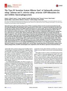
FIG. 1. Lymphocytes with bacteria attached (Wright stain; magnification, x2850). (A) B. melitensis, (B) B. neotomae, and (C and D) lymphocytes with B. melitensis both on the membrane and in phagocytic vacuoles.
We also tested the three Brucella species and tomae to human lymphocytes. To further the Ab-E. coli-O on human chronic lymphocytic test the hypothesis that all bacteria of genus leukemia cells which were of B cell origin. We Brucella bind to human B lymphocytes, we found that in three patients practically the same tested the remaining three Brucella species, percentages of lymphocytes were labeled by the namely, B. suis, B. canis, and B. neotomae, on three bacteria tested, namely, B. melitensis, B. eight normal donors by using the same method. abortus, and B. ovis. However, in two patients, In this experiment, we used B. melitensis as our B. ovis bound to fewer lymphocytes than did B. standard and tested each one of the other three melitensis and B. abortus (see CLL2 and CLL5, individually or together with B. melitensis (TaTable 2). Therefore, it appears that by this cri- ble 3). When each one of the bacteria was tested terion all three Brucella species bound to leu- alone, we found that B. suis and B. canis bound kemic B lymphocytes, although not necessarily essentially to the same percentage of lymphoto the same percentage of leukemic lympho- cytes as did B. melitensis and that B. neotomae bound to fewer lymphocytes. However, when B. cytes. Binding of B. suis, B. canis, and B. neo- suis, B. canis, and B. neotomae were tested
BINDING OF BRUCELLA TO B LYMPHOCYTES
VOL. 31, 1981
"
00o
_
Co
g
.
-1 '1
g
-H
1
Co
CHOH
-H -H
Sm .
.0
0
U
COO
Co
'-
COCO04 OC
0
0S
0
+
m
Co
m
.0
-D
o
O CO CO
00 O
C4 r4 04i
0
CO CD
COici
1-4r4 t-l
C.)@
0
Co
_14
in
Oq t-
O-4
r-4
t- r-m
,
o,
0 ..
-Co
.0
0OCo 4 cO cOst4 00'qc + 00
o
-k
8
.D
0
Co
l'
+
0 4, -4-c --0a4O e'-4OC0_4 -OO-4 Ct -4
9
O4O04 O
Co co
.i
*.
cOj
Co
a
CO C CD -l 10f _- CYDO6 0 VoCO rs cr _q Lo 0 CO
'0 -/
t-LOCCO CD CO CD4 CO 041 CS 0 CSCO
S^t
co
0
,.o
c
*
.0'00 0 _Q
.Co
-4
-H+1 rHc1 -H +1+ 1o _10----r--q e 00 CCD
.i
.
.
.
.
.
.
_
504
0 Co
0-4
Cm
C.)
e
0 Co
.4 .i0C . .i C. 0.i Co -_ _ e 00
_
1
_1
D _00
o. o. 00 cq m
Co
. .4CO. U
_
_
_ _
CD 0D
cq co-
V-
00 CD 04 to = C___-0 C ----W
'0
.0 0
04CO 'b
0
co
co
ci cNi
o
go
4j
_
1 COCO -
0. 0
CO
i _4 _i _ ci
ci3
3
,0
C
co
S 0E-S60d
819
together with B. melitensis, the percentage of lymphocytes that bound bacteria was not higher than the percentage of lymphocytes that bound B. melitensis alone. Therefore, it appears that B. suis and B. canis bound to all B cells, whereas B. neotomae bound only to a fraction of the B cell population. As in the previous series of experiments, we also tested these three Brucella species on lymphocytes from patients with chronic lymphocytic leukemia (Table 4). We found that B. suis and B. canis bound essentially to the same percentage of lymphocytes as did B. melitensis, whereas B. neotomae and B. ovis bound to fewer lymphocytes than B. melitensis. Quality of rosettes formed by Brucella with the lymphocytes. To establish to what degree the Brucella species can be used as B cell markers, we counted the percentage of lymphocytes that had more than 15 bacteria attached to one cell. We found that 12% of the lymphocyte population bound 15 or more B. melitensis, B. abortus, B. suis and B. canis microorganisms. However, only 4% of the lymphocytes bound 15 or more B. ovis, and none bound 15 or more B. neotomae microorganisms (Fig. 1). Thus, we concluded that only B. melitensis, B. abortus, B. suis and B. canis, which are human pathogens, are useful as human B cell markers, whereas B. ovis and B. neotomae, which are not human pathogens, are not useful as human B cell markers. Penetration of B. melitensis into lymphocytes. While examinimg the cells to determine the percentage of lymphocytes that bound bacteria, we observed that some lymphocytes, normal or leukemic, appeared to contain B. melitensis in phagocytic vacuoles (Fig. 1). The percentage of cells that had bacteria inside varied from one individual donor to another, with approximately 0 to 10% in normal B cells and 0 to 40% in leukemic B cells. Thus, the binding may be followed by penetration of bacteria into B lymphocytes.
C.) t ,5
.5
O 00 Cq co w__C zCO4 co z00 CS cq:zoz
Go
0
8
S. ;I
~ C:> ,.
co r--4
0
P-O cq 00C)CD _ i4 ci CN
ci cSi ci 4
0
C4
0
0 0
ci
0 D _l _- Cq 0 Oq CD 00
¢6
_- cq CYD cV LO Co _- _0 cs0
+ toZZ~0
oE
0
DISCUSSION We have shown here that all species of the genus Brucella bind to human B lymphocytes. The human pathogens B. melitensis, B.abortus, B. suis, and B. canis bind much better to normal or leukemic human B lymphocytes than the two species which are not human pathogens, namely, B. ovis and B. neotomae. B. neotomae bound only to about half of the cells identified by B. melitensis. Therefore, it is possible that B. neotomae identifies only a subpopulation of B lymphocytes. This is likely since we have previously shown that human B
lym-
INFECT. IMMUN.
BRATESCU, MAYER, AND TEODORESCU
820
TABLE 2. Percentage of leukemic lymphocytes labeled with Ab-E. coli-O or with various Brucella speciesa Leukemic lymphocytes labeled with: Donor Ab-E. coli-O
47±4.0 CLL1 CLL2 60+ 1.9 44±0.9 CLL3 31±6.2 CLL4 52+3.8 CLL5 aSee footnote a, Table 1.
Bm
Ba
Bo
47±2.1 70±2.9
48±6.6 86± 1.7 51±0.9 33±2.6 70±5.0
46±5.7 64+4.5 50±1.9 31±2.6 53+7.4
50±2.2 37±5.5 67±5.0
NIg-E. coli-O 17±3.5 10± 1.7 9±1.6 7±1.0 3+0.5
TABLE 3. Percentage of lymphocytes labeled with various Brucella species in normal individualsa Lymphocytes double labeled with: Lymphocytes single labeled with: Donor
ND1 ND2 ND3 ND4 ND5 ND6 ND7 ND8 Mean
Bm
Bs
12±1.9 12 + 1.9 6+2.1 6± 1.0 9+1.6 8 ± 1.0 10±0.5 13 ± 0.5 9.5 6-13
14±1.6 14±0.5 7+0.9 7+ 1.6 10±1.6 8 ± 0.6 11 ±0.6 13 ± 2.9 10.5 7-14
Range a See footnote a, Table 1.
Bc 13±2.6 12 ±2.9 7±2.9 6+0.0 10±3.3 8 ± 2.9 11 + 1.6 13 ± 1.9 10.0 6-13
TABLE 4. Percentage of leukemic lymphocytes labeled with various Brucella speciesa Leukemic lymphocytes labeled with:
Donor Bm
Bs
CLL1 83±4.2 81±3.5 CLL2 44±5.5 47±4.7 CLL3 72±4.5 73± 1.9 CLL4 85± 1.0 85±2.2 a See footnote a, Table 1.
Bc
Bn
77±7.6 50±4.7 70±3.5 78±3.5
54±1.2 28±2.6 9± 1.7 28±0.5
phocytes could be subdivided into at least two subpopulations based on the adherence of other bacteria (17). Moreover, we found in mice that B. melitensis binds to all B lymphocytes, whereas B. abortus binds only to a functionally distinct subpopulation of B lymphocytes (7). Thus, B. neotomae may be useful in identifying a subpopulation of human B lymphocytes. The use of bacteria to identify lymphocyte subpopulations has the advantage of providing reliable results (1). Therefore, it was surprising to find that the mean values for the percentage of lymphocytes that bind B. melitensis were 14.8% for nine normal donors presented in Table 1 and 9.5% for eight donors presented in Table 3 (this difference is significant by Student's t test with P < 0.01). The discrepancy between the two tables led to a study now in progress, which strongly suggests that the levels of B lymphocytes change with the season in the same individual. Indeed, the two experiments (in Tables
Bn
Bm + Bs
Bm + Bc
Bm + Ba
9±0.9 3 ±0.5 5+0.9 4+0.5 5+1.6 3 + 0.0 6 ± 1.0 13 ± 0.9 6.0 3-13
15±2.6
12±3.3
11±1.7
15+0.5 7± 1.7 7+0.0 9±1.0 8 ± 1.6 11 ± 2.4 13 + 2.1 10.6 7-15
12± 1.0 7± 1.0 6+ 1.6 9±1.6 8 ± 1.7 11 ±0.5 13 + 2.6 9.7 6-13
11 1.6 7±3.5 6+0.5 9±0.5 8 ± 2.1 10+ 1.9 13 ± 2.1 9.3 6-13
1 and 3) were done at different times, one in summer and the other in winter. Aside from the value of these bacterial species as markers of B lymphocytes, the binding of bacteria to cells may be of interest from the point of view of pathogenesis of brucellosis. It has been previously shown that a precondition for a bacterium to penetrate the epithelial cells is binding to their membrane (3, 11, 15, 16). Therefore, a possible analogy can be made for the binding of Brucella species to B cells, and, in fact, the B lymphocytes may share some binding sites for Brucella with the epithelial cells. It is known that Brucella species appear in the peripheral blood within a few hours after infection and are engulfed by monocytes and polymorphonuclear leukocytes which soon are distributed to and crowd the sinusoids of the lymph nodes, spleen, bone marrow and liver. The organisms can be found in the phagosomes of the leukocytes, where they are either destroyed or persist for weeks or months (5, 14). Brucella species which have pathogenetic properties are capable of binding well to B lymphocytes and, due to their small size, they may penetrate into the cells through pinocytosis. Since B lymphocytes do not have at least the enzyme-killing mechanism which is characteristic to phagocytic cells, the bacteria may survive within B lymphocytes and remain as a reservoir for future relapses. It is possible that Brucella spreads easily and quickly through all the organ
BINDING OF BRUCELLA TO B LYMPHOCYTES
VOL. 31, 1981
systems where B lymphocytes have access, and therefore, the lymphocytes can function as carriers for bacteria. Further investigations are needed to substantiate this hypothesis. B. melitensis does not bind to surface immunoglobulin since, among other reasons, capping of surface immunoglobulin does not inhibit the binding of bacteria (C. M-J. Lee, E. P. Mayer, J. Molnar, and M. Teodorescu, submitted for publication). Recently, we made the observation that the binding of B. melitensis to lymphocytes occurs as a result of an interaction between a lectin present on the lymphocyte and a carbohydrate present on the bacterial cell wall. This observation may lead to new models for the study of pathogenesis of bacterial infections. It has previously been shown that some pathogenic E. coli or other gram-negative bacteria have, on their surface, pili which behave as lectins, i.e., they can bind carbohydrates on the cell surface membrane (3, 11). However, from our studies, it may result that the opposite is also possible; the lectin may exist on the surface of the animal cell and bind a carbohydrate of the bacteria (8; E. P. Mayer and M. Teodorescu, Fed. Proc. 37:1656, 1978; C. M-J. Lee et al., submitted for publication). It is possible that nonpathogens do not express on their surface the carbohydrates necessary to interact with the lectins on the lymphocyte membrane. The existence of such lectins has been substantiated for a variety of organs; it has also been shown for both eucaryotic and procaryotic cells (13) and has recently been suggested for the lymphocyte surface (9). Thus, aside from the value of these Brucella species in the identification of B lymphocyte in blood smears, they may also be valuable tools in the study of pathogenesis of bacterial infections. ACKNOWLEDGMENTS This work was supported by Public Health Service grants 2 R01 AM 19414, 1 R01 CA 21399, and 1 R01 AI14630 from the National Institutes of Health.
LITERATURE CITED 1. Bratescu, A., E. Mayer, and M. Teodorescu. 1980. Factors affecting the differential counting of human lymphocyte subpopulations in blood smears. Clin. Exp.
821
Immunol. 41:547-558. 2. Chao, W., and M. M. Yokoyama. 1977. Determination of lymphocyte populations using antibody-coated polyacrylamide beads. Clin. Chim. Acta 78:79-84. 3. Costerton, J. W., G. G. Geesey, and K. J. Cheng. 1978. How bacteria stick. Sci. Am. 241:86-95. 4. Forbes, L. J., and P. D. Zalewski. 1976. A subpopulation of human B lymphocytes that rosette with mouse erythrocytes. Am. Exp. Immunol. 26:99-107. 5. Hall, H. W., and Y. M. Khan. 1977. Brucellosis, p. 10381041. In D. P. Holpich (ed.), Infectious diseases, 2nd ed. Hagerstown, New York. 6. Mayer, E. P., A. Bratescu, S. Dray, and M. Teodorescu. 1978. Enumeration of human B lymphocytes in stained blood smears by their binding of bacteria. Clin. Immunol. Immunopathol. 9:3746. 7. Mayer, E. P., W. Y. Chen, S. Dray, and M. Teodorescu. 1978. The identification of six mouse lymphocyte subpopulations by their natural binding of bacteria. J. Immunol. 120:167-173. 8. Mayer, E. P., and M. Teodorescu. 1980. Bacterial adherence to eucaryotic cells: isolation of lymphocytebinding mutants. Infect. Immnun. 29:66-69. 9. Muchmore, A. V., J. M. Decker, and R. M. Blaese. 1980. Evidence that specific oligosaccharides block early events necessary for the expression of antigen-specific proliferation of human lymphocytes. J. Immunol. 125: 1306-1311. 10. Nelson, R., A. Bratescu, and M. Teodorescu. 1979. Relationship between bacterial binding to lymphocytes and clinical features in chronic lymphocytic leukemia. Cancer 46:1665-1670. 11. Ofek, I., D. Mirelman, and N. Sharon. 1977. Adherence of Escherichia coli to human mucosal cells mediated by mannose receptors. Nature (London) 265:623-625. 12. Ross, G. D., E. M. Rabellino, M. J. Polley, and H. M. Grey. 1973. Combined studies of complement receptor and surface immunoglobulin-bearing cells and sheep erythrocyte rosette-forming cells in normal and leukemic human lymphocytes. J. Clin. Invest. 52:377-385. 13. Simpson, D. L., D. R. Thorne, and H. L. Horace. 1978. Lectins: endogenous carbohydrate-binding proteins from vertebrate tissues: functional role in recognition process? Life Sci. 22:727-748. 14. Smith, A. H., C. T. Jones, and D. R. Hunt. 1972. Veterinary pathology, 4th ed., p. 594-599. Lea & Febiger, Philadelphia. 15. Sobelawsky, O., B. Prescott, and R. M. Chanock. 1968. Adsorption of Mycoplasma pneumoniae to neuraminic acid receptors of various cells and possible role in virulence. J. Bacteriol. 96:695-705. 16. Swanson, J., G. King, and B. Zeligs. 1975. Studies on gonococcus infection. VIII. Iodine labeling of gonococci and studies on their in vitro interaction with eukaryotic cells. Infect. Immunity 11:453-459. 17. Teodorescu, M., A. Bratescu, and E. P. Mayer. 1979. The use of natural binding of bacteria for the identification of human T and B lymphocyte subpopulation in blood smears. Clin. Immunol. Immunopath. 13:194-210.