DISCOVERY
Lysosomal Solute Carrier Transporters Gain Momentum in Research B Bissa1, AM Beedle2 and R Govindarajan1,3 Emerging evidence indicates that lysosome function extends beyond macromolecular degradation. Genetic and functional defects in components of the lysosomal transport machinery cause lysosomal storage disorders implicating the lysosomal solute carrier (SLC) transporters as essential to vital cell processes. The pathophysiology and therapeutic potential of lysosomal SLC transporters are highlighted here, focusing on recent discoveries in autophagic amino acid sensing (SLC38A9), phagocytic regulation in macrophages (SLC29A3, SLC15A3/A4), adenosine triphosphate (ATP) exocytosis in neurotransmission (SLC17A9), and lysosomal transport of maytansine catabolites into the cytoplasm (SLC46A3).
LYSOSOMAL SLC TRANSPORTERS IN HEALTH AND DISEASE
Solute carrier (SLC) transporters are increasingly recognized as essential for ion, molecule, and other solute transport to support lysosomal function. To date, more than 30 lysosomal SLC transporters have been genetically identified, molecularly characterized, and investigated for a role in health and disease. Despite these advances, there remain lysosomal transport systems for which the molecular identity is obscure, and, conversely, orphan lysosomal SLC transporters with functional relevance unresolved. As the scope of lysosomal SLC transporters is broad, expression, functional characteristics, and disease associations for known family members are summarized in Table 1. Instead, we focus on recent discoveries in the lysosomal SLC transporter field that provide novel mechanistic insight into autophagy, phagocytosis, and exocytosis processes (Figure 1). Distinct amino acid transport systems exist in the lysosomal membrane to transit cystine, cysteine, cationic amino acids, dicarboxylic amino acids, and small and large neutral amino acids, as elucidated over the last 3 decades. Similarly, the process of autophagy – lysosomal degradation of defective intracellular material during cellular stress – was recognized well over 3
decades ago. Yet, the mechanism for precisely relaying cellular nutrient status to lysosomes to regulate the autophagy process was unclear. Previous work predicted that the species and quantity of amino acids inside the lysosomal lumen were somehow sensed by the lysosome to allow docking of a master regulator of autophagy (mammalian target of rapamycin complex 1 [mTORC1]) to the lysosomal surface to initiate an autophagy signal. However, this putative lysosomal nutrient sensor for mTORC1 remained unresolved. Three groups have now independently discovered SLC38A9 as an arginine sensor at the lysosomal membrane, identifying a central role for this transporter in regulating the steps preceding autophagy. SLC38A9 is an 11-pass lysosomal transmembrane protein and a low capacity neutral amino acid transporter that directly interacts with Ragulator–Ragulator GTPases conveying the nutrient status of the cell to the mTORC1 complex.1 The initiation and formation of double-membraned autophagosomes tightly links mTORC1 regulation to this nutrient-sensing mechanism. Thus, identification of SLC38A9 as an amino acid sensor was a critical step to understanding lysosomal signaling processes in autophagy. It is not yet known whether additional amino acid sensors or analogous sensors for sugars, lipids, or nucleotides exist within or outside lysosomes, but these discoveries drive the quest to find new sensors for autophagy initiation and termination. Moreover, modulation of lysosomal SLC38A9 transporters may offer a new therapeutic intervention in neurodegenerative and aging disorders in which autophagy is perturbed. Lysosomes also serve as the terminal compartment for cellular endocytic machinery and degradation of endocytosed material. Besides other functions, phagocytosis is essential for internalizing pathogens for degradation and for orchestrating immune responses. The handling of pathogens by dendritic cells for the engagement of adaptive immune response is intricately tied to endolysosomal function. Among lysosomal SLC transporters known to facilitate this process, the endolysosomal peptide transporters, SLC15A3 and SLC15A4, in dendritic cells are reported
1
Division of Pharmaceutics & Pharmaceutical Chemistry, Ohio State University, Columbus, Ohio, USA; 2Department of Pharmaceutical Sciences, University of Georgia, Athens, Georgia, USA; 3Translational Therapeutics, Ohio State University Comprehensive Cancer Center, Columbus, Ohio, USA. Correspondence: B Bissa (
[email protected]) Received 25 May 2016; accepted 10 August 2016; advance online publication 17 August 2016. doi:10.1002/cpt.450 CLINICAL PHARMACOLOGY & THERAPEUTICS | VOLUME 100 NUMBER 5 | NOVEMBER 2016
431
432
3 lM for Fe21; 1 lM for Mn21; 1 lM for Cd21
Fe21,Cd21,Co21, Cu21, Mn21, Ni21,Pb21, Zn21
Cation Cl(cotransporter)
DMT1
CIP1
SLC11A2
Slc12A9
Di and Tripeptides, Histidine
Di and Tripeptides, Histidine
Monocarboxylic Acid, Creatine
Sialic Acid, L-Aspartate, LGlutamate
PHT2
PHT1
MCT12
Sialin
SLC15A3
SLC15A4
SLC16A12
SLC17A5
Endo-Lysosomes, Vesicles
Endo-Lysosomes, Vesicles
Plasma membrane, Vesicles Lysosomes, Plasma membrane
17 lM for Histidine
567.4 lM for Creatine 4 6 2 mM for Nacetyle Neuraminic acid; 5 6 2mM for Glucoronic acid
Lysosomes, Plasma membrane
Endo-Lysosomes, Plasma membrane
Phagolysosomes
1.3 6 0.5 lM for dipeptides, 43 lM for Histidine
-
4 lM for Fe21
Mn21, Fe21 other divalent ions
NRAMP1
SLC11A1
Endosomes, Mitochondria
5 to 50 mM for Na1
Na1/H1 (Exchanger)
NHE6
SLC9A6
Lysosomes, Plasma membrane
Plasma membrane, Endo-lysosomes
52.7 6 7.5 lM for Lglutamate
1.9 6 0.2 lM for Arginine
CAT
SLC7A14
Fructose
D-Glucose, D-
and
Cationic L-amino acids
GLUT8
SLC2A8
D-aspartate
L-glutamate, L
Localization
Km
Endo-Lysosomes, Endoplasmic Reticulum
EAAT2
SLC1A2
Substrate
2 mM for Glucose
Protein name
Gene name
Table 1 Major lysosomal solute carrier transporters
Ubiquitous
Retina, Lung, Kidney, Testes
Intestine, Eye, Brain
Intestine, Spleen, Lung, Thymus
Ubiquitous
Ubiquitous, Duodenal enterocytes, Erythroid cells, Kidney
Macrophages, Neutrophils
Brain, Skeletal muscle, heart
Brain (mRNA expression)
Testes, Brain, Liver, Lung
Brain (Astrocytes)
Tissue with high expression
Transports sialic acid and glucoronic acid out of lysosomes
Transports monocarboxylic acids and creatine
Transports histidine and oligopeptides from inside the lysosome to the cytosol
Transports histidine and oligopeptides from inside the lysosome to the cytosol
Regulates intracellular chloride concentration
Mediates iron transport from endosomes to cytosol of duodenal enterocytes
Regulates iron homeostasis and host resistance to certain pathogens
Maintains pH homeostasis in organelles
Mediates lysosomal uptake of cationic amino acids
Regulates facilitative glucose transport
Maintains glutamate clearance for synaptic function
Cellular function
10581036 (1999); 20424173 (2010); 23889254 (2013); 23900835 (2014)
18304496 (2008); 23578822 (2013)
9092568 (1997); 25238095 (2014); 25310967 (2015)
11741232 (2000); 24695226 (2014); 24754256 (2014)
10871601 (2000); 23325410 (2013); 24802699 (2014)
16737442 (2006); 22313374 (2012)
7964458 (1994); 19500110 (2009); 26353180 (2015)
9507001 (1998); 11940519 (2002); 26515654 (2016)
22787143 (2012); 24670872 (2014)
10821868 (2000); 19523115 (2009); 22822162 (2012)
9539131 (1998); 10602327 (1999); 25064045 (2015)
References (Pubmed ID)
Table 1 Continued on next page
Salla Disease
Cataract and Glucosuria
SLE
-
Bartter disease
Microcytic Anemia
Mycobacterium Tuberculosis
Angelman Syndrome and Christianson Subtype
Autosomal Recessive Retinitis Pigmentosa
-
Schizophrenia
Associated diseases
DISCOVERY
VOLUME 100 NUMBER 5 | NOVEMBER 2016 | www.wileyonlinelibrary/cpt
CLINICAL PHARMACOLOGY & THERAPEUTICS | VOLUME 100 NUMBER 5 | NOVEMBER 2016
3.6 6 0.8 lM for Copper
11.0 6 2.5 lM for Copper 0.8 mM for GABA; 2.8 mM for Glycine 7.0 6 0.7 mM for Glycine; 7.5 6 0.6 mM for L-Alanine; 2.8 6 0.1 mM for L-Proline; 69 6 5 mM for L-Serine; 6.3 6 0.7 mM for D-Alanine; 3.1 6 0.2 mM for GABA -
-
Zn21
Cu21
Cu21
GABA, L-Glycine
Glycine, Alanine, Proline, Serine, Alanine, GABA
Glucose-6-Phosphate (G6P)
Neutral L-Amino acids
ZnT2
CTR1
CTR2
VIAAT
PAT1/LYAAT1
SPX2
SNAT7
SLC30A2
SLC31A1
SLC31A2
SLC32A1
SLC36A1
SLC37A2
SLC38A7
L-Amino
Low Capacity neutral acid transporter
Lysosomes, Plasma membrane, Endoplasmic Reticulum
14-16 lM for Zn21
Nucleosides
ENT3
SLC29A3
SLC38A9
Vesicles
1.9 6 0.3 mM for Adenosine; 2.0 6 0.4mM for Uridine
HCO3-,Cl-,So4-
KBAT
39mM for Arginine
-
Lysosomes, Late endosomes
Endo-lysosomes
Plasma membrane, Endoplasmic Reticulum
Plasma membrane, Endosomes
Endo-Lysosomes
Vesicles, Lysosomes, Plasma membrane
Endo-Lysosomes
Lysosomes, Plasma membrane
Lysosomes, Secretory Vesicles
SLC26A11
0.61mM for ATP
Purine Nucleotides
Localization
VNUT
Km
SLC17A9
Substrate
Protein name
Gene name
Table 1 Continued
Ubiquitous
Brain, Liver, Muscle Uterus
Ubiquitous
Brain, Intestine, Kidney, Spleen
CNS tissues
Gastro-intestinal Tract, Ubiquitous
Ubiquitous (mRNA expression)
Breast, Pancreas, Kidney, Testis, Small intestine
Ubiquitous
Endothelial cells, Renal cells, Brain
Brain (Neurons), Adrenal, Thyroid
Tissue with high expression
Acts as a sensor for L-arginine in lysosomes
Regulates L-glutamine transport in neurons
Functions as phosphate-linked G6p antiporter
Transports small amino acids L-Glycine, L-Alanine and L-Proline across lysosomes
Mediates GABA and glycine uptake in synaptic vesicles
Mediates low affinity copper uptake
Facilitates copper transport in late endosomes and lysosomes
Functions to secrete zinc into breast milk
Mediates nucleoside efflux across lysosome membrane
Regulates intracellular electrolyte balance
Regulates vesicular ATP storage and exocytosis
Cellular function
25567906 (2015); 25963655 (2015); 25561175 (2015)
18418736 (2008); 21511949 (2011)
17356011 (2007); 21949678 (2011)
12893527 (2003); 16373326 (2005); 25880931 (2015)
12115694 (2002); 19843525 (2009); 25749864 (2015)
17617060 (2007); 24522273 (2014)
26945057 (2016); 12034741 (2002); 9207117 (1997)
8617223 (1996); 9655614 (1998); 24456035 (2014)
15701636 (2005); 20595384 (2010); 22174130 (2012); 24535606 (2014)
12626430 (2003); 17002596 (2007); 25910210 (2015)
22052906 (2008); 18375752 (2008); 25596766 (2015)
References (Pubmed ID)
Table 1 Continued on next page
Upregulated in Lung Cancer
-
-
Iminoglycinuria Digenic
Hyperexcitability Disorders
-
Wilson disease
Acrodermatitis Enteropathica
H Syndrome; PHID; Rosai Dorfman disease
Chronic Myeloid Leukemia
Prokeratosis8
Associated diseases
DISCOVERY
433
434
Uncharacterized
FKSG16
SLC46A3
125 lM
-
-
-
0.3 lM for Zn21; 0.5 lM for Cd21
Km
Endo-Lysosomes
Lysosomes, Plasma membrane
Melanosomes, Plasma membrane
Lysosomes, Plasma membrane
Vesicles, Plasma membrane
Localization
Liver, Heart, Muscle, Small Intestine
Kidney, Liver, Placenta
Melanocytes, Skin, Eye
Macrophages, Liver, Duodenum,
Ubiquitous
Tissue with high expression
Regulates intracellular heme availability through endo-lysosomal compartment
Mediates antibody drug conjugatemaytansine efflux from lysosomes
Regulates melanosome Ph
Mediates iron export from duodenal epithelial cells
Regulates zinc influx
Cellular function
-
-
Oculocutaneous Albinism Type IV
Hemochromatosis
-
Associated diseases
16143108 (2005); 18418376 (2008); 23395172 (2013)
26631267 (2015)
26016411 (2015); 26057890 (2015)
10693807 (2000); 15114483 (2004); 24304836 (2014); 26059880 (2015)
18270315 (2008); 26637978 (2015)
References (Pubmed ID)
CAT1, cationic amino acid transporter 1; CIP1, cation-chloride cotransporter-interacting protein 1; CTR2, copper transporter 2; DMT1, divalent metal transporter 1; EAAT2, excitatory amino-acid transporter 2; ENT3, equilibrative nucleoside transporter; FPN1, ferroportin1; Glut8, glucose transporter 8; HRG-1, heme-responsive gene 1; KBAT, kidney brain anion transporter; Km, apparent substrate affinity LYAAT1, lysosomal amino acid transporter 1; MATP, membrane-associated transporter protein; MCT12, monocarboxylate transporter 12; NHE6, sodium hydrogen exchanger; NRAMP1, natural resistance-associated macrophage protein 1; PAT1, proton/amino acid transporter 1; PHT1, peptide histidine transporter 1; PHT2, peptide histidine transporter 2; SNAT7, sodium-coupled neutral amino acid transporter 7; SPX2, sugar phosphate exchanger 2; URLC11, upregulated in lung cancer 11; VIAAT, vesicular inhibitory amino acid transporter; VNUT, vesicular nucleotide transporter; ZIP8, zinc transporter 8; ZnT2, zinc transporter 2.
Heme
Uncharacterized, transports substances for melanin synthesis
MATP
SLC45A2
HRG-1
Fe21
FPN1
SLC40A1
SLC48A1
Zn21
ZIP8
SLC39A8
Substrate
Protein name
Gene name
Table 1 Continued
DISCOVERY
VOLUME 100 NUMBER 5 | NOVEMBER 2016 | www.wileyonlinelibrary/cpt
DISCOVERY
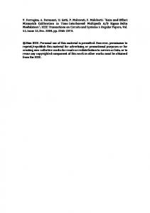
Figure 1 A schematic model of the lysosomal solute carrier (SLC)ome: emerging roles in cellular pathophysiology and pharmacology. A multitude of lysosomal SLCs across lysosomal membrane is involved in the lysosomal transport of solutes. Significant advances in understanding the lysosome proteome has revealed novel roles for lysosomal SLCs, including SLC38A9 as an amino acid sensor (by direct interaction with mTOR complex), SLC17A9 as a mediator of adenosine triphosphate (ATP) exocytosis, SLC15A3/A4 as a participant in toll-like receptor (TLR) signaling, SLC29A3 as a regulator of nucleoside salvage, and SLC46A3 as a facilitator of antibody-drug conjugate maytansine efflux. mTOR, mammalian target of Rapamycin; RagA, Ragulator A; RagC, Ragulator C.
to mediate the intracellular sensing of pathogens after toll-like receptor stimulation.2 Both transporters assist in the egress of bacterially derived components, particularly muramyl dipeptide, to facilitate the NOD2-mediated immune response. Consistently, single nucleotide polymorphism of SLC15A4 is found to associate with autoimmune disease systemic lupus erythematosus. Similarly, SLC11A1 (formerly Nramp1), a divalent metal transporter, is reported in dynamic host-pathogen interactions essential for conferring resistance to certain pathogens. Mutations in SLC11A1 associate with infectious (e.g., tuberculosis, leprosy) and inflammatory (e.g., rheumatoid arthritis) diseases. Recently, the role of an acidic pH-dependent lysosomal nucleoside transporter, SLC29A3, has been linked to macrophage phagocytic function.3 SLC29A3 is predicted to salvage lysosomal nucleobases, nucleosides, and nucleotides, presumably derived from encapsulated pathogens and host macrophage-derived nucleic acids. Accumulating evidence reveals that SLC29A3 mutations can cause a spectrum of human genetic disorders due to abnormal lysosomal nucleoside buildup and increased intralysosomal pH. These diseases include: H syndrome (progressive scleroderma, hyperpigmentation, hypertrichosis, facial telangiectases and dermal and subcutaneous fibrosis); pigmented hypertrichotic dermatosis and insulin-dependent diabetes syndrome; familial Rosai-Dorfman disease; familial histiocytosis; and sinus
histiocytosis with massive lymphadenopathy. SLC29A3 spectrum disorders are allelic, share common mutation(s), and share overlapping manifestations that display an intriguing resemblance to lysosomal storage disorders. Although the basis of these monogenic disorders is only beginning to be appreciated, further studies on the involvement of SLC29A3 in lysosomal and cellular homeostasis should clarify the role of macrophages and other cell types in the pathogenesis of SLC29A3 spectrum disorders. The lysosome participates in membrane trafficking and exocytosis, wherein the lysosome expels its cargo outside the cell. Lysosomal exocytosis is also important in plasma membrane repair and cholesterol homeostasis. Deficiency of lysosomal proteins in Niemann–Pick type C1 disease or Niemann–Pick type C2 diminish cholesterol efflux from the lysosomal compartment, leading to abnormal lysosomal cholesterol accumulation. Correspondingly, a recent finding identified SLC17A9 in lysosomemediated adenosine triphosphate (ATP) release.4 Because ATP is an important neurotransmitter, lysosomal exocytosis maintains regulated ATP release from neurons and/or astrocytes. This lysosomal ATP release is essential as a perturbation of SLC17A9 significantly affected neurotransmission. ATP facilitates synaptic efficiency and plasticity in neurons, and its dysregulation associates with central nervous system pathologies, including brain ischemia, inflammation, and stroke.
CLINICAL PHARMACOLOGY & THERAPEUTICS | VOLUME 100 NUMBER 5 | NOVEMBER 2016
435
DISCOVERY POTENTIAL THERAPEUTIC APPROACHES TARGETING LYSOSOMAL SLC TRANSPORTERS
The localization of SLC transporters on the lysosomal membrane represents a potential class of “druggable” targets for treating lysosomal disorders. However, success, to date, is limited due to the initial requirement of endocytosis for drugs to reach the lysosome. The absence of an SLC transporter crystal structure also slows the drug discovery process. Nevertheless, alternative strategies to effectively utilize or target lysosomal SLCs for development of improved therapeutics are underway. For instance, antibody-drug conjugates are emerging as a cancer-specific treatment to avoid off-target toxicities observed with conventional chemotherapeutic agents. Upon binding of antibody-drug conjugates to surface antigens on cancer cells, they are endocytosed and accumulate in lysosomes. The antibody component is catabolized in the lysosomegenerating active drug. However, the active drug now faces the impending challenge of finding its way out of the lysosome to reach its target site (nucleus, cytoplasm, cytoskeleton, etc.) for action. Because oligonucleotide (siRNA, miRNA) therapeutics and nanoparticle-based therapeutics face the same drug-exit impediment, lysosomal accumulation and degradation have become a widespread concern. Cleavable linker technology, proton-sponge effect, ion pair formation, and hydrophobic modification of vectors or cargos are some of the techniques being currently used for effective drug delivery. In some cases, such strategies are harmful, disrupting (endo) lysosomal membranes. In this regard, lysosomal SLCs offer great promise to aid safe efflux of therapeutic cargos from lysosomes. A recent discovery of SLC46A3 facilitating transport of a noncleavable antibody-drug conjugate catabolite to export cargo from the lysosome validates the feasibility of this strategy.5 Identification and characterization of orphan SLC46A3 identified maytansine as a substrate for SLC46A3, further clarifying the mechanism of noncleavable maytansine-based antibody drug conjugates, including ado-trastuzumab emtansine. The discovery of SLC46A3 as a means for antibody-drug conjugates to target cancer cells suggests future investigation will identify other lysosomal SLC transporters with substrates amenable to therapeutic conjugation for lysosomal exit. In another example, SLC31A2, the lysosomal copper transporter, regulates sensitivity to cisplatin treatment. Measurement of copper flux with new intracellular copper sensors enables testing for copper-dependent sensitivity to chemotherapeutic agents. Enhancement of transporter activity is a treatment option for diseases with loss of transporter function. The lysosomal glutamate transporter, SLC1A2 (EAAT2), is involved in glutamate clearance in astroglial cells, and its protein expression decreases in neurological disorders, like Alzheimer disease, amyotrophic lateral sclerosis, and schizophrenia. The antibiotic ceftriaxone increases transcription of SLC1A2 through modulating nuclear factorkappa B signaling and riluzole, an amyotrophic lateral sclerosis
436
therapeutic, reportedly increases transport activity of SLC1A1 and SLC1A2, supporting this strategy. Furthermore, SLC12A9 is a member of the sodium chloride cotransporter family (SLC12) that regulates ion gradients across renal tubules and maintains cell volume. Mutations in SLC12A9 associate with Bartter disease, characterized by a defect in the thick ascending limb of the loop of Henle, hypokalemia, and alkalosis. Diuretic drugs, like bumetanide and furosemide, inhibit multiple SLC12 transporters; thus, directed development of novel diuretics that target specific SLC12 transporters may offer more effective disease management. Finally, various lysosomal storage disorders are caused by an aberrant accumulation of undigested material within lysosomes. Classic examples involving lysosomal transporters include SLC17A5 and MFSD8 in Salla disease and neuronal ceroid lipofuscinosis, respectively. Although there are no curative treatments yet, enzyme replacement therapy and gene therapy are promising options. Some treatment options for lysosomal storage disorders, like Niemen-Pick disease, Gaucher disease, and alphamannosidosis using enzyme replacement therapy are already approved or are under active (pre)clinical evaluations. Overall, targeting and utilization of SLC transporters for better treatment and management of lysosomal disorders or drug delivery is becoming a reality and research on lysosomal SLCs is gaining momentum to realize new therapeutic possibilities. ACKNOWLEDGMENTS Space limitation restricted us from acknowledging all research groups who contributed to this subject of research. The authors acknowledge support from the National Institutes of Health (NIH) (1R01CA188464 [to R.G.] and R03AR063326 [to R.G. & A.M.B.]) and the Ohio State University Fund (to R.G.). CONFLICT OF INTEREST The authors declared no conflict of interest. C 2016 The Authors Clinical Pharmacology & Therapeutics published by V
Wiley Periodicals, Inc. on behalf of American Society for Clinical Pharmacology and Therapeutics This is an open access article under the terms of the Creative Commons Attribution-NonCommercial-NoDerivs License, which permits use and distribution in any medium, provided the original work is properly cited, the use is non-commercial and no modifications or adaptations are made.
1. Wang, S. et al. Metabolism. Lysosomal amino acid transporter SLC38A9 signals arginine sufficiency to mTORC1. Science 347, 188– 194 (2015). 2. Nakamura, N. et al. Endosomes are specialized platforms for bacterial sensing and NOD2 signalling. Nature 509, 240–244 (2014). 3. Hsu C.L. et al. Equilibrative nucleoside transporter 3 deficiency perturbs lysosome function and macrophage homeostasis. Science 335, 89–92 (2012). 4. Ho, T. et al. Vesicular expression and release of ATP from dopaminergic neurons of the mouse retina and midbrain. Front. Cell. Neurosci. 9, 389 (2015). 5. Hamblett, K.J. et al. SLC46A3 is required to transport catabolites of noncleavable antibody maytansine conjugates from the lysosome to the cytoplasm. Cancer Res. 75, 5329–5340 (2015).
VOLUME 100 NUMBER 5 | NOVEMBER 2016 | www.wileyonlinelibrary/cpt