SHORT COMMUNICATION
Mammary gland biopsy does not affect lactation performance in sows1 R. N. Kirkwood2, J. Pérez Laspiur3, N. K. Ames2, J. B. Moore3, A. Cegielski4, and N. L. Trottier3,5 2Department 3Department
of Large Animal Clinical Sciences, Michigan State University, East Lansing, MI 48824, USA; of Animal Science, Michigan State University, East Lansing, MI 48824, USA; 4Meadowbrook Animal Clinic, Edmonton, Alberta, Canada T6T 1L5. Received 15 November 2006, accepted 9 March 2007. Kirkwood, R. N., Pérez Laspiur, J., Ames, N. K., Moore, J. B., Cegielski, A. and Trottier, N. L. 2007. Mammary gland biopsy does not affect lactation performance in sows. Can. J. Anim. Sci. 87: 281–284. To determine morphological and molecular characteristics of porcine mammary tissue in vivo, mammary tissue was collected from 18 sows at 3 to 6 d of lactation and 17 to 19 d of lactation using a biopsy technique. The success of the technique was determined by monitoring lactation performance, as evidenced by sow rectal temperature, voluntary feed intake, milk somatic cell count, and piglet average daily gain. Up to 1.7 g of mammary tissue was collected at each biopsy without decreasing sow feed intake or piglet growth. Key words: Biopsy, mammary gland, lactation, sow Kirkwood, R. N., Pérez Laspiur, J., Ames, N. K., Moore, J. B., Cegielski, A. et Trottier, N. L. 2007. Les biopsies mammaires n’affectent pas les performances de lactation chez la truie. Can. J. Anim. Sci. 87: 281–284. Afin d’étudier la morphologie ainsi que les charactéristiques moléculaires du tissue mammaire in vivo chez la truie en lactation, du tissue mammaire fut echantillonné sur un total de 18 truies. Les échantillons mammaires ont été prélevé par biopsie entre les jours 3 et 6 de lactation, et encore entre les jours 17 et 19 de lactation. Le succès de la biopsie fut basé sur la quantité de tissue obtenue et le bien-être de la truie. Le bien-être de la truie fut évalué à partir des performances de lactation (la prise alimentaire et gain de poids quotidien), de la température corporelle, et du taux de cellules somatiques dans le lait. Jusqu’à 1.7 g de tissue mammaire ont été prélevé pour chaque biopsie sans affecter la prise alimentaire des truies et le gain de poids quotidien des porcelets. Mots clés: Biopsie, glande mammaire, lactation, truie
relatively large tissue samples. Cerveza et al. (2000) reported the collection of relatively large (1.5 g) sow mammary tissue samples in early and late lactation, but few details of the procedure were provided. A mammary gland biopsy procedure was adequately described by Richert et al. (1998), but only single samples of mammary tissue were obtained. Serial biopsy samples to monitor changes in mammary composition or metabolism may be necessary, but subsequent biopsies may be impacted by inflammatory responses to the first biopsy, affecting sow lactation performance. In none of the earlier studies were potential adverse effects on sow lactation performance investigated. Therefore, our primary goal was to obtain mammary tissue from the same sow at two stages of lactation in sufficient quantity to perform RNA and protein abundance assays, morphometric analysis and immunohistochemistry. Our secondary goal was to determine any effects on the welfare of the sow and litter as evidenced by piglet growth. The All-University Committee on Animal Use and Care of Michigan State University approved all procedures performed in this study. At 110 d of gestation, 18 multiparous sows (12 Landrace × Yorkshire and 6 Yorkshire) were moved to individual crates within an environmentally controlled farrowing room. Room temperature was maintained
Investigating developmental changes in pig mammary tissue composition and metabolism during lactation has historically been achieved by partial or total mammary tissue collection following the serial slaughter of sows (e.g., Kim et al. 1999). Serial slaughter is costly and wasteful of animals, in particular because it requires a relatively large number of sows to account for variation. Furthermore, post-mortem decay of cellular components can occur in only minutes (Wilusz et al. 2001), so time of tissue collection after euthanasia, compared with tissue collection in vivo, may alter the integrity of these components. A mammary needle biopsy approach has been used in different animal species, including lactating goats (Cvek et al. 1998) and sows (Magnusson 1999; Thiel et al. 2005). However, experimental requirements may arise necessitating the collection of 1Presented at the Midwestern Section of the American Dairy
Science Association and American Society of Animal Science Annual Meeting, Des Moines, Iowa, March 21-23, 2005. J. Anim. Sci. 83(Suppl.2):43. http://www.asas.org/ midwest/2005. 5To whom correspondence should be addressed (e-mail:
[email protected]). 281
282
CANADIAN JOURNAL OF ANIMAL SCIENCE
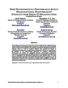
at 20°C and photoperiod controlled at 10 h of light and 14 h of darkness. Litters were equalized to eight piglets per sow by cross-fostering within 36 h of birth. Sows were fed corn-soybean meal-based diets formulated to meet all nutrient requirements according to National Research Council (1998) for lactating sows housed in a thermoneutral environment (20°C) and nursing eight piglets with a predicted average daily gain of 225 g d–1. Throughout the study, sows were fed twice daily at 0800 and 1600. Prior to farrowing, sows received a total of 2.5 kg d–1. After farrowing, on days 1, 2, and 3 of lactation sows were fed a total of 2, 4, and 6 kg, respectively. Thereafter, sows were fed to appetite. Fresh water was freely available throughout the experimental period. Sow feed intake was recorded daily from 1 to 20 d of lactation. Orts were collected before the morning meal, weighed and discarded. Sows were weighed on 1 and 17 d of lactation and piglets were weighed individually on days 1, 4, and 18 of lactation. Milk samples were collected manually at 3 and 17 d of lactation from the first and second thoracic mammary glands on each side of the udder (four glands total) and pooled within sow. Milk letdown was stimulated by intramuscular injection of 10 IU oxytocin (Vet Tek, Phoenix Scientific, St. Joseph, MO). Milk samples were preserved with 8 mg of 2-bromo-2-nitropropane-,3diol (bronopol) and 0.3 mg of natamycin (Broad Spectrum Microtabs II, D&F Control Systems, Inc., Son Ramon, CA) and kept refrigerated for determination of somatic cell counts (Dairy Herd Improvement Association, East Lansing, MI). Mammary gland tissue was collected between 3 and 6 d and again between 17 and 19 d of lactation from the first and second thoracic mammary glands, respectively. On the day of biopsy sows were fed 2 kg of their morning meal and then from 60 min after feeding, piglets were isolated for 45 min. Isolated piglets were kept in a pen adjacent to the sow equipped with a heat lamp and ample bedding. Following the 45-min isolation period, piglets were returned to sows and allowed to nurse. Piglets suckling the glands to be biopsied were identified on their back using a permanent marker. Immediately upon cessation of milk letdown, sows were rapidly anesthetized by intramuscular injection 1.0 mL per 34 kg BW of TKX [(250 mg tiletamine and 250 mg zolazapam (Telozol®; Fort Dodge Animal Health, Fort Dodge, IA), dissolved in 2.5 mL ketamine (VetaKet®; Lloyd Laboratories, Shenandoah, IA) and 2.5 mL xylazine-00 (AnaSed®; Lloyd Laboratories)]. Piglets were removed to their isolation pen, and the sow remained in her farrowing crate. While under anesthesia, sows were positioned in lateral recumbency to expose one entire lateral side of the udder. One mammary gland was prepared for biopsy using Povidone-iodine scrub, USP, 7.5 % (Novaplus, Novation, Irving, TX) followed by rinsing with 70% alcohol and cleaning with Povidone-iodine solution, USP, 10% (Novaplus, Novation) (Fig. 1). Following subcutaneous and intramammary injection of 1.0 mL of 2% lidocaine (Lidoject®; Butler Animal Health supply, Dublin, OH) (Fig. 2), a 2-cm incision was made vertical to the plica lateralis, aligned with the nipple and approximately 5 cm
dorsal to the perimeter of the nipple areola. Hemorrhage was minimal but, if evident, was controlled by pressure with gauze. Two pieces of mammary tissue (~400 to 850 mg each) were exteriorized by gently grasping a small piece of mammary tissue with forceps and using this to elevate a much larger piece of tissue, which was then excised with a scalpel in a circular motion (Fig. 3). Mammary tissue was rapidly flash-frozen in liquid nitrogen and stored at –80ºC to determine the relative transcript abundance of key genes of interest or incubated in 10% buffered formalin for histological preparation and in situ hybridization. The incision was closed with simple interrupted sutures using size 0 nylon (CP-1 ethilon suture, Ethicon, Inc., Piscataway, NJ) (Fig. 4). Time from incision to closing was approximately 5 min. Sows received an intramuscular injection of Banamine® (1.1 mg kg–1 BW) (flunixin meglumine, Schering-Plough Animal Health, Omaha, NE) immediately after biopsy and again at 24 and 48 h later. Following full recovery from anesthesia, sows were fed the remainder of their morning meal and piglets were returned to the sow and allowed to suckle normally. Rectal temperature was recorded after biopsy and again at 24 and 48 h to monitor health status of the sow. Sutures were removed 7 d after biopsy. Analysis of variance was conducted to determine differences in average daily gain for piglets suckling the mammary glands subjected to biopsy compared with piglets suckling the intact glands, and to determine differences in sow daily feed intake, rectal temperature and milk somatic cell count one day prior to biopsy (pre) compared to 2 d following biopsy (post). Sources of variation in the model included effect of sow, stage of lactation, and mammary gland treatment (biopsy vs. intact). Statistical analysis was performed using the MIXED procedure of SAS (SAS Institute, Inc., Cary, NC). Repeated measures analysis was used for repeated measures over days of lactation based on different covariance structures (Littell et al. 1998). The best fitting repeated measures covariance structure was determined using the Akaike information criterion. Separation of means was performed by least squares estimates. Least square means were considered different at P < 0.05. Preand post-test power calculations were performed as previously described by Stroup (1999, 2002). Up to 1.7 g of mammary tissue was obtained from live lactating sows at both early and late stages of lactation without apparent effect on sow welfare as indicated by sow feed intake, rectal temperature, and milk somatic cell count (Table 1). One sow developed a local infection at the biopsy site and was treated with a topical triple antibiotic containing neomycin, polymyxin B sulfates, and bacitracin zinc (Fougera, Altana Inc, Melville, NY) for 5 d until infection subsided. No local or systemic infections were observed for the remaining 17 sows. Average sow rectal temperature was lower (P < 0.001) on the 2 d following biopsy compared with that of the day of biopsy, reflecting the efficacy of the Banamine. Sow feed intake was not different on the day following biopsy than that on the day prior to biopsy. On average, sows gained 3.3 kg of body weight during the 18-d lactation period. Average daily gain from days 4 to 18 of lactation of the piglets suckling the glands subjected to biop-
KIRKWOOD ET AL. — MAMMARY BIOPSY IN SOWS
283
Fig. 1. One mammary gland (thoracic) was prepared for biopsy with a combination of povidone-iodine scrub and 70 % ethanol.
Fig. 3. Mammary tissue was exteriorized with forceps and excised with a scalpel in a circular motion.
Fig. 2. Lidocaine was injected sub-cutaneously and intramuscularly 1 min prior to incision.
Fig. 4. The incision was closed using simple interrupted sutures. Three to four sutures were sufficient to close the incision and ensuring space for drainage.
Table 1. Sow feed intake, rectal temperature and milk somatic cell count pre and post mammary biopsyz Item
Time relative to biopsy
Sow feed intake (kg d–1) Sow rectal temperature (°C) Milk somatic cell count (1000 mL–1) zData
Before
After
P value
5.13 ± 0.32 39.2 ± 0.07 3677 ± 781
5.02 ± 0.33 38.7 ± 0.07 4969 ± 918
0.79