341 Original Article
Management of Primary Central Nervous System Lymphoma Francois G. El Kamar, MD, Lauren E. Abrey, MD, New York, New York
Key Words Lymphoma, brain, methotrexate, radiotherapy Abstract Primary central nervous system lymphoma (PCNSL) is a unique subtype of non-Hodgkin’s lymphoma (NHL). Although PCNSL is both chemosensitive and radiosensitive, it is very difficult to treat and outcomes are substantially inferior when compared with similar extranodal NHL. Furthermore, as a subtype of brain tumors and NHL, the overall incidence has been increasing over the past several decades. This review summarizes the salient clinical features and approach to management of the immunocompetent patient with PCNSL. (JNCCN 2004;2:341–349).
Primary central nervous system lymphomas (PCNSL)
are a group of high- or intermediate-grade extranodal lymphomas arising in and confined to the central nervous system (CNS).1,2 This definition excludes angiotropic lymphomas and metastatic lymphomas.3 PCNSL was described as early as 1929 by Bailey in his description of the “perivascular sarcomatous tumour,”4 but modern immunohistochemical techniques allowed the definitive identification as a B-cell type of non-Hodgkin’s lymphoma (NHL). The incidence of PCNSL in the immunocompetent population has increased significantly over the past 3 decades.5 However, despite a threefold increase, the absolute risk remains very low, and the incidence approximates 0.3/100,000/y.6 PCNSL represents 2.7% of all CNS tumors and 1% to 2% of all lymphomas.7 It has a slight From the Department of Neurology, Memorial Sloan-Kettering Cancer Center, New York, New York. Submitted February 5, 2004; accepted for publication April 20, 2004. Dr. Abrey receives grant support from Schering Plough, Inc., and Genentech, Inc. Correspondence: Lauren E. Abrey, MD, Department of Neurology, Memorial Sloan-Kettering Cancer Center, 1275 York Avenue, New York, NY 10021. E-mail:
[email protected]
male predominance, with a gender ratio of 1.5 and a peak incidence at 60 years of age. It is regarded as a highly treatable disease, but overall prognosis is poor, with a 5-year overall survival rate of 25%.1,8,9
Clinical Presentation Symptomatology in PCNSL is dictated primarily by the location of the tumor within the CNS as well as by the rapidity of disease progression. Personality changes and cognitive dysfunction are the most common presenting symptoms, followed by symptoms of increased intracranial pressure such as headaches and nausea. Focal neurologic symptoms (sensory or motor) can be present and vary according to the location of the disease. To a lesser degree, psychomotor slowing, disorientation, and seizures may be present.10,11 The average time to diagnosis from the beginning of symptoms is 2 to 3 months.10,11
Imaging Magnetic resonance imaging (MRI) is the gold standard and typically shows a solitary (60% to 70%) hypointense nodular lesion with indistinct borders on T1-weighted images. Dense and homogenous enhancement can be seen after injection of gadolinium. On T2-weighted images, lesions are hyperintense with moderate if any surrounding vasogenic edema.12 In AIDS patients, ring enhancement caused by central tumor necrosis may be seen. Perifocal edema can be seen, which is less prominent than the edema seen with malignant glioma or metastases.13 PCNSL is usually supratentorial rather than infratentorial, and is localized to the cerebral hemispheres, frontal lobes, corpus callosum, basal ganglia, or thalamus, often in a periventricular location. Spinal cord involvement is rare.12 The high cell density and metabolic rate of PCNSL may make discriminating it from other
© Journal of the National Comprehensive Cancer Network | Volume 2 Number 4 | July 2004
Original Article
342
El Kamar and Abrey
high-grade brain tumors on 18fluorodeoxyglucose positron emission tomography possible.14,15
Staging
More than 90% of cases are B-cell lymphomas CD19+ CD20+, monoclonal with either k or l light chain restriction, and morphologically indistinguishable from systemic lymphoma. The pattern of expression of adhesion molecules is identical to NHL.16,17 According to the Revised European American Classification (REAL) system for extranodal lymphomas, most are diffuse large cell, immunoblastic, or lymphoblastic cell type. Approximately 3% are T-cell lymphomas, which are more commonly infratentorial.2,18,19 The growth pattern shows angiocentric, infiltrating lesions forming cuffs around the arterioles and venules, sometimes invading the vessel walls, and a centrifugal infiltration of the surrounding parenchyma.2
After the diagnosis is made, disease work-up is critical to evaluate ocular or leptomeningeal involvement and to rule out systemic dissemination (Table 1). Ocular involvement is present in 20% to 25%, and a slit lamp ophthalmologic examination for ocular involvement is always required. Cytologic examination of vitreal aspirates may be necessary in patients with isolated ocular lymphoma (PIOL).24–26 CSF cytology reveals tumor cells in about 15% and nonspecific abnormalities in up to 75%.24,27 b2-microglobulin levels, immunophenotyping, and clonal immunoglobulin gene rearrangement can be performed to increase the yield of the CSF analysis. 24 The rest of the work up comprises a computed tomography (CT) scan of the chest, abdomen, and pelvis, and a bone marrow aspiration. Systemic lymphoma is found in fewer than 4% of cases.28 Serology for HIV is imperative to rule out AIDS.
Prognostic Factors
Therapy
The most important and widely recognized prognostic factors are age 60 years or younger and Eastern Cooperative Oncology Group (ECOG) performance status 0 to 2.20,21 More recently, the International Extranodal Lymphoma Study Group identified age, performance status, lactate dehydrogenase (LDH) serum level, cerebrospinal fluid (CSF) protein concentration, and involvement of the deep structures of the brain as independent prognostic variables. They were able to use these variables to construct a prognostic index in which each variable was assigned a value of 0 if favorable and 1 if not. The 2-year overall survival was 80% for patients with a score of 0 to 1, 48% for a score of 2 to 3, and 15% for a score of 4 to 5.21
NHLs are exquisitely chemosensitive and radiosensitive malignancies, with an initial response in excess of 90% for each modality. This is also true for PCNSL, but PCNSL has lower response rates of 60% per modality and a high risk of early relapse if radiotherapy is used as a single modality. Therefore, numerous clinical trials have sought to improve outcome with various combinations of chemotherapy and radiotherapy.
Pathology
Table 1 Recommended Baseline Evaluation Brain MRI with and without gadolinium Clinical examination including a baseline performance status Serum LDH levels LP for CSF analysis; protein, cytology, b2-microglobulin
Diagnosis and Staging
Ophthalmologic examination including a slit lamp examination
Biopsy
CT Scan with IV contrast of the chest, abdomen and pelvis
When the diagnosis of PCNSL is suspected, steroids should be avoided if possible because their potent cytolytic effect on the lymphoma cells may obscure the diagnosis. Stereotactic or open biopsy of the contrastenhancing lesions is the most appropriate diagnostic procedure.22 Resection is rarely performed because it provides no therapeutic benefit, and the risk of surgical complications is high.23
Bilateral bone marrow biopsy Testicular examination and ultrasound examination in males older than 65 years. MRI of the spine with and without gadolinium if symptomatic Abbreviations: MRI, magnetic resonance imaging; LDH, lactate dehydrogenase; LP, lumbar puncture; CSF, cerebrospinal fluid; CT, computed tomography; IV, intravenous.
© Journal of the National Comprehensive Cancer Network | Volume 2 Number 4 | July 2004
Original Article
Primary Central Nervous System Lymphoma
Corticosteroids
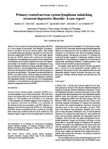
Corticosteroids induce apoptosis via a direct cytotoxic effect in lymphoid cells. Dexamethasone may lead to complete remission in 15% or partial remissions in 25% of PCNSL patients (Fig. 1).29 This correlates with clinical and radiologic improvement that may occasionally last long after discontinuation of the corticosteroid.30 More commonly, this is temporary, and relapse typically occurs shortly after discontinuation of dexamethasone. Resistance may develop after re-exposure to corticosteroids in previously documented sensitive disease or during withdrawal therapy. Therefore, the corticosteroid may be restarted after biopsy to prevent symptom recurrence and to decrease associated vasogenic edema.23 Long-term steroid therapy should be avoided to minimize secondary complications.31 Radiotherapy
Whole brain radiotherapy (WBRT) has a response rate close to 65%, but median survival is only 12 to 18 months, and 5-year survival is 4%.32,33 In comparison, radiotherapy alone for equivalent stages IE or IIE extranodal systemic NHL has a local control rate in excess of 90%, and a 5-year survival of 43% to 77%. Thus, WBRT is unacceptable as a single modality treatment for PCNSL.34 Furthermore, there is no role for dose intensification, as was shown by the prospective RTOG 8315
trial, in which a total dose of 40 Gy to the whole brain with a boost of 20 Gy to the tumor bed did not yield improved disease control (39%) or median survival (12.2 months). In this study, 79% of the recurrences occurred in the boosted field.32 In cases of primary ocular lymphoma or ocular dissemination, treatment should include radiotherapy to the globe, with doses of 35 to 40 Gy fractionated over 5 weeks. Both eyes should be treated even in cases with only evidence of monocular involvement. Most patients will experience a symptomatic improvement and vitreal clearing.23,35 However, some patients show vitreal clearing without improved vision and others may not respond to radiotherapy. Accelerated cataract, optic neuropathy, conjunctivitis, dry eyes, corneal epithelial defects, retinal atrophy, and vitreous hemorrhage are possible complications of ocular irradiation.36 Chemotherapy
The addition of chemotherapy to WBRT has generally resulted in improved response rates and survival; however, the optimal chemotherapy regimen has yet to be identified, and numerous agents have shown activity in PCNSL (Table 2).37 A standard NHL regimen such as CHOP or the slightly modified CHOD (cyclophosphamide, Adriamycin, vincristine, and Decadron) combined with WBRT has a very short-lived response, with distant CNS treatment failure or florid leptomeningeal
Figure 1 Contrast-enhanced scan of PCNSL before (left) and after (right) 1 week of steroids shows dramatic partial resolution
of tumor mass.
© Journal of the National Comprehensive Cancer Network | Volume 2 Number 4 | July 2004
343
Original Article
344
El Kamar and Abrey
Table 2 Chemotherapeutic Agents with Known Activity in PCNSL Agent
Properties and use in PCNSL
Methotrexate
Antimetabolite, intravenous high dose single agent or in combination. Intrathecal, intravitreous or intraarterial administration possible. Requires aggressive hydration and alkalinization with leucovorin rescue.
Cytarabine
Antimetabolite, prolonged intra-CSF activity, intravenous and intrathecal administration as a single agent or in combination.
Corticosteroids
Steroid hormones, oral or intravenous, single agent or in combination rapid response as trigger oncolytic response, numerous side effects.
Procarbazine
Oral methyl-hydrazine derivative in conjunction with HD-MTX, neurotoxicities usually reversible.
Vincristine
Mitotic spindle inhibitor, intravenous administration in combination regimen, has cumulative neurotoxicity.
Thiotepa
Alkylating agent, IV in combination regimen or HD intensification prior to stem cell transplant, also intrathecal and intravitreal administration.
Temozolomide
Oral imidazotetrazines single agent or in combination regimen.
Rituximab
Human/murine chimeric anti CD-20 monoclonal antibody, IV, intrathecal or ventricular administration in combination regimen, may prolong post chemotherapy granulocytic recovery.
Topotecan
Oral or IV topoisomerase I inhibitor, few reports of activity in PCNSL and dose limiting myelosuppression.
Lomustine
Oral nitrosourea used in combination regimen, has cumulative pulmonary and renal toxicities.
Abbreviations: PCNSL, primary central nervous system lymphoma; CSF, cerebrospinal fluid; HD-MTX, high dose methotrexate; HD, high dose.
recurrences. Randomized studies failed to show any survival advantage when compared with WBRT alone.38–42 MACOP and MACOP-B combined with WBRT PCNSL showed excellent initial responses that were comparable to the responses in systemic NHL. However,
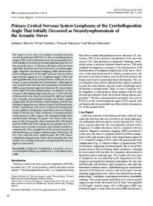
no survival advantage was seen over WBRT alone.43 Methotrexate is the most effective and extensively used drug in PCNSL (Fig. 2). Its activity against CNS lymphoma was recognized with the observation that NHL patients treated with methotrexate experienced fewer
Figure 2 Sagittal MRI of the brain with gadolinium before (left) and after (right) completion of 5 cycles of HD-methotrexate
show a complete radiographic response.
© Journal of the National Comprehensive Cancer Network | Volume 2 Number 4 | July 2004
Original Article
345
Primary Central Nervous System Lymphoma
CNS relapses.44,45 This efficacy of MTX in improving survival outcomes was first noted by Loeffler et al.46 In a group of 5 patients who received intravenous or intrathecal methotrexate followed by WBRT, the median survival rate seen was 44 months.46 Similar observations by other investigators led to a wide range of reported methotrexate-based regimens with varying degrees of success (Table 3). A prospective trial by the RTOG confirmed the observation that methotrexatebased chemotherapy in combination with WBRT improves disease control and overall survival. The role of intrathecal chemotherapy in treating PCNSL is controversial. Rapid intravenous administration of both methotrexate and cytarabine
produce tumoricidal levels within the CSF and may obviate intrathecal administration of drug. Furthermore, at least two retrospective studies have failed to show any advantage in terms of disease control or survival for patients treated with intrathecal chemotherapy.60–62 However, disease recurrence is common, and treatment-related neurotoxicity is a recognized complication of the combination of high-dose methotrexate and WBRT. Therefore, current efforts to improve outcomes have focused on strategies that seek to intensify the chemotherapy regimen and minimize or defer WBRT.37,53 Administration of chemotherapy after disruption of the blood-brain barrier in 111 patients
Table 3 Survival and Response Rates to Various Methotrexate Based Chemotherapy for PCNSL Author year published
N
Regimen
IT CHT
RT and dose
Response
Median Survival
Gabbai et al.47 1989
13
MTX 3.5 g/m2
—
30-44 Gy
92%
9.5 months
Glass et al. 1994
25
MTX 3.5 g/m2
—
30-44 Gy
88%
33 months
DeAngelis et al. 1992
31
MTX 1 g/m
MTX 12 mg 3 6
40 Gy + 14.4 Gy boost
64%
41 months
Sandor et al.50 1998
14
MTV (MTX 8.4 g/m2 ) IT Ara-C
MTX 12 mg 3 2 and ARA-C 15mg/ d/33d 3 2
—
100%
16.5 months PFS
Cheng et al.51 1998
19
BOMES (MTX 1.5 g/m2)
MTX 12 mg 3 4 If CSF involved
—
84%
6 months PFS
Guha-Thakurta et al.52 1999
31
MTX 8 g/m2 and consolidation with 3.5 g/m2 every 3rd month.
—
—
100%
30+ months
Abrey et al.53 2000
52
MPV (MTX 3.5 g/m2 ) Ara-C
MTX 12 mg 3 3
45 Gy in 35 Patients
90%
60 months
McAllister et al.54 2000
74
IA MTX (2.5 g) + cyclophosphamide and etoposide
—
—
65% complete
40.7 months
Ferreri et al.55 2001
13
MPV (MTX 3 g/m2)
—
39.6 Gy
92%
25+ months
MPV (MTX 2.5 g/m2 )
MTX 12 mg 3 5
45 Gy
94%
30+ months
—
—
74%
22.8+
48
49
DeAngelis et al.56 RTOG 2002
102
2
Batchelor et al.57 2003
25
MTX 8 g/m2
Poortmans et al. 2003 - EORTC 20962
52
MBVP (MTX 3 g/m )
MTX 15 mg – ARA-C40 mg and hydrocortisone 25mg 3 2
30 Gy + 10 Gy boost
81%
46 months
Pels et al.59 2003
65
MTX 5 g/m2 and ARA-C 3 g/m2 wit ifosfamide VCR cyclophosphamide and dexamethasone
MTX 3 mg, ARA-C 30 mg and prednisolone and 2.5 mg 3 3
—
71%
>60 year old MS 34 months.