Instruction Manual. LIbrary PrEParaTION. NEbNext® Multiplex Small rNa. Library
Prep Set for Illumina® (Set 1). NEb #E7300S/L. 24/96 reactions ...
LIBRARY PREPARATION
NEBNext® Multiplex Small RNA Library Prep Set for Illumina® (Set 1) Instruction Manual
NEB #E7300S/L 24/96 reactions
Sign up for the NEBNext e-newsletter Scan this code or visit www.neb.com/ NEBNextnews2 to sign up for the NEBNext bimonthly e-newsletter to learn about new NEBNext products, recent publications and advances in library prep for next gen sequencing.
ISO 9001
ISO 14001
ISO 13485
Registered
Registered
Registered
Quality Management
Environmental Management
Medical Devices
Methods for avoidance of adaptor dimer formation are covered by pending patent (New England Biolabs, Inc.). NEW ENGLAND BIOLABS®, LONGAMP®, NEBNEXT® and QUICK-LOAD® are registered trademarks of New England Biolabs, Inc. AGENCOURT® and AMPURE® are registered trademarks of Beckman Coulter, Inc. AGILENT® and BIOANALYZER® are registered trademarks of Agilent Technologies, Inc. ILLUMINA® is a registered trademark of Illumina, Inc. CORNING®, COSTAR® and SPIN-X® are registered trademarks of Corning, Inc. NOVEX®, FIRSTCHOICE® and SYBR® are registered trademarks of Life Technologies, Inc. FLASHPAGE™ is a registered trademark of Life Technologies, Inc. PELLET PESTLE® is a registered trademark of Kimble Kontes Asset Management, Inc. QIAGEN® and QIAQUICK® are registered trademarks of The Qiagen Group.
NEBNext Multiplex Small RNA Library Prep Set for Illumina (Set 1)
Table of Contents Set 1 Includes . . . . . . . . . . . . . . . . . . . . . . . . . . . . . . . . . . . . . . . . . . . . . . . . . . . . . . . . . . . . . . . . . . . . . . . . . . 2 Required Materials Not Included . . . . . . . . . . . . . . . . . . . . . . . . . . . . . . . . . . . . . . . . . . . . . . . . . . . . 3 Applications . . . . . . . . . . . . . . . . . . . . . . . . . . . . . . . . . . . . . . . . . . . . . . . . . . . . . . . . . . . . . . . . . . . . . . . . . . . . 4 Multiplex Small RNA Library Prep Workflow . . . . . . . . . . . . . . . . . . . . . . . . . . . . . . . . . . . . . . . 4 Protocols . . . . . . . . . . . . . . . . . . . . . . . . . . . . . . . . . . . . . . . . . . . . . . . . . . . . . . . . . . . . . . . . . . . . . . . . . . . . . . . 5 NEBNext 3´ Ligation Reaction Buffer (2X) . . . . . . . . . . . . . . . . . . . . . . . . . . . . . . . . . . . . . . . . 14 NEBNext 3´ Ligation Enzyme Mix . . . . . . . . . . . . . . . . . . . . . . . . . . . . . . . . . . . . . . . . . . . . . . . . . . 15 NEBNext 3´ SR Adaptor for Illumina . . . . . . . . . . . . . . . . . . . . . . . . . . . . . . . . . . . . . . . . . . . . . . 16 NEBNext 5´ SR Adaptor for Illumina . . . . . . . . . . . . . . . . . . . . . . . . . . . . . . . . . . . . . . . . . . . . . . 17 NEBNext 5´ Ligation Reaction Buffer (10X) . . . . . . . . . . . . . . . . . . . . . . . . . . . . . . . . . . . . . . 18 NEBNext 5´ Ligation Enzyme Mix . . . . . . . . . . . . . . . . . . . . . . . . . . . . . . . . . . . . . . . . . . . . . . . . . . 19 NEBNext SR RT Primer for Illumina . . . . . . . . . . . . . . . . . . . . . . . . . . . . . . . . . . . . . . . . . . . . . . . 20 NEBNext First Strand Synthesis Reaction Buffer . . . . . . . . . . . . . . . . . . . . . . . . . . . . . . . . 21 ProtoScript II Reverse Transcriptase . . . . . . . . . . . . . . . . . . . . . . . . . . . . . . . . . . . . . . . . . . . . . . 22 Murine RNase Inhibitor . . . . . . . . . . . . . . . . . . . . . . . . . . . . . . . . . . . . . . . . . . . . . . . . . . . . . . . . . . . . . 23 ® LongAmp Taq 2X Master Mix . . . . . . . . . . . . . . . . . . . . . . . . . . . . . . . . . . . . . . . . . . . . . . . . . . . . . 24 NEBNext SR Primer for Illumina . . . . . . . . . . . . . . . . . . . . . . . . . . . . . . . . . . . . . . . . . . . . . . . . . . . 25 Gel Loading Dye, Blue (6X) . . . . . . . . . . . . . . . . . . . . . . . . . . . . . . . . . . . . . . . . . . . . . . . . . . . . . . . . . 26 ® Quick-Load pBR322 DNA-MspI Digest . . . . . . . . . . . . . . . . . . . . . . . . . . . . . . . . . . . . . . 27–28 DNA Gel Elution Buffer, 1X . . . . . . . . . . . . . . . . . . . . . . . . . . . . . . . . . . . . . . . . . . . . . . . . . . . . . . . . . 29 Linear Acrylamide (10 mg/ml) . . . . . . . . . . . . . . . . . . . . . . . . . . . . . . . . . . . . . . . . . . . . . . . . . . . . . 30 TE Buffer . . . . . . . . . . . . . . . . . . . . . . . . . . . . . . . . . . . . . . . . . . . . . . . . . . . . . . . . . . . . . . . . . . . . . . . . . . . . . . 31 Nuclease-free Water . . . . . . . . . . . . . . . . . . . . . . . . . . . . . . . . . . . . . . . . . . . . . . . . . . . . . . . . . . . . . . . . . 32 NEBNext Index 1–12 Primers for Illumina . . . . . . . . . . . . . . . . . . . . . . . . . . . . . . . . . . . . 33–34 Frequently Asked Questions (FAQs) . . . . . . . . . . . . . . . . . . . . . . . . . . . . . . . . . . . . . . . . . . . . . . 35 Revision History . . . . . . . . . . . . . . . . . . . . . . . . . . . . . . . . . . . . . . . . . . . . . . . . . . . . . . . . . . . . . . . . . . . . . 36
1
The NEBNext Multiplex Small RNA Library Prep Set for Illumina (Set 1) Includes: The volumes provided are sufficient for preparation of up to 24 reactions (NEB #E7300S) and 96 reactions (NEB #E7300L). All reagents should be stored at –20°C.
• (green) NEBNext 3´ Ligation Reaction Buffer (2X) • (green) NEBNext 3´ Ligation Enzyme Mix • (green) NEBNext 3´ SR Adaptor for Illumina • (yellow) NEBNext 5´ SR Adaptor for Illumina • (yellow) NEBNext 5´ Ligation Reaction Buffer (10X) • (yellow) NEBNext 5´ Ligation Enzyme Mix • (pink) NEBNext SR RT Primer for Illumina • (red) NEBNext First Strand Synthesis Reaction Buffer • (red) ProtoScript II Reverse Transcriptase • (red) Murine RNase Inhibitor • (blue) LongAmp Taq 2X Master Mix • (blue) NEBNext SR Primer for Illumina • (blue) NEBNext Index 1 Primer for Illumina • (blue) NEBNext Index 2 Primer for Illumina • (blue) NEBNext Index 3 Primer for Illumina • (blue) NEBNext Index 4 Primer for Illumina • (blue) NEBNext Index 5 Primer for Illumina • (blue) NEBNext Index 6 Primer for Illumina • (blue) NEBNext Index 7 Primer for Illumina • (blue) NEBNext Index 8 Primer for Illumina • (blue) NEBNext Index 9 Primer for Illumina • (blue) NEBNext Index 10 Primer for Illumina • (blue) NEBNext Index 11 Primer for Illumina • (blue) NEBNext Index 12 Primer for Illumina • (orange) Gel Loading Dye, Blue (6X) • (orange) Quick-Load pBR322 DNA-MspI Digest • (white) DNA Gel Elution Buffer, 1X • (white) Linear Acrylamide (10 mg/ml) • (white) TE Buffer • (white) Nuclease-free Water
2
Required Materials Not Included: 3 M Sodium Acetate, pH 5.5 100% Ethanol 80% Ethanol Corning®, Costar®, Spin-X® Centrifuge Tube Filters (Cellulose Acetate Filters) (Sigma Aldrich # CLS8162)
Size Selection Materials: for gel size selection: 6% Novex® TBE PAGE gel 1.0 mM 10-well (Life Technologies, Inc. #EC6265BOX) SYBR® Gold Nucleic Acid Gel Stain (Life Technologies, Inc. #S-11494) RNase-free Disposable Pellet Pestles® (Kimble Kontes Asset Management, Inc. #749521-1590) Dry Ice/Methanol Bath or –80°C freezer for bead selection: Agencourt® AMPure® XP Beads (Beckman Coulter, Inc. #A63881) for Pippin Prep™ selection: 3% Agarose Dye Free Gel (Sage Science #CDF 3010)
QIAquick® PCR Purification Kit (Qiagen® #28104) Bioanalyzer® (Agilent® Technologies, Inc.)
3
Applications The NEBNext Multiplex Small RNA Library Prep Set for Illumina (Set 1) contains adaptors, primers, enzymes and buffers that are ideally suited to convert small RNA transcripts into barcoded cDNA libraries for next-generation sequencing on the Illumina (Illumina, Inc.) platform. Each of these components must pass rigorous quality control standards and is lot controlled, both individually and as a set of reagents. Lot Control: The lots provided in the NEBNext Multiplex Small RNA Library Prep Set for Illumina (Set 1) are managed separately and are qualified by additional functional validation. Individual reagents undergo standard enzyme activity and quality control assays, and also meet stringent criteria in the additional quality controls listed on each individual component page. Functionally Validated: Each set of reagents is functionally validated together through construction and sequencing of barcoded small RNA libraries on the Illumina sequencing platform. For larger volume requirements, customized and bulk packaging is available by purchasing through the OEM/Bulks department at NEB. Please contact OEM@ neb.com for further information.
Multiplex Small RNA Library Prep Workflow
STEPS
This kit includes a novel protocol that results in higher yields and lower adaptor-dimer contamination.
3´ Ligation
Primer Hybridization
5´ Ligation
1st Strand Synthesis
PCR Amplification
Size Selection
Kit Optimization The components of this kit were collectively optimized with FirstChoice® Human Brain Reference RNA (Life Technologies, Inc. #AM6050). High quality total RNA should be used as starting material. An RNA Integrity Number (RIN) > 7 is recommended. Assess the quality and quantity of your sample using the Agilent 2100 Bioanalyzer using an Agilent RNA 6000 Nano Chip.
4
Protocols Please refer to revision history for a summary of protocol updates Symbols SAFE STOP
This is a point where you can safely stop the protocol and store the samples prior to proceeding to the next step in the protocol.
! This caution sign signifies a step in the protocol that has two paths
leading to the same end point but is dependent on a user variable, like the type of RNA input.
•
Colored bullets indicate the cap color of the reagent to be added
Libraries prepared by this method are compatible with Illumina paired-end flow cells. Starting Material: 100 ng–1 µg Total RNA. Ligate the 3´ SR Adaptor
Note: For total RNA inputs of 100 ng, dilute the • (green) 3´ SR Adaptor for Illumina 1:2 in nuclease free water.
1. Mix the following components in a sterile nuclease-free PCR tube: Input RNA • (green) 3´ SR Adaptor for Illumina Nuclease-Free Water Total volume
1–6 µl 1 µl variable 7 µl
2. Incubate in a preheated thermal cycler for 2 minutes at 70°C. Transfer tube to ice. 3. Add the following Components:
• (green) 3´ Ligation Reaction Buffer (2X) • (green) 3´ Ligation Enzyme Mix
Total volume
10 µl 3 µl 20 µl
4. Incubate for 1 hour at 25°C in a thermal cycler.
Note: Longer incubation times and reduced temperatures (18 hours; 16°C) increase ligation efficiency of methylated RNAs such as piwiinteracting RNAs (piRNAs) (if present in the sample). However, some concatamerization products might be formed.
Hybridize the Reverse Transcription Primer This step is important to prevent adaptor-dimer formation. The SR RT Primer hybridizes to the excess of 3´ SR Adaptor (that remains free after the 3´ ligation reaction) and transforms the single stranded DNA adaptor into a
5
double-stranded DNA molecule. dsDNAs are not substrates for ligation mediated by T4 RNA Ligase 1 and therefore do not ligate to the 5´ SR Adaptor in the subsequent ligation step.
Note: For total RNA inputs of 100 ng, dilute the • (pink) SR RT Primer for Illumina 1:2 in nuclease free water.
5. Add the following components to the ligation mixture from step 4 and mix well: Nuclease-Free Water
• (pink) SR RT Primer for Illumina Total volume now should be
4.5 µl 1 µl 25.5 µl
6. Heat samples for 5 minutes at 75°C. Transfer to 37°C for 15 minutes, followed by 15 minutes at 25°C. Ligate the 5´ SR Adaptor 7. With 5 minutes remaining, resuspend the • (yellow) 5´ SR adaptor in 120 µl of nuclease free water.
Note: For total RNA inputs of 100 ng, dilute the • (yellow) 5´ SR Adaptor for Illumina 1:2 in nuclease free water.
8. Aliquot 1.1 N µl of the • (yellow) 5´ SR Adaptor into a separate, nucleasefree 200 µl PCR tube, with N equal to the number of samples being processed for the current experiment. 9. Incubate the adaptor in the thermal cycler at 70°C for 2 minutes and then immediately place the tube on ice. Keep the tube on ice and use the denatured adaptor within 30 minutes of denaturation.
Note: Store the remaining resuspended 5´ SR adaptor at –80°C.
10. Add the following components to the ligation mixture from step 6 and mix well:
• (yellow) 5´ SR Adaptor for Illumina (denatured) • (yellow) 5´ Ligation Reaction Buffer (10X) • (yellow) 5´ Ligation Enzyme Mix
Total volume
1 µl 1 µl 2.5 µl 30 µl
11. Incubate for 1 hour at 25°C in a thermal cycler. Perform Reverse Transcription 12. Mix the following components in a sterile, nuclease-free tube: Adaptor Ligated RNA from Step 11 • (red) First Strand Synthesis Reaction Buffer • (red) Murine RNase Inhibitor • (red) ProtoScript II Reverse Transcriptase Total volume 6
30 µl 8 µl 1 µl 1 µl 40 µl
13. Incubate for 60 minutes at 50°C. 14. Immediately proceed to PCR amplification. SAFE STOP
Safe Stopping Point: If you do not plan to proceed immediately to PCR amplification, then heat inactivate the RT reaction at 70°C for 15 minutes. Samples can be safely stored at –15°C to –25°C.
Perform PCR Amplification 15. Add the following components to the RT reaction mix from step 14 and mix well:
• (blue) LongAmp Taq 2X Master Mix • (blue) SR Primer for Illumina • (blue) Index (X) Primer*
50 µl 2.5 µl 2.5 µl 5 µl 100 µl
Nuclease free water Total volume now should be
*Note: The NEBNext Multiplex Small RNA Library Prep Set for Illumina Set 1 contains 1–12 PCR primers, each with a different index. For each reaction, only one of the 12 PCR primer indices is used during the PCR step.
PCR Cycling conditions:
CYCLE STEP
TEMP
TIME
CYCLES
Initial Denaturation
94°C
30 sec
1
Denaturation
94°C
15 sec
Annealing
62°C
30 sec
Extension
70°C
15 sec
Final Extension
70°C
5 min
Hold
4°C
∞
12–15* 1
*Amplification conditions may vary based on RNA input amount, tissue, and species. This protocol was optimized using 1 µg of total RNA from human brain and 12 PCR cycles. The number of PCR cycles can be adjusted if clear and distinct bands are not observed in the gel image. Run only between 12 and 15 cycles. For 100 ng total RNA input run 15 cycles of PCR.
Perform Library QC and Size Selection Note: There are several different methods for performing size selection. It is recommended to choose the appropriate method based on the QC check of the library using the Bioanalyzer. Size selection using AMPure XP Beads does not remove small fragments. If you perform the QC check and your sample contains Adaptor dimer (127 bp peak) or excess primers (70-80 bp) it is recommended to use gel or Pippin Prep for size selection.
7
QC Check and Size Selection using AMPure XP Beads 1. Purify the PCR amplified cDNA construct (100 µl) using a QIAQuick PCR Purification Kit. IMPORTANT: Before eluting the DNA from the column, centrifuge the column with the lid of the spin column open for 5 minutes at 13,200 rpm. Centrifugation with the lid open ensures that no ethanol remains during DNA elution. It is important to dry the spin column membrane of any residual ethanol that may interfere with the correct loading of the sample on the PAGE gel. 2. Elute amplified DNA in 27.5 µl Nuclease-free Water. 3. Load 1 µl of the purified PCR reaction on the Bioanalyzer using a DNA 1000 chip according to the manufacturer's instructions (Figure 1). 4. To the purified PCR reaction (25 µl), add 32.5 μl (1.3X) of resuspended AMPure XP beads and mix well on a vortex mixer or by pipetting up and down at least 10 times. 5. Incubate for 5 minutes at room temperature. 6. Place the tube on an appropriate magnetic stand to separate beads from supernatant. After the solution is clear (about 5 minutes), carefully transfer the supernatant (57.5 µl) to a new tube (Caution: do not discard the supernatant). Discard beads that contain the large DNA fragments. 7. Add 92.5 μl (3.7X) of resuspended AMPure XP beads to the supernatant (57.5 μl), mix well and incubate for 5 minutes at room temperature. 8. Place the tube on an appropriate magnetic stand to separate beads from supernatant. After the solution is clear (about 5 minutes), carefully remove and discard the supernatant. Be careful not to disturb the beads that contain DNA targets (Caution: do not discard beads). 9. Add 200 μl of freshly prepared 80% ethanol to the tube while in the magnetic stand. Incubate at room temperature for 30 seconds, and then carefully remove and discard the supernatant. 10. Repeat Step 9 once. 11. Briefly spin the tube, and put the tube back in the magnetic stand. 12. Completely remove the residual ethanol, and air dry beads for 10 minutes while the tube is on the magnetic stand with lid open. 13. Elute the DNA target from the beads with 15 μl nuclease-free water. Mix well on a vortex mixer or by pipetting up and down, and put the tube in the magnetic stand until the solution is clear. 14. Transfer the supernatant to a clean PCR tube.
8
Figure 1: Typical results from (A) human brain and (B) rat testis total RNA libraries before size selection. A. [FU] 70
3
14
00
15
60 50 40
1 19 3 15 5 07 16 2
15
30 20
1
27
10 0 -10 15
200
100
300
400
700
1,500
[nt]
B. [FU] 120
3
15
100 80 00
60
15
40 20
6
15
18
3
27
0 15
100
300
500
1,500
[nt]
The 143 and 153 bp bands correspond to miRNAs and piRNAs, respectively. The bands on the Bionalyzer electropherograms resolve in sizes ~ 6-8 nucleotides larger than sizes observed on PAGE gels and can shift from sample to sample due to an incorrect identification of the marker by the bioanalyzer software. miRNA peak should be ~ 143-146 bp.
9
15. Run 1 µl on the Bioanalyzer High Sensitivity chip. Check peak distribution and molarity of the small RNA library.
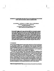
Figure 2: Electropherogram trace of the bead size selected purified library from human brain total RNA.
QC Check and Size Selection using 6% PolyAcrylamide Gel 1. Purify the PCR amplified cDNA construct (100 µl) using a QIAQuick PCR Purification Kit. IMPORTANT: Before eluting the DNA from the column, centrifuge the column with the lid of the spin column open for 5 minutes at 13,200 rpm. Centrifugation with the lid open ensures that no ethanol remains during DNA elution. It is important to dry the spin column membrane of any residual ethanol that may interfere with the correct loading of the sample on the PAGE gel. 2. Elute amplified DNA in 27.5 µl Nuclease-free Water. 3. Load 1 µl of the purified PCR reaction on the Bioanalyzer using a DNA 1000 chip according to the manufacturer's instructions (Figure 1). 4. Mix the purified PCR product (25 µl) with 5 µl of Gel Loading Dye, Blue (6X).
Note: Vortex the Gel Loading Dye, Blue throughly to mix well before using.
5. Load 5 µl of Quick-Load pBR322 DNA-MspI Digest in one well on the 6% PAGE 10-well gel. 6. Load two wells with 15 µl each of mixed amplified cDNA construct and loading dye on the 6% PAGE 10-well gel. 7. Run the gel for 1 hour at 120 V or until the blue dye reaches the bottom of the gel. Do not let the blue dye exit the gel. 8. Remove the gel from the apparatus and stain the gel with SYBR Gold nucleic acid gel stain in a clean container for 2–3 minutes and view the gel on a UV transiluminator (Figure 3). 10
A.
bp
Molecular Human Marker Brain
B.
bp
622
622
527
527
Molecular Marker
Rat Testis
404
404
307 307 242 217 190 160 147 123 110
miRNA (~140 bp)
242 217 190 160 147 123 110
piRNA (~150 bp)
90 76 67
90 76 67
Figure 3: Shows typical results from Human Brain (A) and Rat Testis (B) Total RNA libraries. The 140 and 150 bp bands correspond to miRNAs (21 nt) and piRNAs (30 nt), respectively.
9. The 140 and 150 nucleotide bands correspond to adapter-ligated constructs derived from the 21 and 30 nucleotide RNA fragments, respectively. For miRNAs, isolate the bands corresponding to ~140 bp. For piRNAs, isolate the band corresponding to ~150 bp. 10. Place the two gel slices from the same sample in one 1.5 ml tube and crush the gel slices with the RNase-free Disposable Pellet Pestles and then soak in 250 µl DNA Gel Elution buffer (1X). 11. Rotate end-to-end for at least 2 hours at room temperature. 12. Transfer the eluate and the gel debris to the top of a gel filtration column. 13. Centrifuge the filter for 2 min at > 13,200 rpm. 14. Recover eluate and add 1 µl Linear Acrylamide, 25 µl 3M sodium acetate, pH 5.5 and 750 µl of 100% ethanol. 15. Vortex well. 16. Precipitate in a dry ice/methanol bath or at –80°C for at least 30 minutes. 17. Spin in a microcentrifuge @ > 14,000 x g for 30 minutes at 4°C. 18. Remove the supernatant taking care not to disturb the pellet. 19. Wash the pellet with 80% ethanol by vortexing vigorously. 11
20. Spin in a microcentrifuge @ > 14,000 x g for 30 minutes at 4°C. 21. Air dry pellet for up to 10 minutes at room temperature to remove residual ethanol. 22. Resuspend pellet in 12 µl TE Buffer. 23. Load 1 µl of the size selected purified library on a 2100 Bioanalyzer using a DNA 1000 or High Sensitivity DNA chip according to the manufacturer's instructions (Figure 4). 24. Check the size, purity, and concentration of the sample.
Figure 4: Electropherogram trace of the gel size selected purified library from human brain total RNA. [FU]
7
14
1,000
500
0
,38
35
10
40
50
60
70
80
90
100
110
4
,63
12
0
120
130
[s]
QC Check and Size Selection Using Pippin Prep Size selection of the Small RNA library (147 bp) can done on Pippin Prep instrument using the 3% Agarose, dye free gel with internal standards (Sage Science # CDF3010). 1. Purify the PCR amplified cDNA construct (100 μl) using a QIAQuick PCR Purification Kit. IMPORTANT: Before eluting the DNA from the column, centrifuge the column with the lid of the spin column open for 5 minutes at 13,200 rpm. Centrifugation with the lid open ensures that no ethanol remains during DNA elution 2. Elute amplified DNA in 32 μl nuclease-free water. It is recommended to QC your library before performing size selection: 1. Load 1 μl of the purified PCR reaction on the Bioanalyzer using a DNA 1000 chip according to the manufacturer's instructions (Figure 1). 12
2. miRNA library should appear as a peak at 147 bp peak (that correspond for 21 nucleotide insert). Program the protocol for size selection on Pippin Prep Instrument as follow: 1. In the Pippin Prep software, go to the Protocol Editor Tab. 2. Click “Cassette” folder, and select “3% DF Marker F”. 3. Select the collection mode as “Range” and enter the size selection parameters as follow: BP start (105) and the BP end (155). BP Range Flag should indicate “broad”. Note: This protocol is optimized to select for 147–149 bp peak. 4. Click the “Use of Internal Standards” button. 5. Make sure the “Ref Lane” values match the lane numbers. 6. Press “Save As” and name and save the protocol. Prepare sample for size selection as follow: 1. Bring loading solution to room temperature 2. For each sample, combine 30 µl sample with 10 µl of DNA marker F (labeled F). 3. Mix samples thoroughly (vortex mixer). Briefly centrifuge to collect. 4. Load 40 µl (DNA plus marker) on one well of the 3% agarose cassette. 5. Run the program with the settings indicated above. 6. After sample has been eluted, collect 40 µl sample from elution well. Run 1 µl in a Bioanalzyer using the high sensitivity chip. Note: If the Ethidium Bromide free cassettes was used, no purification is required before running sample on the bioanalyzer.
Figure 5: Electropherogram trace of Pippin Prep size selected library from human brain total RNA.
13
NEBNext 3´ Ligation Reaction Buffer #E7301A: #E7301AA:
0.24 ml 0.96 ml
Concentration: 2X
Store at –20°C
Quality Control Assays 16-Hour Incubation: 50 μl reactions containing 3´ Ligation Reaction Buffer at 1X concentration and 1 μg of HindIII digested Lambda DNA incubated for 16 hours at 37°C results in no detectable non-specific nuclease degradation as determined by agarose gel electrophoresis. 50 μl reactions containing this reaction buffer at 1X concentration and 1 μg T3 DNA incubated for 16 hours at 37°C also results in no detectable non-specific nuclease degradation as determined by agarose gel electrophoresis. Endonuclease Activity: Incubation of a 50 μl reaction containing 1X 3´ Ligation Reaction Buffer with 1 μg of φX174 RF I DNA in assay buffer for 4 hours at 37°C results in less than 10% conversion to RF II as determined by agarose gel electrophoresis. Phosphatase Activity: Incubation of 1X 3´ Ligation Reaction Buffer in protein phosphatase assay buffer (1 M diethanolamine @ pH 9.8 and 0.5 mM MgCl2) containing 2.5 mM p-nitrophenyl phosphate at 37°C for 4 hours yields no detectable p-nitrophenylene anion as determined by spectrophotometric analysis at 405 nm. RNase Activity: Incubation of a 10 μl reaction of 3´ Ligation Reaction Buffer at 1X concentration with 40 ng of a FAM-labeled RNA transcript for 16 hours at 37°C results in no detectable RNase activity as determined by polyacrylamide gel electrophoresis. Lot Controlled
14
NEBNext 3´ Ligation Enzyme Mix #E7288A: 0.072 ml #E7288AA: 0.288 ml Store at –20°C Description: NEBNext 3´ Ligation Enzyme Mix has been optimized to ligate short single-stranded RNAs (17–30 nucleotide length) to a 5´-adenylated, 3´-blocked single-stranded DNA adaptor in 1X NEBNext 3´ Ligation Reaction Buffer at 25°C.
Quality Control Assays SDS-PAGE Purity: SDS-PAGE analysis of this enzyme indicates > 95% enzyme purity. 16-Hour Incubation: 50 μl reactions containing 1 μl of 3´ Ligation Enzyme Mix and 1 μg of HindIII digested Lambda DNA incubated for 16 hours at 37°C results in no detectable non-specific nuclease degradation as determined by agarose gel electrophoresis. 50 μl reactions containing 1 μl of 3´ Ligation Enzyme Mix and 1 μg T3 DNA incubated for 16 hours at 37°C also results in no detectable non-specific nuclease degradation as determined by agarose gel electrophoresis. Endonuclease Activity: Incubation of a 50 μl reaction containing 1 μl 3´ Ligation Enzyme Mix with 1 μg of φX174 RF I DNA for 4 hours at 37°C results in less than 10% conversion to RF II as determined by agarose gel electrophoresis. Phosphatase Activity: Incubation of a minimum of 1 μl 3´ Ligation Enzyme Mix in protein phosphatase assay buffer (1 M diethanolamine @ pH 9.8 and 0.5 mM MgCl2) containing 2.5 mM p-nitrophenyl phosphate at 37°C for 4 hours yields no detectable p-nitrophenylene anion as determined by spectrophotometric analysis at 405 nm. RNase Activity: Incubation of a 10 μl reaction containing 1 μl 3´ Ligation Enzyme Mix with 40 ng of a FAM-labeled RNA transcript for 16 hours at 37°C results in no detectable RNase Activity as determined by polyacrylamide gel electrophoresis. Functional Activity: 200 units of T4 RNA Ligase2, truncated, ligates 80% of a 31-mer RNA to the pre-adenylated end of a 17-mer DNA [Universal miRNA Cloning Linker (NEB #S1315)] in a total reaction volume of 10 µl in 1 hour at 25°C. Unit assay conditions: 1X Reaction Buffer (50 mM Tris-HCl, 10 mM MgCl2, 1 mM DTT, pH 7.5 @ 25°C) supplemented to 10% (w/v) PEG MW 4000, 5 pmol of 5´-FAM labeled RNA, and 10 pmol preadenylated DNA linker. After incubation at 25°C for 1 hour, the ligated product is detected on a 15% denaturing polyacrylamide gel. One unit of Murine RNase Inhibitor inhibits the activity of the 5 ng of RNase A by 50%. Activity is measured by the inhibition of hydrolysis of cytidine 2, 3´ – cyclic monophosphate by RNase A. Lot Controlled
15
NEBNext 3´ SR Adaptor for Illumina #E7332A: #E7332AA:
0.024 ml 0.096 ml
Store at –20°C Description: 5´ adenylated, 3´ blocked oligodeoxynucleotide Sequence: 5´-rAppAGATCGGAAGAGCACACGTCT-NH2-3´
Quality Control Assays 16-Hour Incubation: 50 µl reactions containing 1 µl NEBNext 3´ SR Adaptor and 1 µg of HindIII digested Lambda DNA incubated for 16 hours at 37°C results in no detectable non-specific nuclease degradation as determined by agarose gel electrophoresis. 50 µl reactions containing 1 µl NEBNext 3´ SR Adaptor and 1 µg of T3 DNA incubated for 16 hours at 37°C results in no detectable nonspecific nuclease degradation as determined by agarose gel electrophoresis. Endonuclease Activity: Incubation of a 50 µl reaction containing 1 µl NEBNext 3´ SR Adaptor with 1 µg of φX174 RF I supercoiled DNA for 4 hours at 37°C results in less than 10% conversion to RF II as determined by agarose gel electrophoresis. RNase Activity: Incubation of a 10 µl reaction containing 1 µl NEBNext 3´ SR Adaptor with 40 ng of RNA transcript for 16 hours at 37°C resulted in no detectable degradation of RNA as determined by gel electrophoresis. Phosphatase Activity: Incubation of 1 µl NEBNext 3´ SR Adaptor in protein phosphatase assay buffer (1 M diethanolamine @ pH 9.8 and 0.5 mM MgCl2) containing 2.5 mM p-nitrophenyl phosphate at 37°C for 4 hours yields no detectable p-nitrophenylene anion as determined by spectrophotometric analysis at 405 nm. Lot Controlled
16
NEBNext 5´ SR Adaptor for Illumina #E7328A:
1350 pmol
Store at –20°C Sequence: 5´- rGrUrUrCrArGrArGrUrUrCrUrArCrArGrUrCrCrGrArCrGrArUrC-3´
Quality Control Assays 16-Hour Incubation: 50 µl reactions containing 1 µl NEBNext 5´ SR Adaptor and 1 µg of HindIII digested Lambda DNA incubated for 16 hours at 37°C results in no detectable non-specific nuclease degradation as determined by agarose gel electrophoresis. 50 µl reactions containing 1 µl NEBNext 5´ SR Adaptor and 1 µg of T3 DNA incubated for 16 hours at 37°C results in no detectable nonspecific nuclease degradation as determined by agarose gel electrophoresis. Endonuclease Activity: Incubation of a 50 µl reaction containing 1 µl NEBNext 5´ SR Adaptor with 1 µg of φX174 RF I supercoiled DNA for 4 hours at 37°C results in less than 10% conversion to RF II as determined by agarose gel electrophoresis. RNase Activity: Incubation of a 10 µl reaction containing 1 µl NEBNext 5´ SR Adaptor with 40 ng of RNA transcript for 16 hours at 37°C resulted in no detectable degradation of RNA as determined by gel electrophoresis. Phosphatase Activity: Incubation of 1 µl NEBNext 5´ SR Adaptor in protein phosphatase assay buffer (1 M diethanolamine @ pH 9.8 and 0.5 mM MgCl2) containing 2.5 mM p-nitrophenyl phosphate at 37°C for 4 hours yields no detectable p-nitrophenylene anion as determined by spectrophotometric analysis at 405 nm. Lot Controlled
17
NEBNext 5´ Ligation Reaction Buffer #E7304A: #E7304AA:
0.024 ml 0.096 ml
Store at –20°C
Quality Control Assays 16-Hour Incubation: 50 µl reactions containing 1X 5´ Ligation Reaction Buffer and 1 µg of HindIII digested Lambda DNA incubated for 16 hours at 37°C results in no detectable non-specific nuclease degradation as determined by agarose gel electrophoresis. 50 µl reactions containing 1X 5´ Ligation Reaction Buffer and 1 µg of T3 DNA incubated for 16 hours at 37°C results in no detectable nonspecific nuclease degradation as determined by agarose gel electrophoresis. Endonuclease Activity: Incubation of a 50 µl reaction containing 1X 5´ Ligation Reaction Buffer with 1 µg of φX174 RF I supercoiled DNA for 4 hours at 37°C results in less than 10% conversion to RF II as determined by agarose gel electrophoresis. Phosphatase Activity: Incubation of 1X 5´ Ligation Reaction Buffer in protein phosphatase assay buffer (1 M diethanolamine @ pH 9.8 and 0.5 mM MgCl2) containing 2.5 mM p-nitrophenyl phosphate at 37°C for 4 hours yields no detectable p-nitrophenylene anion as determined by spectrophotometric analysis at 405 nm. RNase Activity: Incubation of a 10 µl reaction containing 1X 5´ Ligation Reaction Buffer with 40 ng of a FAM-labeled RNA transcript for 16 hours at 37°C resulted in no detectable degradation of RNA as determined by polyacrylamide gel electrophoresis. Lot Controlled
18
NEBNext 5´ Ligation Enzyme Mix #E7305A: #E7305AA:
0.06 ml 0.24 ml
Store at –20°C Description: NEBNext 5´ Ligation Enzyme Mix has been optimized to ligate short single-stranded RNAs (17–30 nucleotide length) in 1X NEBNext 5´ Ligation Reaction Buffer at 25°C.
Quality Control Assays SDS-PAGE Purity: SDS-PAGE analysis of this enzyme indicates > 95% enzyme purity. 16-Hour Incubation: 50 µl reactions containing 1 µl of 5´ Ligation Enzyme Mix and 1 µg of HindIII digested Lambda DNA incubated for 16 hours at 37°C results in no detectable non-specific nuclease degradation as determined by agarose gel electrophoresis. 50 µl reactions containing 1 µl of 5´ Ligation Enzyme Mix and 1 µg of T3 DNA incubated for 16 hours at 37°C results in no detectable nonspecific nuclease degradation as determined by agarose gel electrophoresis. Endonuclease Activity: Incubation of a 50 µl reaction containing 1 µl 5´ Ligation Enzyme Mix with 1 µg of φX174 RF I supercoiled DNA for 4 hours at 37°C results in less than 10% conversion to RF II as determined by agarose gel electrophoresis. Phosphatase Activity: Incubation of a minimum of 1 µl 5´ Ligation Enzyme Mix in protein phosphatase assay buffer (1 M diethanolamine @ pH 9.8 and 0.5 mM MgCl2) containing 2.5 mM p-nitrophenyl phosphate at 37°C for 4 hours yields no detectable p-nitrophenylene anion as determined by spectrophotometric analysis at 405 nm. Functional Activity: One unit of T4 RNA Ligase 1 is defined as the amount of enzyme required to convert 1 nmol of 5´-[32P] rA16 into a phosphatase-resistant form in 30 minutes at 37°C. One unit of Murine Inhibitor inhibits the activity of 5 ng of RNase A by 50%. Activity is measured by the inhibition of hydrolysis of cytidine 2, 3´-cyclic monophosphate by RNase A. Lot Controlled
19
NEBNext SR RT Primer for Illumina #E7333A: #E7333AA:
0.024 ml 0.096 ml
Store at –20°C Sequence: 5´-AGACGTGTGCTCTTCCGATCT-3´
Quality Control Assays 16-Hour Incubation: 50 µl reactions containing 1 µl NEBNext SR RT Primer and 1 µg of HindIII digested Lambda DNA incubated for 16 hours at 37°C results in no detectable non-specific nuclease degradation as determined by agarose gel electrophoresis. 50 µl reactions containing NEBNext SR RT Primer and 1 µg of T3 DNA incubated for 16 hours at 37°C results in no detectable non-specific nuclease degradation as determined by agarose gel electrophoresis. Endonuclease Activity: Incubation of a 50 µl reaction containing 1 µl NEBNext SR RT Primer with 1 µg of φX174 RF I supercoiled DNA for 4 hours at 37°C results in less than 10% conversion to RF II as determined by agarose gel electrophoresis. RNase Activity: Incubation of a 10 µl reaction containing 1 µl NEBNext SR RT Primer with 40 ng of RNA transcript for 16 hours at 37°C resulted in no detectable degradation of RNA as determined by gel electrophoresis. Phosphatase Activity: Incubation of 1 µl NEBNext SR RT Primer in protein phosphatase assay buffer (1 M diethanolamine @ pH 9.8 and 0.5 mM MgCl2) containing 2.5 mM p-nitrophenyl phosphate at 37°C for 4 hours yields no detectable p-nitrophenylene anion as determined by spectrophotometric analysis at 405 nm. Lot Controlled
20
NEBNext First Strand Synthesis Reaction Buffer #E7334A: #E7334AA:
0.192 ml 0.768 ml
Concentration: 5X
Store at –20°C
Quality Control Assays 16-Hour Incubation: 50 µl reactions containing 1X First Strand Synthesis Reaction Buffer and 1 µg of HindIII digested Lambda DNA incubated for 16 hours at 37°C results in no detectable non-specific nuclease degradation as determined by agarose gel electrophoresis. 50 µl reactions containing 1X First Strand Synthesis Reaction Buffer and 1 µg of T3 DNA incubated for 16 hours at 37°C results in no detectable non-specific nuclease degradation as determined by agarose gel electrophoresis. Endonuclease Activity: Incubation of a 10 μl reaction containing 1X First Strand Synthesis Reaction Buffer with 1 µg of φX174 RF I supercoiled DNA for 4 hours at 37°C results in < 10% conversion to RF II (nicked molecules) as determined by gel electrophoresis. RNase Activity: Incubation of a 10 μl reaction containing 1X First Strand Synthesis Reaction Buffer with 40 ng of RNA transcript for 16 hours at 37°C resulted in no detectable degradation of RNA as determined by gel electrophoresis. Phosphatase Activity: Incubation of 1X First Strand Synthesis Reaction Buffer in protein phosphatase assay buffer (1 M diethanolamine @ pH 9.8 and 0.5 mM MgCl2) containing 2.5 mM p-nitrophenyl phosphate at 37°C for 4 hours yields no detectable p-nitrophenylene anion as determined by spectrophotometric analysis at 405 nm. Lot Controlled
21
ProtoScript II Reverse Transcriptase #E7355A: 0.024 ml #E7355AA: 0.096 ml
Concentration: 200,000 U/ml
Store at –20°C Description: ProtoScript II Reverse Transcriptase is a recombinant M-MuLV reverse transcriptase with reduced RNase H activity and increased thermostability. It can be used to synthesize first strand cDNA at higher temperatures than the wild type M-MuLV. The enzyme is active up to 50°C, providing higher specificity, higher yield of cDNA and more full-length cDNA product up to 12 kb. Source: The gene encoding a mutant M-MuLV Reverse Transcriptase (RNase H–) is expressed in E. coli and purified to near homogeneity. Supplied in: 20 mM Tris-HCl (pH 7.5), 100 mM NaCl, 0.1 mM EDTA, 1 mM DTT, 0.01% (v/v) IGEPAL® CA-630, 50% (v/v) glycerol
Quality Control Assays 16-Hour Incubation: A 50 μl reaction containing 1 μg of φX174 DNA and 100 units of ProtoScript II Reverse Transcriptase incubated for 16 hours at 37°C resulted in a DNA pattern free of detectable nuclease degradation as determined by agarose gel electrophoresis. Exonuclease Activity: Incubation of a 50 μl reaction containing 100 units of ProtoScript II Reverse Transcriptase with 1 μg of a mixture of single and double-stranded [3H] E. coli DNA (105 cpm/μg) for 4 hours at 37°C released 95% pure as determined by SDS PAGE analysis using Coomassie blue detection. Lot Controlled
22
Murine RNase Inhibitor #E7308A: #E7308AA:
0.024 ml 0.096 ml
Store at –20°C Description: Murine RNase inhibitor is a 50 kDa recombinant protein of murine origin. The inhibitor specifically inhibits RNases A, B and C. It inhibits RNases by binding noncovalently in a 1:1 ratio with high affinity. It is not effective against RNase 1, RNase T1, S1 Nuclease, RNase H or RNase from Aspergillus. In addition, no inhibition of polymerase activity is observed when RNase Inhibitor is used with Taq DNA Polymerase, AMV or M-MuLV Reverse Transcriptases, or Phage RNA Polymerases (SP6, T7, or T3). Recombinant murine RNase inhibitor does not contain the pair of cysteines identified in the human version that is very sensitive to oxidation, which causes inactivation of the inhibitor (1). As a result, Murine RNase Inhibitor has significantly improved resistance to oxidation compared to the human/ porcine RNase inhibitors, even under conditions where the DTT concentration is low. Therefore, it is advantageous to use murine RNase inhibitor in reactions where high concentration DTT is adverse to the reaction (eg. Real-time RT-PCR). Source: An E. coli strain that carries the ribonuclease inhibitor gene from mouse Supplied in: 20 mM HEPES-KOH, 50 mM KCl, 8 mM DTT and 50% glycerol
Quality Control Assays 16-Hour Incubation: 50 µl reactions containing a minimum of 40 units of Murine RNase Inhibitor and 1 µg of HindIII digested Lambda DNA incubated for 16 hours at 37°C results in no detectable non-specific nuclease degradation as determined by agarose gel electrophoresis. 50 µl reactions containing a minimum of 40 units of Murine RNase Inhibitor and 1 µg of T3 DNA incubated for 16 hours at 37°C results in no detectable non-specific nuclease degradation as determined by agarose gel electrophoresis. Exonuclease Activity: Incubation of a 50 μl reaction containing 200 units of Murine RNase Inhibitor with 1 μg of a mixture of single and double-stranded [3H] E. coli DNA (205 cpm/μg) for 4 hours at 37°C released < 0.5% of the total radioactivity. Latent RNase Assay: Heating the Murine RNase Inhibitor for 20 minutes at 65°C, followed by incubation of a 10 μl reaction containing 40 units of RNase Inhibitor with 40 ng of RNA transcript for 4 hours at 37°C resulted in no detectable degradation of RNA as determined by gel electrophoresis. RNase Activity: Incubation of a 10 μl reaction containing 40 units of Murine RNase Inhibitor with 40 ng of RNA transcript for 16 hours at 37°C resulted in no detectable degradation of RNA as determined by gel electrophoresis. Endonuclease Activity: Incubation of a 10 μl reaction containing 40 units of Murine RNase Inhibitor with 1 µg of φX174 RF I supercoiled DNA for 4 hours at 37°C results in < 10% conversion to RF II (nicked molecules) as determined by gel electrophoresis. Phosphatase Activity: Incubation of a minimum of 40 units of Murine RNase Inhibitor in protein phosphatase assay buffer (1 M diethanolamine @ pH 9.8 and 0.5 mM MgCl2) containing 2.5 mM p-nitrophenyl phosphate at 37°C for 4 hours yields no detectable p-nitrophenylene anion as determined by spectrophotometric analysis at 405 nm. Functional Activity: One unit of Murine RNase Inhibitor inhibits the activity of 5 ng of RNase A by 50%. Activity is measured by the inhibition of hydrolysis of cytidine 2, 3'-cyclic monophosphate by RNase A. Lot Controlled References: 1. Kim, B.M. et al. (1999). Protein Science, 8, 430–434.
23
LongAmp Taq 2X Master Mix #E7309A: #E7309AA:
1.2 ml 4.8 ml
Concentration: 2X
Store at –20°C
Quality Control Assays SDS-PAGE Purity: SDS-PAGE analysis of this enzyme indicates > 95% enzyme purity. 16-Hour Incubation: 50 µl reactions containing 1 µl of LongAmp Taq 2X Master Mix and 1 µg of HindIII digested Lambda DNA incubated for 16 hours at 37°C results in no detectable non-specific nuclease degradation as determined by agarose gel electrophoresis. 50 µl reactions containing 1 µl of LongAmp Taq 2X Master Mix and 1 µg of T3 DNA incubated for 16 hours at 37°C results in no detectable non-specific nuclease degradation as determined by agarose gel electrophoresis. Endonuclease Activity: Incubation of a minimum of 10 µl of LongAmp Taq 2X Master Mix with 1 µg of φX174 RF I supercoiled DNA for 4 hours at 37°C results in less than 10% conversion to RF II as determined by agarose gel electrophoresis. RNase Activity: Incubation of a 10 µl reaction containing 1 µl of LongAmp Taq 2X Master Mix with 40 ng of a FAM-labeled RNA transcript for 16 hours at 37°C resulted in no detectable degradation of RNA as determined by polyacrylamide gel electrophoresis. Phosphatase Activity: Incubation of a minimum of 10 µl of LongAmp Taq 2X Master Mix in protein phosphatase assay buffer (1 M diethanolamine @ pH 9.8 and 0.5 mM MgCl2) containing 2.5 mM p-nitrophenyl phosphate at 37°C for 4 hours yields no detectable p-nitrophenylene anion as determined by spectrophotometric analysis at 405 nm. Functional Activity: One unit of LongAmp Taq DNA Polymerase is defined as the amount of enzyme that will incorporate 10 nmol of dNTP into acid-insoluble material in 1X ThermoPol Reaction Buffer, 200 µM dNTPs including [3H]-dTTP and 200 µg/ml activated Calf Thymus DNA; in 30 minutes at 65°C. Lot Controlled
24
NEBNext SR Primer for Illumina #E7310A: 0.060 ml #E7310AA: 0.240 ml Store at –20°C Sequence: 5´-AATGATACGGCGACCACCGAGATCTACACGTTCAGAGTTCTACAGTCCG-s-A-3´ Where -s- indicates phosphorothioate bond.
Quality Control Assays 16-Hour Incubation: 50 µl reactions containing 1 µl NEBNext SR Primer and 1 µg of HindIII digested Lambda DNA incubated for 16 hours at 37°C results in no detectable non-specific nuclease degradation as determined by agarose gel electrophoresis. 50 µl reactions containing NEBNext SR Primer for Illumina and 1 µg of T3 DNA incubated for 16 hours at 37°C results in no detectable nonspecific nuclease degradation as determined by agarose gel electrophoresis. Endonuclease Activity: Incubation of a 50 µl reaction containing 1 µl NEBNext SR Primer with 1 µg of φX174 RF I supercoiled DNA for 4 hours at 37°C results in less than 10% conversion to RF II as determined by agarose gel electrophoresis. RNase Activity: Incubation of a 10 µl reaction containing 1 µl NEBNext SR Primer with 40 ng of RNA transcript for 16 hours at 37°C resulted in no detectable degradation of RNA as determined by gel electrophoresis. Phosphatase Activity: Incubation of 1 µl NEBNext SR Primer in protein phosphatase assay buffer (1 M diethanolamine @ pH 9.8 and 0.5 mM MgCl2) containing 2.5 mM p-nitrophenyl phosphate at 37°C for 4 hours yields no detectable p-nitrophenylene anion as determined by spectrophotometric analysis at 405 nm.
25
Gel Loading Dye, Blue #E6138A: 0.2 ml #E6138AA: 1 ml
Concentration: 6X
Store at 25°C Description: Gel Loading Dye, Blue (6X) is a pre-mixed loading buffer with a tracking dye for agarose and non-denaturing poylacrylamide gel electrophoresis. This solution contains SDS, which often results in sharper bands, as some enzymes are known to remain bound to their DNA substrates following cleavage. EDTA is also included to chelate magnesium (up to 10 mM) in enzymatic reactions, thereby stopping the reaction. Bromophenol Blue migrates at approximately 300 bp on a standard 1% TBE agarose gel.
Quality Control Assays 16-Hour Incubation: 50 µl reactions containing 1X Gel Loading Dye and 1 µg of HindIII digested Lambda DNA incubated for 16 hours at 37°C results in no detectable non-specific nuclease degradation as determined by agarose gel electrophoresis. 50 µl reactions containing 1X Gel Loading Dye and 1 µg of T3 DNA incubated for 16 hours at 37°C results in no detectable non-specific nuclease degradation as determined by agarose gel electrophoresis. Endonuclease Activity: Incubation of a 50 µl reaction containing 1X Gel Loading Dye with 1 µg of φX174 RF I supercoiled DNA for 4 hours at 37°C results in less than 10% conversion to RF II (nicked molecules) as determined by agarose gel electrophoresis. RNase Activity: Incubation of a 10 µl reaction containing 1X Gel Loading Dye with 40 ng of RNA transcript for 16 hours at 37°C resulted in no detectable degradation of RNA as determined by gel electrophoresis. Phosphatase Activity: Incubation of 1X Gel Loading Dye in protein phosphatase assay buffer (1 M diethanolamine @ pH 9.8 and 0.5 mM MgCl2) containing 2.5 mM p-nitrophenyl phosphate at 37°C for 4 hours yields no detectable p-nitrophenylene anion as determined by spectrophotometric analysis at 405 nm. Lot Controlled
26
Quick-Load pBR322 MspI-DNA Digest #E7323A: #E7323AA:
0.24 ml 0.96 ml
Concentration: 50 µg/ml
Store at –20°C or 4°C Description: Quick-Load pBR322 DNA-MspI Digest is a pre-mixed, ready to load molecular weight marker containing Bromophenol Blue as a tracking dye. The MspI Digest of pBR322 DNA yields 26 fragments.The double-stranded DNA is digested to completion with MspI, phenol extracted, and equilibrated to 10 mM Tris-HCl (pH 8.0) and 1 mM EDTA. Fragment
Size (bp)
1
622
2
527
3
404
4
307
5
242
6
238
7
217
8
201
9
190
10
180
11,12
160
13,14
147
15
123
16
110
17
90
18
76
19
67
20,21
34
22,23
26
24
15
25,26
9
27
Quick-Load pBR322 MspI-DNA Digest (Cont.) Quality Control Assays 16-Hour Incubation: 50 µl reactions containing 1 µl Quick-Load pBR322 MspI DNA Digest and 1 µg of HindIII digested Lambda DNA incubated for 16 hours at 37°C results in no detectable non-specific nuclease degradation as determined by agarose gel electrophoresis. 50 µl reactions containing 1 µl Quick-Load pBR322 MspI DNA Digest and 1 µg of T3 DNA incubated for 16 hours at 37°C results in no detectable non-specific nuclease degradation as determined by agarose gel electrophoresis. Endonuclease Activity: Incubation of a 50 µl reaction containing 1 µl QuickLoad pBR322 MspI DNA Digest with 1 µg of φX174 RF I supercoiled DNA for 4 hours at 37°C results in less than 10% conversion to RF II (nicked molecules) as determined by agarose gel electrophoresis. RNase Activity: Incubation of a 10 µl reaction containing 1 µl Quick-Load pBR322 MspI DNA Digest with 40 ng of RNA transcript for 16 hours at 37°C resulted in no detectable degradation of RNA as determined by gel electrophoresis. Phosphatase Activity: Incubation of Quick-Load pBR322 MspI DNA Digest in protein phosphatase assay buffer (1 M diethanolamine @ pH 9.8 and 0.5 mM MgCl2) containing 2.5 mM p-nitrophenyl phosphate at 37°C for 4 hours yields no detectable p-nitrophenylene anion as determined by spectrophotometric analysis at 405 nm. Lot Controlled
28
DNA Gel Elution Buffer #E7324A: #E7324AA:
12 ml 48 ml
Concentration: 1X
Store at –20°C or 4°C Description: DNA Gel Elution Buffer is provided for the extraction of the size selected amplified cDNA library from the polyacrylamide gel.
Quality Control Assays 16-Hour Incubation: 50 µl reactions containing 10 µl DNA Gel Elution Buffer and 1 µg of HindIII digested Lambda DNA incubated for 16 hours at 37°C results in no detectable non-specific nuclease degradation as determined by agarose gel electrophoresis. 50 µl reactions containing 10 µl DNA Gel Elution Buffer and 1 µg of T3 DNA incubated for 16 hours at 37°C results in no detectable non-specific nuclease degradation as determined by agarose gel electrophoresis. Endonuclease Activity: Incubation of a 50 µl reaction containing 10 µl DNA Gel Elution Buffer with 1 µg of φX174 RF I supercoiled DNA for 4 hours at 37°C results in less than 10% conversion to RF II (nicked molecules) as determined by agarose gel electrophoresis. RNase Activity: Incubation of a 10 µl reaction containing 1 µl DNA Gel Elution Buffer with 40 ng of RNA transcript for 16 hours at 37°C resulted in no detectable degradation of RNA as determined by gel electrophoresis. Phosphatase Activity: Incubation of DNA Gel Elution Buffer in protein phosphatase assay buffer (1 M diethanolamine @ pH 9.8 and 0.5 mM MgCl2) containing 2.5 mM p-nitrophenyl phosphate at 37°C for 4 hours yields no detectable p-nitrophenylene anion as determined by spectrophotometric analysis at 405 nm. Lot Controlled
29
Linear Acrylamide #E7325A: 0.048 ml #E7325AA: 0.192 ml
Concentration: 10 mg/ml
Store at –20°C or 4°C
Quality Control Assays 16-Hour Incubation: 50 µl reactions containing 1 µg Linear Acrylamide and 1 µg of HindIII digested Lambda DNA incubated for 16 hours at 37°C results in no detectable non-specific nuclease degradation as determined by agarose gel electrophoresis. 50 µl reactions containing 1 µg Linear Acrylamide and 1 µg of T3 DNA incubated for 16 hours at 37°C results in no detectable non-specific nuclease degradation as determined by agarose gel electrophoresis. Endonuclease Activity: Incubation of a 50 µl reaction containing 1 µg Linear Acrylamide with 1 µg of φX174 RF I supercoiled DNA for 4 hours at 37°C results in less than 10% conversion to RF II (nicked molecules) as determined by agarose gel electrophoresis. RNase Activity: Incubation of a 10 µl reaction containing 1 µg Linear Acrylamide with 40 ng of RNA transcript for 16 hours at 37°C resulted in no detectable degradation of RNA as determined by gel electrophoresis. Phosphatase Activity: Incubation of 1 µg Linear Acrylamide in protein phosphatase assay buffer (1 M diethanolamine @ pH 9.8 and 0.5 mM MgCl2) containing 2.5 mM p-nitrophenyl phosphate at 37°C for 4 hours yields no detectable p-nitrophenylene anion as determined by spectrophotometric analysis at 405 nm. Lot Controlled
30
TE Buffer #E7326A: #E7326AA:
0.48 ml 1.92 ml
Store at –20°C or 4°C
Quality Control Assays 16-Hour Incubation: 50 µl reactions containing 10 µl TE Buffer and 1 µg of HindIII digested Lambda DNA incubated for 16 hours at 37°C results in no detectable non-specific nuclease degradation as determined by agarose gel electrophoresis. 50 µl reactions containing 10 µl TE Buffer and 1 µg of T3 DNA incubated for 16 hours at 37°C results in no detectable non-specific nuclease degradation as determined by agarose gel electrophoresis. Endonuclease Activity: Incubation of a 50 µl reaction containing 10 µl TE Buffer with 1 µg of φX174 RF I supercoiled DNA for 4 hours at 37°C results in less than 10% conversion to RF II (nicked molecules) as determined by agarose gel electrophoresis. RNase Activity: Incubation of a 10 µl reaction containing 1 µl TE Buffer with 40 ng of RNA transcript for 16 hours at 37°C resulted in no detectable degradation of RNA as determined by gel electrophoresis. Phosphatase Activity: Incubation of TE Buffer in protein phosphatase assay buffer (1 M diethanolamine @ pH 9.8 and 0.5 mM MgCl2) containing 2.5 mM p-nitrophenyl phosphate at 37°C for 4 hours yields no detectable p-nitrophenylene anion as determined by spectrophotometric analysis at 405 nm. Lot Controlled
31
Nuclease-free Water #E7327A: 5.0 ml #E7327AA: 20.0 ml Store at –20°C or 4°C Description: Nuclease-free Water is free of detectable DNA and RNA nucleases and phosphatases and is suitable for use in DNA and RNA applications.
Quality Control Assays 16-Hour Incubation: 50 µl reactions containing Nuclease-free Water and 1 µg of HindIII digested Lambda DNA incubated for 16 hours at 37°C results in no detectable non-specific nuclease degradation as determined by agarose gel electrophoresis. 50 µl reactions containing Nuclease-free Water and 1 µg of T3 DNA incubated for 16 hours at 37°C results in no detectable non-specific nuclease degradation as determined by agarose gel electrophoresis. Endonuclease Activity: Incubation of a 10 μl reaction containing Nuclease-free Water with 1 µg of φX174 RF I supercoiled DNA for 4 hours at 37°C produced no nicked molecules as determined by gel electrophoresis. RNase Activity: Incubation of a 10 μl reaction containing Nuclease-free Water with 40 ng of RNA transcript for 16 hours at 37°C resulted in no detectable degradation of RNA as determined by gel electrophoresis. Phosphatase Activity: Incubation of Nuclease-free Water in protein phosphatase assay buffer (1 M diethanolamine @ pH 9.8 and 0.5 mM MgCl2) containing 2.5 mM p-nitrophenyl phosphate at 37°C for 4 hours yields no detectable p-nitrophenylene anion as determined by spectrophotometric analysis at 405 nm.
32
NEBNext Index 1–12 Primers for Illumina Description: 12 Index Primers are included for producing barcoded libraries. #E7311A: 0.010 ml #E7311AA: 0.040 ml
NEBNext Index 1 Primer for Illumina
5´-CAAGCAGAAGACGGCATACGAGATCGTGATGTGACTGGAGTTCAGACGTGTGCTCTTCCGATC-s-T-3´
#E7312A: 0.010 ml #E7312AA: 0.040 ml
NEBNext Index 2 Primer for Illumina
5´-CAAGCAGAAGACGGCATACGAGATACATCGGTGACTGGAGTTCAGACGTGTGCTCTTCCGATC-s-T-3´
#E7313A: 0.010 ml #E7313AA: 0.040 ml
NEBNext Index 3 Primer for Illumina
5´-CAAGCAGAAGACGGCATACGAGATGCCTAAGTGACTGGAGTTCAGACGTGTGCTCTTCCGATC-s-T-3´
#E7314A: 0.010 ml #E7314AA: 0.040 ml
NEBNext Index 4 Primer for Illumina
5´-CAAGCAGAAGACGGCATACGAGATTGGTCAGTGACTGGAGTTCAGACGTGTGCTCTTCCGATC-s-T-3´
#E7315A: 0.010 ml #E7315AA: 0.040 ml
NEBNext Index 5 Primer for Illumina
5´-CAAGCAGAAGACGGCATACGAGATCACTGTGTGACTGGAGTTCAGACGTGTGCTCTTCCGATC-s-T-3´
#E7316A: 0.010 ml #E7316AA: 0.040 ml
NEBNext Index 6 Primer for Illumina
5´-CAAGCAGAAGACGGCATACGAGATATTGGCGTGACTGGAGTTCAGACGTGTGCTCTTCCGATC-s-T-3´
#E7317A: 0.010 ml #E7317AA: 0.040 ml
NEBNext Index 7 Primer for Illumina
5´-CAAGCAGAAGACGGCATACGAGATGATCTGGTGACTGGAGTTCAGACGTGTGCTCTTCCGATC-s-T-3´
#E7318A: 0.010 ml #E7318AA: 0.040 ml
NEBNext Index 8 Primer for Illumina
5´-CAAGCAGAAGACGGCATACGAGATTCAAGTGTGACTGGAGTTCAGACGTGTGCTCTTCCGATC-s-T-3´
#E7319A: 0.010 ml #E7319AA: 0.040 ml
NEBNext Index 9 Primer for Illumina
5´-CAAGCAGAAGACGGCATACGAGATCTGATCGTGACTGGAGTTCAGACGTGTGCTCTTCCGATC-s-T-3´
#E7320A: 0.010 ml #E7320AA: 0.040 ml
NEBNext Index 10 Primer for Illumina
5´-CAAGCAGAAGACGGCATACGAGATAAGCTAGTGACTGGAGTTCAGACGTGTGCTCTTCCGATC-s-T-3´
#E7321A: 0.010 ml #E7321AA: 0.040 ml
NEBNext Index 11 Primer for Illumina
5´-CAAGCAGAAGACGGCATACGAGATGTAGCCGTGACTGGAGTTCAGACGTGTGCTCTTCCGATC-s-T-3´
#E7322A: 0.010 ml #E7322AA: 0.040 ml
NEBNext Index 12 Primer for Illumina
5´-CAAGCAGAAGACGGCATACGAGATTACAAGGTGACTGGAGTTCAGACGTGTGCTCTTCCGATC-s-T-3´
Where -s- indicates phosphorothioate bond.
33
NEBNext Index 1–12 Primers for Illumina (Cont.) Table 1: Index Sequences Index
Sequence
Index 1
ATCACG
Index 2
CGATGT
Index 3
TTAGGC
Index 4
TGACCA
Index 5
ACAGTG
Index 6
GCCAAT
Index 7
CAGATC
Index 8
ACTTGA
Index 9
GATCAG
Index 10
TAGCTT
Index 11
GGCTAC
Index 12
CTTGTA
Quality Control Assays 16-Hour Incubation: 50 µl reactions containing 1 µl NEBNext Index [X] Primer and 1 µg of HindIII digested Lambda DNA incubated for 16 hours at 37°C results in no detectable non-specific nuclease degradation as determined by agarose gel electrophoresis. 50 µl reactions containing NEBNext Index [X] Primer for Illumina and 1 µg of T3 DNA incubated for 16 hours at 37°C results in no detectable nonspecific nuclease degradation as determined by agarose gel electrophoresis. Endonuclease Activity: Incubation of a 50 µl reaction containing 1 µl NEBNext Index [X] Primer with 1 µg of φX174 RF I supercoiled DNA for 4 hours at 37°C results in less than 10% conversion to RF II (nicked molecules) as determined by agarose gel electrophoresis. RNase Activity: Incubation of a 10 µl reaction containing 1 µl NEBNext Index [X] Primer with 40 ng of RNA transcript for 16 hours at 37°C resulted in no detectable degradation of RNA as determined by gel electrophoresis. Phosphatase Activity: Incubation of 1 µl NEBNext Index [X] Primer in protein phosphatase assay buffer (1 M diethanolamine @ pH 9.8 and 0.5 mM MgCl2) containing 2.5 mM p-nitrophenyl phosphate at 37°C for 4 hours yields no detectable p-nitrophenylene anion as determined by spectrophotometric analysis at 405 nm. Lot Controlled
34
Frequently Asked Questions (FAQs) Q: Can I use Total RNA to make small RNA libraries or do I have to isolate or enrich the sample for small RNA? A. Small RNA libraries can be done using Total RNA. It is not necessary to enrich the sample for small RNA if small RNA species are in a concentration higher than 0.5%. Total RNA with a RIN (RNA Integrity Number) higher than 7 is recommended. Q: Why does the RT primer hybridization occur before 5´ adaptor ligation? A. The small RNA library prep protocol has been improved to prevent adaptordimer formation. The RT primer is added to anneal with the un-ligated 3´ adaptor and transform a single-stranded DNA adaptor in to a double-stranded DNA molecule that is no longer a substrate for T4 RNA ligase 1. Q: Do I have to hybridize the RT primer again after 5´ ligation? A. No, RT primer hybridization occurs before the 5´ adaptor ligation. After the 5´ ligation reaction, keep the sample on ice and do not heat the sample to avoid denaturation of the RT annealed primer. Q: How are the barcodes introduced in the Multiplex libraries? A. A six-base indices are introduced during the PCR step. This design allows for the indexes to be read using a second read and significantly reduces bias compared to designs which include the index within the first read. Q: During size selection on 6% PAGE gel, which bands should I cut out of the gel? A. The 140 and 150 bp bands correspond to adaptor-ligated constructs derived from the 21 and 30 nucleotide RNA fragments, respectively. The miRNA libraries run at 140 bp corresponding to 21 nucleotides and piRNA libraries run at 150 bp corresponding to 30 nucleotides. Q: Are libraries prepared by this method compatible with paired-end flowcells for cluster generation? A. Yes, libraries prepared by this method can be loaded on paired-end flowcells. Q: Can I use the small RNA sample preparation kit for Directional-RNA sequencing? A. Yes, the Multiplex small RNA library preparation kit can be used for Directional RNA-Seq. For this application use purified and fragmented messenger RNA as input. Depending on the amount of mRNA used as input, dilution of the adaptors and reverse transcription primer and PCR primers might be required.
35
Revision History Revision #
Description
2.0
Replaced M-MuLV Reverse Transcriptase RNase H– with ProtoScript II Reverse Transcriptase.
2.1
Formatted components with cap color information. Added Pippin Prep as an alternative method for size selection. Adding more recommendations on starting material and how to choose the method for size selection. Removed AMPure Bead Protocol that did not require the Qiagen column cleanup. Provided more clarification on how to choose size selection method based on Library QC. Added note to vortex loading dye prior to use. Added FAQ section to the manual.
36
2.2
Updated marker settings for size selection using the Pippin Prep. Marker F is replacing Marker M.
3.0
Change catalog number for NEBNext 3´ Ligation Enzyme Mix to NEB #E7288.
37
DNA CLONING DNA AMPLIFICATION & PCR EPIGENETICS RNA ANALYSIS LIBRARY PREP FOR NEXT GEN SEQUENCING PROTEIN EXPRESSION & ANALYSIS CELLULAR ANALYSIS
USA
New England Biolabs, Inc. 240 County Road Ipswich, MA 01938-2723 Telephone: (978) 927-5054 Toll Free: (USA Orders) 1-800-632-5227 Toll Free: (USA Tech) 1-800-632-7799 Fax: (978) 921-1350 e-mail:
[email protected] www.neb.com
GERMANY & AUSTRIA
New England Biolabs GmbH Telephone: +49/(0)69/305 23140 Free Call: 0800/246 5227 (Germany) Free Call: 00800/246 52277 (Austria) Fax: +49/(0)69/305 23149 Free Fax: 0800/246 5229 (Germany) e-mail:
[email protected] www.neb-online.de JAPAN
CANADA
New England Biolabs, Ltd. Telephone: (905) 665-4632 Toll Free: 1-800-387-1095 Fax: (905) 665-4635 Fax Toll Free: 1-800-563-3789 e-mail:
[email protected] www.neb.ca CHINA, PEOPLE’S REPUBLIC
New England Biolabs (Beijing), Ltd. Telephone: 010-82378265/82378266 Fax: 010-82378262 e-mail:
[email protected] www.neb-china.com FRANCE
New England Biolabs France Free Call: 0800-100-632 Free Fax: 0800-100-610 e-mail:
[email protected] www.neb-online.fr
New England Biolabs Japan, Inc. Telephone: +81 (0)3 5669 6191 Fax: +81 (0)3 5669 6192 e-mail:
[email protected] www.nebj.jp SINGAPORE
New England Biolabs Pte. Ltd. Telephone: +65 6776 0903 Fax: +65 6778 9228 e-mail:
[email protected] www.neb.sg UNITED KINGDOM
New England Biolabs (UK) Ltd. Telephone: (01462) 420616 Call Free: 0800 318486 Fax: (01462) 421057 Fax Free: 0800 435682 e-mail:
[email protected] www.neb.uk.com
Version 3.0
11/15