Hindawi Publishing Corporation BioMed Research International Volume 2013, Article ID 137985, 11 pages http://dx.doi.org/10.1155/2013/137985
Review Article Materials and Manufacturing Technologies Available for Production of a Pediatric Bioabsorbable Stent Ryan D. Alexy and Daniel S. Levi Mattel Children’s Hospital, University of California, Los Angeles, CA 90095, USA Correspondence should be addressed to Ryan D. Alexy;
[email protected] Received 23 May 2013; Accepted 6 August 2013 Academic Editor: Florence Siepmann Copyright © 2013 R. D. Alexy and D. S. Levi. This is an open access article distributed under the Creative Commons Attribution License, which permits unrestricted use, distribution, and reproduction in any medium, provided the original work is properly cited. Transcatheter treatment of children with congenital heart disease such as coarctation of the aorta and pulmonary artery stenosis currently involves the use of metal stents. While these provide good short term results, there are long term complications with their use. Children outgrow metal stents, obligating them to future transcatheter dilations and eventual surgical removal. A bioabsorbable stent, or a stent that goes away with time, would solve this problem. Bioabsorbable stents are being developed for use in coronary arteries, however these are too small for use in pediatric congenital heart disease. A bioabsorbable stent for use in pediatric congenital heart disease needs to be low profile, expandable to a diameter 8 mm, provide sufficient radial strength, and absorb quickly enough to allow vessel growth. Development of absorbable coronary stents has led to a great understanding of the available production techniques and materials such as bioabsorbable polymers and biocorrodable metals. Children with congenital heart disease will hopefully soon benefit from the current generation of bioabsorbable and biocorrodable materials and devices.
1. Introduction Transcatheter treatment of children with congenital heart disease using angioplasty and stenting has evolved in recent years. While rare lesions are successfully treated with angioplasty, there is a significant incidence of acute vessel recoil and late aneurysm formation. The availability of metal stents can prevent recoil and lessen aneurysm formation. However, vessels containing a metal stent have a fixed vessel diameter, and this becomes a problem when placed in rapidly growing neonates or small children. The stented vessel is unable to grow with the child leading to a relative restenosis of the vessel. This obligates the child to require serial redilations, “unzipping procedures” or even surgical removal. A bioabsorbable stent (BAS) would allow for stent placement in small children without these issues, and it would revolutionize the treatment of many infants with common forms of congenital heart disease.
2. Need Although bioresorbable materials have been used in medicine for several decades, these materials only recently have been
utilized for pediatric transcatheter and surgical vascular and cardiac devices. Metallic stents are critical in the treatment of many types of congenital heart lesions such as coarctation of the aorta and pulmonary artery stenosis, though in most cases, these devices only need to serve as temporary scaffoldings. Their permanent presence can lead to complications such as thrombosis, late restenosis, and stent fracture. A biodegradable stent would provide the mechanical support required to prevent vessel recoil. The stent would then disappear as the vessel heals and no longer requires structural support. Once the stent absorbs, the vessel would be able to grow with child, eliminating the need for future surgery [1, 2]. Ideally the vessel would regain its vasoreactivity. The ABSORB trial of the Bioresorbable Vascular Scaffold (BVS) everolimus-eluting stent, a bioabsorbable stent developed for use in coronary arteries, showed restoration of a functionally normal endothelium at the stented site in some patients [3]. Even in the absence of growth in the target vessel a bioabsorbable stent would facilitate future intervention such as angioplasty and would allow for improved access to previously jailed side branches [4, 5]. Bioabsorbable stents are also compatible with noninvasive imaging such as MRI and CT angiography, and they would not hamper future treatment
2 options [5–8]. The ideal bioabsorbable stent would provide enough radial force to resist recoil, would be acceptably flexible for deployment into the distal pulmonary arteries of a small child, and should absorb without creating a significant local inflammatory response or systemic toxicities [4, 5]. It has been hypothesized that structural vascular support from a pediatric BAS for three to six months will be sufficient for long term healing [1, 9]. The stent would then lose structural integrity in nine to 12 months, allowing for vessel growth. It should be low profile, requiring less than a 6F sheath for delivery [10]. The ideal pediatric stent length and diameter are 15 mm–20 mm and 6 mm–9 mm, respectively [10]. The diameter of stents being developed for coronary arteries is 3-4 mm (1), so they are too small for reliable treatment of most pulmonary artery stenosis and aortic coarctations. Reduction or avoidance of stent restenosis is a huge priority in the treatment of coronary lesions with bioabsorbable stents, though this could be much less of a concern for treatment of congenital heart disease. Even in newborns, these vessels are much larger than coronary arteries and less prone to severe neointimal hyperplasia and restenosis. Thus, it is likely that bioabsorbable stents designed specifically for the aorta and pulmonary arteries in children with congenital heart disease will be more successful and will not require drug-eluting agents to be imbedded within the stent matrix. This paper will review the congenital heart lesions most commonly treated with stents, the materials and manufacturing techniques available for production of bioabsorbable stents, and the bioabsorbable stents currently under development and testing.
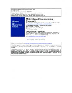
3. Coarctation of the Aorta Coarctation is a narrowing of the aorta typically just after or close to the origin of the left subclavian artery. Many coarctations are associated with “ductal” tissue that constricts the lumen of the aorta as the ductus arteriosus closes. In some newborns, severe coarctation causes immediate congestive symptoms, left ventricular failure and can be life threatening. Mild coarctation can cause progressive hypertension, left ventricular hypertrophy, and claudication. With an incidence of 1.5 to 3 per 10,000 live births, coarctation is one of the most common forms of congenital heart disease [11]. In older children, stenting of coarctation is very simple as the narrowing can be easily accessed and stented with bare metal stents from the femoral artery (Figure 1). Stenting in newborns and smaller children (90% magnesium, zirconium, yttrium, and rare earth metals), and different design with a square cross sectional shape of the strut and a reduced strut thickness of 120 𝜇m [22]. It is a premounted stent that is compatible with 6F introducer systems [9]. Research has shown that coating magnesium with biodegradable polymer is an effective method to slow magnesium’s corrosion and loss of mechanical strength [41]. A study was performed comparing the degradation under cell culture condition of magnesium to magnesium covered in different types of biodegradable polymers [42]. The high molecular weight PLLA coated magnesium had a significantly lower corrosion rate compared to uncoated magnesium, and it showed the most uniform corrosion compared to low molecular weight PLLA coating and PCL coating. They concluded high molecular weight PLLA coating is a suitable option for slowing the corrosion of magnesium [42]. Another study showed PLLA and PCL can be applied to magnesium by spin coating. This study showed low molecular weight PLLA film had better adhesion strength to magnesium than the high molecular weight PLLA or PCL but concluded that both PLLA and PCLA are promising materials for protective coating [43]. The redesign of the AMS stent from Biotronik, the AMS3 or DREAMS stent (Drug Eluting Absorbable Magnesium Scaffold), is covered with a layer of polymer to slow the degradation time and allow for Paclitaxel release (Figure 8). It is 6F compatible. The available diameters are 3 and 3.5 mm; however, it can be overexpanded up to 5 mm and still provide mechanical support [27]. During the first 3 months after implantation the polymer layer remains stable while the magnesium gradually begins to degrade. At 6 months the magnesium degradation is complete and the polymer absorption is ongoing. At 9 months the polymer is completely absorbed and structural disintegration begins. The Biosolve study is the first in-man study, and it showed improved mechanical properties with less early restenosis compared to the AMS-1 stent at 6-month follow-up [40, 44–46]. Biotronik is currently developing a second generation DREAMS stent that has an improved 6-crown 2-link design and is coated in PLLA carrying sirolimus. The new design provides greater radial stiffness and mechanical strength for a longer period of time. It also allows for increased postdilatation capabilities [27]. The radial stiffness of the 2nd generation DREAMS stent is comparable to metal stents, at 1.38 N/mm [27]. It
8 contains radio-opaque markers and elutes sirolimus [27]. A clinical trial, BIOSOLVE-II, will commence in 2013 pending preclinical results [27]. More importantly, magnesium based biocorrodable stents are already being used in select cases in Europe for the treatment of pulmonary artery stenosis and aortic stenosis in infants. There are already several published case reports describing the use of magnesium stents in the pediatric population. Zartner et al. reported the first placement of a magnesium stent in the left pulmonary (LPA) artery of a 1.7 kg preterm baby born at 26-week gestation. The LPA had been inadvertently ligated, and a 3 mm × 10 mm AMS-I stent was successfully placed in the LPA in a hybrid fashion with a surgical cut-down of the pulmonary bifurcation. At 4-month follow-up the stent had completely degraded and the left lung continued to be well perfused. Despite the baby’s small size, the stent was well tolerated without signs of local or systemic toxicity [47]. About 5 months after stent implantation the patient died from severe pneumonia. On autopsy the inner surface of the vessel was endothelialized with a smooth surface. The inner diameter of the lumen where the stent had been placed measured 3.7 mm. There were no visible or palpable pieces of the magnesium stent. Neointimal proliferation measured 100 𝜇m at its thickest, but there was no evidence of inflammatory reactions [2]. Schranz et al. reported the first use of a magnesium stent for treatment of coarctation of the aorta. A 15-dayold newborn developed severe long segment recoarctation after surgical coarctation repair. The baby was too unstable to tolerate repeat surgery. A 3.5 mm × 15 mm magnesium stent (AMS-1, Biotronik, Germany) was deployed with a 16 atm inflation which resulted in a final diameter of 4 mm. The coarctation reoccurred as the stent degraded, so a second magnesium stent was placed. Serum magnesium levels remained within normal limits even after placement of the second stent. At 3 months of age the baby had surgical closure of a ventricular septal defect. The aorta appeared widely patent at that time, but the surgeon decided to patch augment the previously stented aortic segment. For this reason no long-term follow-up is available [48]. 6.2. Iron. Iron was the first metal used to make a biocorrodable stent. Iron stents have been placed in animals; however, none have reached clinical trials. The NOR-I stent (Devon Medical, Hamburg, Germany) is a balloon expandable stent that was placed in the descending aorta of New Zealand white rabbits [49]. Commercially available tubes consisting of 99.8% iron (Goodfellow, Cambridge, UK) were laser cut with a stent design similar to a stainless steel coronary stent (PUVA-AS16). To prevent corrosion, the tubes were laser cut in a nitrogen atmosphere. They were then electropolished to a strut thickness of 100–120 𝜇m. The expanded diameter of the stents ranged from 3 to 6 mm. No significant inflammation or neointimal formation was seen, there was minimal stent recoil, and there was no in-stent stenosis or thrombus formation at 18-month follow-up [49]. At 18 months the stent struts were still intact; however, there was some sign of degradation on histological evaluation [49].
BioMed Research International A second similar iron stent was developed from 99.5% iron tubes purchased from Goodfellow Inc. (Huntingdon, UK). Additional contents of the tube included Aluminium, Calcium, Cobalt, Chromium, Copper, Mangenese, Nickel, Selenium, Carbon Phosphate, and Sulphur. The tubes were laser cut to a similar slotted tube design used in the Saxx stent (CR Bard, Tempe, AZ, USA) and were electropolished to a strut thickness of 120 𝜇m. The author’s intention was to test an iron stent that could be implanted in a pediatric patient with congenital heart disease, so a larger stent diameter was chosen. The expanded diameters of the stents ranged from 6 mm to 12 mm. They were placed in the descending aortas of swine which were followed for up to one year. Results were similar to the NOR-I stent: the stents performed well without significant recoil or in-stent stenosis. There was no more local inflammation or neointimal formation compared to traditional stainless steel stents, and histopathological examination of all major organs showed no signs of iron related toxicity or overload. At one-year follow-up, large portions of the stent remained intact [50]. There are several advantages to using iron as a material for biocorrodable stents. Its mechanical properties are similar to those of traditional stainless steel stents. Iron is radioopaque, so addition of markers to make the stent visible by fluoroscopy is not necessary. Compared to magnesiumbased alloys, iron has a higher ductility so the laser cutting is less complicated. Iron is less brittle than magnesium, so iron stents can be made with thinner struts. The interaction of iron with the body, including its transportation and storage, is well understood. These stents contain only 40 mg of pure iron, about the recommended monthly intake of iron, so it should have good biocompatibility [51]. In the published animal studies, iron stents did not cause local or systemic toxicity from corrosion products, and they did not cause significant neointimal proliferation. They also performed as well as traditional stainless steel stents without significant restenosis. The iron stents degraded very slowly; however, design modification would be needed to expedite the degradation process for use in the pediatric population. Possible modifications include using iron based alloys, thinner struts, or a stent with designed areas of weakness resulting in stent fragmentation after endothelialization [49, 50]. A stent made from an iron-manganese alloy is being developed by powder metallurgy. In vitro studies show that this alloy has good mechanical properties and degrades faster than pure iron [52]. Electroformed iron is a material that may be very suitable for a bioabsorbable stent. Electroforming is a process that uses electrodeposition to produce metallic parts. The structure is formed atom layer by atom layer, so it is ideal for creating thin walled structures with dimensional precision [53]. A study was done to evaluate the feasibility of using electroformed iron to create a bioabsorbable stent. Flat sheets of electroformed iron were created, and the mechanical properties, microstructure, and corrosion behavior of these sheets were compared to pure iron and stainless steel. The electroformed foils measured 100 𝜇m thick and yielded strength comparable to that of stainless steel. It exhibited a higher corrosion rate compared to CTT-Fe pure iron, and a lower corrosion rate compared to magnesium.
BioMed Research International
9
DREAMS crimped (3 mm nominal)
Expansion to 3 mm
(a)
(b)
Overexpansion to 5 mm
(c)
Figure 8: Biotronik DREAMS stent shown while crimped, expanded to 3 mm, and overexpanded to 5 mm.
These properties make it a good candidate for application as a biocorrodable stent material [54]. A numerical model has been developed to predict the effect of corrosion on the mechanical properties of metallic bioabsorbable stents. Metallic foils were studied, but the model is applied in such a way to allow analysis of complex three-dimensional structures. The model is able to predict the performance of a bioabsorbable metallic stent as it corrodes over time [55].
7. Conclusion There is a need for a bioabsorbable stent for use in children with congenital heart disease. We currently have absorbable stents for use in adult coronary artery disease which are not yet FDA approved. The largest expanded diameter of these stents is generally 4 mm. Treatment of coarctation of the aorta or pulmonary artery stenosis in a newborn would require a stent which can be expanded to 6–8 mm. The stent needs to be low profile to minimize vessel injury. While the bioabsorbable coronary stents are relatively low profile, a larger version of the same stent design that could be expanded to 8 mm would be higher profile. The stents need to be redesigned to maintain adequate radial strength and have a larger expanded diameter without a larger crossing profile. It needs to have the mechanical strength to prevent vessel recoil, while also absorbing in a short enough time to allow vessel growth. Development of absorbable coronary stents has led to a great understanding of the available materials and production techniques, and children with congenital heart disease will hopefully soon benefit from the current
generation of bioabsorbable and biocorrodable materials and devices.
References [1] J. A. Ormiston and P. W. S. Serruys, “Bioabsorbable coronary stents,” Circulation, vol. 2, no. 3, pp. 255–260, 2009. [2] P. Zartner, M. Buettner, H. Singer, and M. Sigler, “First biodegradable metal stent in a child with congenital heart disease: evaluation of macro and histopathology,” Catheterization and Cardiovascular Interventions, vol. 69, no. 3, pp. 443–446, 2007. [3] P. W. Serruys, J. A. Ormiston, Y. Onuma et al., “A bioabsorbable everolimus-eluting coronary stent system (ABSORB): 2-year outcomes and results from multiple imaging methods,” The Lancet, vol. 373, no. 9667, pp. 897–910, 2009. [4] N. Gonzalo and C. Macaya, “absorbable stent: focus on clinical applications and benefits,” Journal of Vascular Health and Risk Management, vol. 8, pp. 125–132, 2012. [5] Y. Onuma, J. Ormiston, and P. W. Serruys, “Bioresorbable scaffold technologies,” Circulation Journal, vol. 75, no. 3, pp. 509–520, 2011. [6] J. A. Ormiston and P. W. S. Serruys, “Bioabsorbable coronary stents,” Circulation, vol. 2, no. 3, pp. 255–260, 2009. [7] H. Eggebrecht, J. Rodermann, P. Hunold et al., “Images in cardiovascular medicine. Novel magnetic resonance-compatible coronary stent: the absorbable magnesium-alloy stent,” Circulation, vol. 112, no. 18, pp. e303–e304, 2005. [8] P. W. Serruys, H. M. Garcia-Garcia, and Y. Onuma, “From metallic cages to transient bioresorbable scaffolds: change in paradigm of coronary revascularization in the upcoming decade?” European Heart Journal, vol. 33, no. 1, pp. 16–25, 2012.
10 [9] P. Erne, M. Schier, and T. J. Resink, “The road to bioabsorbable stents: reaching clinical reality?” CardioVascular and Interventional Radiology, vol. 29, no. 1, pp. 11–16, 2006. [10] D. S. Levi and Cheng, “Biodegradable implants,” in Pediatric and Congenital Cardiology, Cardiac Surgery and Intensive Care, E. M. Dacruz, D. Ivy, V. Hraska, and J. Jagger, Eds. [11] C. A. Loffredo, “Epidemiology of cardiovascular malformations: prevalence and risk factors,” Journal of Medical Genetics, vol. 97, no. 4, pp. 319–325, 2000. [12] T. J. Forbes, D. W. Kim, W. Du et al., “Comparison of surgical, stent, and balloon angioplasty treatment of native coarctation of the aorta: an observational study by the CCISC (Congenital cardiovascular interventional study consortium),” Journal of the American College of Cardiology, vol. 58, no. 25, pp. 2664–2674, 2011. [13] S. Kaushal, C. L. Backer, J. N. Patel et al., “Coarctation of the aorta: midterm outcomes of resection with extended end-toend anastomosis,” Annals of Thoracic Surgery, vol. 88, no. 6, pp. 1932–1938, 2009. [14] J. A. Noonan, “Noonan syndrome: an update and review for the primary pediatrician,” Clinical Pediatrics, vol. 33, no. 9, pp. 548– 555, 1994. [15] D. Alagille, A. Estrada, M. Hadchouel et al., “Syndromic paucity of interlobular bile ducts (Alagille syndrome or arteriohepatic dysplasia): review of 80 cases,” Journal of Pediatrics, vol. 110, no. 2, pp. 195–200, 1987. [16] P. C. Painter and M. M. Coleman, Fundamentals of Polymer Science: An Introductory Text, CRC Press, Boca Raton, Fla, USA, 2nd edition, 2000. [17] A. G¨opferich, “Mechanisms of polymer degradation and erosion,” Biomaterials, vol. 17, no. 2, pp. 103–114, 1996. [18] A. C. Vieira, J. C. Vieira, J. M. Ferra, F. D. Magalh˜aes, R. M. Guedes, and A. T. Marques, “Mechanical study of PLA-PCL fibers during in vitro degradation,” Journal of the Mechanical Behavior of Biomedical Materials, vol. 4, no. 3, pp. 451–460, 2011. [19] D. Garlotta, “A literature review of poly(lactic acid),” Journal of Polymers and the Environment, vol. 9, no. 2, pp. 63–84, 2001. [20] J. O. Hollinger and G. C. Battistone, “Biodegradable bone repair materials. Synthetic polymers and ceramics,” Clinical Orthopaedics and Related Research, no. 207, pp. 290–305, 1986. [21] H. Tamai, K. Igaki, E. Kyo et al., “Initial and 6-month results of biodegradable poly-l-lactic acid coronary stents in humans,” Circulation, vol. 102, no. 4, pp. 399–404, 2000. [22] S. Garg and P. Serruys, “Biodegradable and non-biodegradable stents,” Minerva Cardioangiologica, vol. 57, no. 5, pp. 537–565, 2009. [23] T. Yamawaki, H. Shimokawa, T. Kozai et al., “Intramural delivery of a specific tyrosine kinase inhibitor with biodegradable stent suppresses the restenotic changes of the coronary artery in pigs in vivo,” Journal of the American College of Cardiology, vol. 32, no. 3, pp. 780–786, 1998. [24] J. A. Ormiston, P. W. Serruys, E. Regar et al., “A bioabsorbable everolimus-eluting coronary stent system for patients with single de-novo coronary artery lesions (ABSORB): a prospective open-label trial,” The Lancet, vol. 371, no. 9616, pp. 899–907, 2008. [25] Y. Onuma, P. W. Serruys, L. E. L. Perkins et al., “Intracoronary optical coherence tomography and histology at 1 month and 2, 3, and 4 years after implantation of everolimus-eluting bioresorbable vascular scaffolds in a porcine coronary artery model: An attempt to decipher the human optical coherence
BioMed Research International
[26]
[27]
[28]
[29] [30]
[31]
[32]
[33]
[34]
[35]
[36]
[37]
[38]
[39]
[40]
tomography images in the ABSORB trial,” Circulation, vol. 122, no. 22, pp. 2288–2300, 2010. P. W. Serruys, Y. Onuma, D. Dudek et al., “Evaluation of the second generation of a bioresorbable everolimus-eluting vascular scaffold for the treatment of de Novo Coronary Artery stenosis: 12-month clinical and imaging outcomes,” Journal of the American College of Cardiology, vol. 58, no. 15, pp. 1578–1588, 2011. Euro PCR Focus Group, “Bioresorbable scaffold,” March 2013, http://www.pcronline.com/PCR-focusgroup/PCR-FOCUSGROUP-ON-BIORESORBABLE-VASCULAR-SCAFFOLDSMARCH-2012. Y. Onuma and P. W. Serruys, “Bioresorbable scaffold: the advent of a new era in percutaneous coronary and peripheral revascularization?” Circulation, vol. 123, no. 7, pp. 779–797, 2011. Abbott Vascular, Temecula, California Laboratory. “Press Release- New Data Reinforce Strong Long-Term Clinical Performance of Abbott’s Absorb Bioresorbable Vascular Scaffold,” 2013, http://www.abbott.com/press-release/ new-data-reinforce-strong-longterm-clinical-performance-ofabbotts-absorb-bioresorbable-vascular.htm. R. Waksman, “Update on bioabsorbable stents: from bench to clinical,” Journal of Interventional Cardiology, vol. 19, no. 5, pp. 414–421, 2006. C. G. Pitt, M. M. Gratzl, and A. R. Jeffcoat, “Sustained drug delivery systems. II: factors affecting release rates from poly(𝜀caprolactone) and related biodegradable polyesters,” Journal of Pharmaceutical Sciences, vol. 68, no. 12, pp. 1534–1538, 1979. S.-J. Liu, F.-J. Chiang, C.-Y. Hsiao, Y.-C. Kau, and K.-S. Liu, “Fabrication of balloon-expandable self-lock drug-eluting polycaprolactone stents using micro-injection molding and spray coating techniques,” Annals of Biomedical Engineering, vol. 38, no. 10, pp. 3185–3194, 2010. R. Jabara, N. Chronds, and K. Robinson, “Novel bioabsorbable salicylate-based polymer as a drug-eluting stent coating,” Catheterization and Cardiovascular Interventions, vol. 72, no. 2, pp. 186–194, 2008. P. W. Serruys, H. M. Garcia-Garcia, and Y. Onuma, “From metallic cages to transient bioresorbable scaffolds: change in paradigm of coronary revascularization in the upcoming decade?” European Heart Journal, vol. 33, no. 1, pp. 16–25, 2012. G. Colotti, A. Ilari, A. Boffi, and V. Morea, “Metals and metal derivatives in medicine,” Mini Reviews in Medicinal Chemistry, vol. 13, no. 2, pp. 211–221, 2013. M. Watanabe, A. Shinohara, T. Matsukawa et al., “Chronic magnesium deficiency decreases tolerance to hypoxia/reoxygenation injury in mouse heart,” Life Sciences, vol. 88, no. 15-16, pp. 658–663, 2011. B. Heublein, R. Rohde, V. Kaese, M. Niemeyer, W. Hartung, and A. Haverich, “Biocorrosion of magnesium alloys: a new principle in cardiovascular implant technology?” Heart, vol. 89, no. 6, pp. 651–656, 2003. K. Sternberg, M. Gratz, K. Koeck et al., “Magnesium used in bioabsorbable stents controls smooth muscle cell proliferation and stimulates endothelial cells in vitro,” Journal of Biomedical Materials Research B, vol. 100, no. 1, pp. 41–50, 2012. P. Barlis, J. Tanigawa, and C. Di Mario, “Coronary bioabsorbable magnesium stent: 15-Month intravascular ultrasound and optical coherence tomography findings,” European Heart Journal, vol. 28, no. 19, p. 2319, 2007.
BioMed Research International [41] J. E. Gray-Munro, C. Seguin, and M. Strong, “Influence of surface modification on the in vitro corrosion rate of magnesium alloy AZ31,” Journal of Biomedical Materials Research A, vol. 91, no. 1, pp. 221–230, 2009. [42] L. Xu and A. Yamamoto, “In vitro degradation of biodegradable polymer-coated magnesium under cell culture condition,” Applied Surface Science, vol. 258, no. 17, pp. 6353–6358, 2012. [43] L. Xu and A. Yamamoto, “Characteristics and cytocompatibility of biodegradable polymer film on magnesium by spin coating,” Colloids and Surfaces B, vol. 93, pp. 67–74, 2012. [44] R. Waksman, R. Erbel, C. Di Mario et al., “Early- and longterm intravascular ultrasound and angiographic findings after bioabsorbable magnesium stent implantation in human coronary arteries,” Journal of the American College of Cardiology, vol. 2, no. 4, pp. 312–320, 2009. [45] Biotronik Press Release, “BIOTRONIK Announces Positive 6-Month Results for DREAMS, the Pioneering Drug-Eluting Absorbable Metal Scaffold,” 2011, http://www.biotronik.com/ wps/wcm/connect/int web/biotronik/newsroom/press releases?p=http://www.biotronik.com/wps/wcm/connect/int web/biotronik/newsroom/press releases/press release biosolve i&pw=770&pt. [46] M. Haude, R. Erbel, P. Erne et al., “Safety and performance of the drug-eluting absorbable metal scaffold (DREAMS) in patients with de-novo coronary lesions: 12 month results of the prospective, multicenter first-in-man BIOSOLVE-1 trial,” The Lancet, vol. 381, pp. 836–844, 2013. [47] P. Zartner, R. Cesnjevar, H. Singer, and M. Weyand, “First successful implantation of a biodegradable metal stent into the left pulmonary artery of a preterm baby,” Catheterization and Cardiovascular Interventions, vol. 66, no. 4, pp. 590–594, 2005. [48] D. Schranz, P. Zartner, I. Michel-Behnke, and H. Akint¨urk, “Bioabsorbable metal stents for percutaneous treatment of critical recoarctation of the aorta in a newborn,” Catheterization and Cardiovascular Interventions, vol. 67, no. 5, pp. 671–673, 2006. [49] M. Peuster, P. Wohlsein, M. Br¨ugmann et al., “A novel approach to temporary stenting: degradable cardiovascular stents produced from corrodible metal - Results 6-18 months after implantation into New Zealand white rabbits,” Heart, vol. 86, no. 5, pp. 563–569, 2001. [50] M. Peuster, C. Hesse, T. Schloo, C. Fink, P. Beerbaum, and C. von Schnakenburg, “Long-term biocompatibility of a corrodible peripheral iron stent in the porcine descending aorta,” Biomaterials, vol. 27, no. 28, pp. 4955–4962, 2006. [51] M. Auerbach and H. Ballard, “Clinical use of intravenous iron: administration, efficacy, and safety,” Hematology, vol. 2010, pp. 338–347, 2010. [52] H. Hermawan, H. Alamdari, D. Mantovani, and D. Dub´e, “Iron-manganese: new class of metallic degradable biomaterials prepared by powder metallurgy,” Powder Metallurgy, vol. 51, no. 1, pp. 38–45, 2008. [53] H. D. Merchant, W. C. Liu, L. A. Giannuzzi, and J. G. Morris, “Grain structure of thin electrodeposited and rolled copper foils,” Materials Characterization, vol. 53, no. 5, pp. 335–360, 2004. [54] M. Moravej, F. Prima, M. Fiset, and D. Mantovani, “Electroformed iron as new biomaterial for degradable stents: development process and structure-properties relationship,” Acta Biomaterialia, vol. 6, no. 5, pp. 1726–1735, 2010.
11 [55] J. A. Grogan, B. J. O’Brien, S. B. Leen, and P. E. McHugh, “A corrosion model for bioabsorbable metallic stents,” Acta Biomaterialia, vol. 7, no. 9, pp. 3523–3533, 2011.